This document provides an overview of pancreatitis, including its epidemiology, pathophysiology, etiology, clinical presentation, workup, severity scoring systems, treatment, prognosis, and complications. It defines acute and chronic pancreatitis and describes the reversible inflammation of the pancreas that occurs in acute pancreatitis. Key points include that the annual incidence is 13-45 per 100,000 people, the pathophysiology involves premature activation of digestive enzymes within the pancreas rather than the intestines, and treatment depends on the severity but generally involves IV rehydration and pain management for mild cases and more aggressive monitoring and support in an ICU for severe cases.
3. Introduction
• Inflammation in pancreas associated with
injury to exocrine parenchyma.
• Types:
1. Acute: Emergency condition.
2. Chronic: Prolonged & frequently lifelong
disorder resulting from the development of
fibrosis within the pancreas
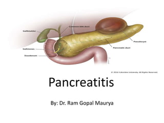
4. Acute Pancreatitis
• Definition:
Acute condition of diffuse pancreatic
inflammation & autodigestion, presents with
abdominal pain, and is usually associated with
raised pancreatic enzyme levels in the blood &
urine.
• Reversible inflammation of the pancreas
5. Epidemiology
• The annual incidence ranges from 13–
45/100,000 persons.
• Acute pancreatitis results in >250,000
hospitalizations per year.
• higher among males than females.
• It may occur at any age, peak incidence is
between 50 and 60 years.
6. Pathophysiology
• Pathologically, acute pancreatitis varies from interstitial
pancreatitis (pancreas blood supply maintained), which
is generally self-limited to necrotizing pancreatitis
(pancreas blood supply interrupted).
• Autodigestion is a currently accepted pathogenic
theory; according to this theory, pancreatitis results
when proteolytic enzymes (e.g., trypsinogen,
chymotrypsinogen, proelastase, and lipolytic enzymes
such as phospholipase A2) are activated in the
pancreas acinar cell rather than in the intestinal lumen.
7.
8. • A number of factors (e.g., endotoxins,
exotoxins, viral infections, ischemia, oxidative
stress, lysosomal calcium, and direct trauma)
are believed to facilitate premature activation
of trypsin. Activated proteolytic enzymes,
especially trypsin, not only digest pancreatic
and peripancreatic tissues but also can
activate other enzymes, such as elastase and
phospholipase A2.
10. Clinical Presentation
• Symptoms: Upper Abdominal pain, sudden
onset, sharp, severe, continuous, radiates to
the back, reduced by leaning forward.
• Patient lies very still.
• Nausea, non-projactile vomiting, retching
• Anorexia
• Fever, weakness
11. ABDOMEN PAIN-Cardinal Symptom
• SITE: usually experienced first in the epigastrium – but may be localized to
either upper quadrant or felt diffusely throughout the abdomen or lower
chest
• ONSET: characteristically develops quickly, generally following substantial
meal and precedes N&V
• SEVERITY: frequently severe, reaching max. intensity within minutes
rather than hours
• NATURE: “boring through”, “knifing” (illimitable agony)
• DURATION: hours-days
• COURSE: constant (refractory to usual doses of analgesics, not relieved by
vomiting)
• RADIATION: directly to back(50%), chest or flanks
• RELEIVING FACTOR: sitting or leaning/stooping forward (MUHAMMEDAN
PRAYER SIGN) – due to shifting forward of abdominal contents and taking
pressure off from inflamed pancreas
• AGGRAVATING FACTOR: food/alcohol intake, walking, lying supine
12. General Physical Examination
• Appearance: well to critically ill with profound
shock, toxicity and confusion
• Vitals: Tachypnea(and dyspnea-10%),
Tachycardia(65%), Hypotension, temp :
high(76%)/normal/low
• Icterus(28%) – gallstone pancreatitis or due to
edema of pancreatic head
• Cyanosis – Improper lung perfusion
• Pallor, cold clammy skin,diaphoretic
13. ABDOMEN EXAMINATION
• Tenderness + Rebound tenderness: – epigastrium/upper
abdomen.
• Distension: – Ileus(BS decreased or absent) – ascites
with shifting dullness.
• Guarding(also called “defense musculaire” )-upper
abdomen – tensing of the abdominal wall muscles to
guard inflamed organs within the abdomen from the
pain of pressure upon them(i.e. during palpation).
• Rigidity(involuntary stiffness)-unusual – Tensing of the
abdominal wall muscles to guard inflamed organs even
if patient not touched.
14. Cutaneous Ecchymosis(1 % cases)
• Acute Hemorrhagic Necrotizing/fulminant
Pancreatitis--Periperitoneal/retroperitoneal
Hemorrhage.
• Methemalbumin formed from digested blood
tracks around.
• GREY TURNER’S SIGN
• CULLEN’S SIGN
• FOX SIGN
15. GREY TURNER’S SIGN
hemorrhagic spots and ecchymosis in flanks, ruddy erythema in flanks
due to extravasated pancreatic exudate.
20. HEMATOLOGICAL investigations
Blood tests:
Complete Blood Count
Serum amylase & lipase
C-reactive Protein
Serum electrolytes
Blood glucose
Renal Function Tests
Liver Function Tests
LDH
Coagulation profile
Arterial Blood Gas Analysis
21. Serum Amylase
• Sensitivity: 72% Specificity: 99%
• Released within 6-12 hours of the onset, &
Remains elevated for 3-5 days.
• Elevation ˃ 3X normal is
significant.
• Undergoes renal clearance.
After its serum levels decline,
its urinary level remains elevated.
• Its level doesn't correlate with
the disease activity
22. Serum Lipase
• More pancreatic-specific than s. Amylase.
• Sensitivity: about 100% Specificity: 96%
• Remains elevated longer than amylase (up to
week).
• Useful in patients presenting late to the
physician.
• S. Amylase tends to be higher in gallstone
pancreatitis.
• S. Lipase tend to be higher in alcoholic
pancreatitis.
23.
24. • Patients with more severe disease may show
hemoconcentration with hematocrit values
>44% and/or prerenal azotemia with a blood
urea nitrogen (BUN) level >22 mg/dL resulting
from loss of plasma into the retroperitoneal
space and peritoneal cavity.
• Hyperglycemia is common d/t including
decreased insulin release, increased glucagon
release, and an increased output of adrenal
glucocorticoids and catecholamines.
• Hypocalcemia occurs in ~25% of patients.
25. • Hyperbilirubinemia (serum bilirubin >68
mmoL or >4.0 mg/dL) occurs in ~10% of
patients.
• Hypertriglyceridemia occurs in 5–10% of
patients.
• Approximately 5–10% of patients have
hypoxemia (arterial PO2≤60 mmHg), which
may herald the onset of ARDS.
26. Imaging Investigations
• Plain erect chest X-ray: not diagnostic on
pancreatitis, but to rule out other D/D
• Pleural effusion, diffuse alveolar infiltrate
(ARDS)
• Pleural effusion, common
on left side.
31. CT Scan
Not indicated in every patient, only in:
1. Diagnostic uncertainty.
2. Severe acute pancreatitis.
3. Clinical deterioration, with multi-organ
failure, sepsis, progressive deterioration.
4. Local complications occurs (fluid collection,
pseuodocyst, pseudo-aneurysm).
32. • Axial CT Scan: Peripancreatic stranding
(arrow). Multiple gallstones in the gallbladder
33. Contrast-enhanced CT: acute necrotising
pancreatitis. Pancreatic area of reduced
enhancement, peripancreatic edema and
stranding of the fatty tissue
34.
35. Magnetic Resonant
Cholangiopancreatography
• INDICATION:
– diagnosis of suspected biliary and pancreatic duct
obstruction in the setting of pancreatitis.
– Repeated attacks of idiopathic acute pancreatitis
• Merit
– used if choledocholithiasis is suspected but there is
concern that pancreatitis might worsen is ERCP is
performed
– Provide non-invasive/fast/safe high-quality (Heavily T2–
weighted) imaging for diagnostic and/or severity purposes
37. Endoscopic Retrograde
Cholangiopancreatography
INDICATION
Severe gallstone AP or AP with concurrent acute
cholangitis/biliary obstruction/ biliary
sepsis/jaundice (due to persistent stone)
NOT INDICATED
• Not needed early in most patients with gallstone
pancreatitis who lack laboratory or clinical
evidence of ongoing biliary obstruction
• As most of gallstones causing AP readily pass to
duodenum and are lost in stool
• MRCP /EUS as accurate as diagnostic ERCP
41. MISCELLANEOUS
• Peritoneal(sensitivity 54%,specificity
93%)/Pleural fluid tap – High
amylase/lipase/protein
• Urine Complete Examination – Proteinuria,
granular cast, glycosuria
• Electrocardiography – ST-T wave changes
• Bile Aspiration – Crystal analysis, if suspicion
of microlithiasis
42.
43.
44.
45. SEVERITY SCORING SYSTEMS
• Ranson score
• Glagsow score
• Bedside Index for Severity in Acute
Pancreatitis(BISAP) score
• CT severity index
• Acute Physiology And Chronic Health
Evaluation(APACHE) II score
46. Each system has merits and demerits, and none is
currently recognized as a criterion standard.
Although amylase/lipase are used in diagnosing
pancreatitis, they are NOT use for predicting severity
of disease
47. RANSON SCORE-1974 (for alcohol
pancreatitis)
ON ADMISSION
• Age > 55 yrs
• TLC > 16,000/mm3
• BSR> 200 mg/dL
• AST > 250 IU/L
• LDH > 350 IU/L
WITHIN 48 HOURS
• BUN rise >5 mg/dL
• Pa02 < 60 mmHg ( 8 KPa)
• Serum Calcium < 8 mg/dL
• Base deficit > 4 meq/L
• Fluid Sequestration > 6000
mL
• Hct fall > 10 %
NOTE: Disease classified as SEVERE when 3 or more factors are
present
48. Revised RANSON SCORE-1979 (for
Gallstone pancreatitis)
ON ADMISSION
• Age > 70 years
• TLC > 18000/mm3
• BSR > 220 mg/dL
• AST> 250 IU/L
• LDH >400 IU/L
WITHIN 48 HOURS
• BUN rise >5 mg/dL
• Pa02 < 60 mmHg ( 8 KPa)
• Serum Calcium < 8 mg/dL
• Base deficit > 5 meq/L
• Fluid Sequestration > 4 litres
• Hct fall > 10 %
NOTE: Disease classified as SEVERE when 3 or more factors are
present
49. RANSON SCORE
Ranson
score
Mortality rate SEVERITY Interpretation
0-2 ≈ 0-2 %i.e. minimal mortality Mild AP Admit in regularward
3-5 10-20 %
SevereAP
Admit in ICU/HDU
6-7 40 % Associated with moresystemic
complications
>7 >50% Sameas above
50. Glasgow-Imrie score
ON ADMISSION
• Age > 55 yrs
• TLC > 15 x 109 l-1
• BSR>180 mg/dL (10 mmol l-
1) (no H/O diabetes)
• BUN > 16 mmol l-1 (no
response to IV fluids)
• Pa02 < 60 mmHg ( 8 KPa)
WITHIN 48 HOURS
• Serum Calcium < 2.0 mmol
l-1
• Serum albumin <32 g l-1
• LDH > 600 units l-1
• AST/ALT > 200 units l-1
NOTE: Disease classified as SEVERE when 3 or more factors are
present
51. Modified Glasgow/PANCREAS score
• PaO2 < 8kPa (60mmhg)
• Age > 55 years
• Neutrophils: WBC >15 x109/l
• Calcium < 2mmol/l
• Renal function: (Urea > 16mmol/l)
• Enzymes: (AST/ALT > 200 iu/L or LDH > 600 iu/L)
• Albumin < 32g/l
• Sugar: (Glucose >10mmol/L)
• *Applicable for both gallstone and alcohol induced
pancreatitis within 48 hours of admission
• *Omission of age/serum transaminase increases the
predictive valve of scoring system as serum
transaminase did not differ significantly between mild
and severe pancreatitis.
54. • NOTE: Disease classified as SEVERE if clinical impression of
very ill patient with APACHE II score above 8
55. APACHE II SCORE
MERIT
• Immediate assessment of the severity of
pancreatitis possible.
• Can be used even after 48 hours.
• Unlike ALL pancreatic specific scoring systems,
APACHE (and SOFA) also includes clinical features
of patient besides laboratory values.
(Clinical findings are more important than lab
findings in predicting SIRS,sepsis and other
complications)
56. MANAGEMENT
• Mild acute pancreatitis
– Conservative Approach
– Admit in general ward
– Non invasive monitoring
• (Moderate)Severe acute pancreatitis
– Aggressive Approach
– Admit in HDU/ICU
– Invasive monitoring
__________________________________________________
Recognizing patients with severe acute pancreatitis ASAP is
critical for achieving optimal outcomes
57. Mild Acute Pancreatitis
• mild and self-limiting, needing only brief hospitalization.
• Rehydration by IV fluids
• Frequent non-invasive observation/monitoring(atleast 8 hrly)
• Brief period of fasting till pain/vomiting settles – Little physiological
justification for prolonged NPO
• No medication required other than analgesics(important) and anti-emetics
– Antibiotics not indicated in absence of signs or documented sources of
infection
– Pain results in ongoing cholinergic discharge, stimulating gastric and
pancreatic secretions
– Analgesics: Avoid Morphine-cause sphincter of Oddi spasm
• Metabolic support – Correction of electrolyte imbalance
58. Severe Acute Pancreatitis
• P:– Pain relief
– Proton pump inhibitors-
omeprazole
– Peritoneal lavage
• A:– Admit in HDU/ICU
– Antibiotics
• N:– Nasogastric intubation
– Nasal oxygen
– Nutrition support
• C:– Calcium gluconate
– CVP line
– Catherisation- Foley
• R:– Rehydration by IV
fluids,plasma,blood
– Ranitidine(for stress ulcer)
– Radiology: CT scan, USG
– Resuscitation when required
• E:-Endotracheal intubation
– Electrolytes managemen
– ERCP
• A – Antacids
• S:– Suction-in case of aspiration
– Steroids in case of ARDS
– Supportive therapy for organ
failure • Inotropes •
Hemofiltration •
Ventilator(PEEP)
59. ACG 2013 Recommendations
• Despite dozens of randomized trials, no
medication has been shown to be effective in
treating AP.
• However, an effective intervention has been
well described: EARLY AGRESSIVE IV
hydration.
60. • Rationale for EARLY AGRESSIVE IV hydration
• Frequent hypovolemia due to
– vomiting,
– reduced oral intake,
– third spacing of fluids(increased vascular
permeability)
– increased respiratory losses, and
– diaphoresis.
61. Kon
sa?
Lactated Ringer ’s solution may be the preferred isotonic crystalloid
replacement fluid
•Ringer lactate is better electrolyte balance and more pH-balanced
•Normal saline given in large volumes may lead to the development of a
non-anion gap, hyperchloremic metabolic acidosis and increased
chances of SIRS
•Low pH activates the trypsinogen, makes the acinar cells more
susceptible to injury and increases the severity of established AP
Kab? Early aggressive IV hydration is most beneficial during the first 12 – 24 h,
and may have little benefit beyond this time period
Kitna? Aggressive hydration, defined as 250 – 500 ml per hour of isotonic
crystalloid solution should be provided to all patients, unless
cardiovascular, renal, or other related comorbid factors exist.
• In a patient with severe volume depletion, manifest as hypotension and
tachycardia, more rapid repletion (bolus) may be needed
• Fluid requirements should be reassessed at frequent intervals within 6
h of admission and for the next 24 – 48 h.
EARLY AGRESSIVE IV hydration
62. Antibiotics
• Routine use* NOT recommended(ACG 2013) as –
Prophylaxis in severe AP – Preventive measure in
sterile necrosis to prevent development of
infected necrosis
• Indicated in
– Established infected pancreatic necrosis or
– Extraperitoneal infections
-- Cholangitis,
-- catheter-acquired infections, bacteremia, UTIs,
pneumonia
63. • As SIRS may be indistinguishable from sepsis
syndrome, so if infection is suspected, antibiotics
should be given while source of infection is being
investigated – Once blood and other cultures are
found negative, antibiotics should be
discontinued.
• Few antibiotics penetrate due to consistency of
pancreatic necrosis – cefuroxime, or imipenem,
or ciprofloxacin plus metronidazole.
64. • Relatively stable patients with infected
necrosis can be managed conservatively on
antibiotics without needing
surgery(necrosectomy) or intervention
(percutaneous or endoscopic drainage)
– Surgical debridement recommended if no
response to conservative treatment or
deteriorates clinically
65. Nutrition
• In mild AP
– oral feedings can be started immediately if there is no
nausea/vomiting, and the abdominal pain/tenderness/Ileus has
resolved(amylase return to normal, patient feel hunger).
– Initiation of feeding with a small and slowly increasing low-fat
(low-protein) soft diet appears as safe as a clear liquid diet,
providing more calories.
-- Stepwise manner increase from clear liquids to soft diet NOT
necessary.
• In severe AP
– Enteral route is recommended to prevent infectious complications
– Parenteral nutrition should be avoided, unless enteral route is not
available, not tolerated, or not meeting caloric requirements
66. RATIONALE OF EARLY ENTERAL
NUTRITION
• The need to place pancreas at rest until complete
resolution of AP no longer recommended
– Bowel rest associated with intestinal mucosal atrophy and
bacterial translocation from gut and increased infectious
complications
• Early enteral feeding maintains the gut mucosal
barrier, prevents disruption, and prevents
translocation of bacteria that seed pancreatic
necrosis
– Decrease in infectious complications, organ failure and
mortality
67. Rather than using antibiotics to prevent infected
necrosis………….start early enteral feeding to
prevent translocation of bacteria
68. Route of enteral Nutrition
• Traditionally nasojejunal route has been
preferred to avoid the gastric phase of
stimulation BUT
– Nasogastric route appears comparable in
efficacy and safety
MERITS OF NASOGASTRIC ROUTE DEMERITS OF NASOGASTRIC ROUTE
NG tube placement is far easier than
nasojejunal tube placement( requiring
interventional radiology or endoscopy,
thus expensive) especially in HDU/ICU
setting
Slight increased risk of aspiration
(Can be overcome by placing patient in
upright position and be placed on
aspiration precautions)
69. FEEDING REGIMEN
• The jejunum does not have the reservoir capacity of
the stomach, so jejunal feedings should be advanced
more slowly than gastric feedings
• The diluting effect of gastric secretions is also lost in
the jejunum, so isotonic feeding formulas are preferred
to hypertonic formulas
• Standard (polymeric) tube feedings can be used for
jejunal feedings, but in patients with diarrhea,
elemental feeding formulas may be preferred.
(Elemental formulas are low in fat, and the protein is
available as individual amino acids, which are
presumably easier to digest.)
70. Role of Surgery in AP
• In case of mild gallstone AP, cholecystectomy
should be performed before discharge to prevent
a recurrence of AP.
• In case of necrotizing biliary AP, in order to
prevent infection, cholecystectomy is to be
deferred until active inflammation subsides and
fluid collections resolve or stabilize.
• If patient unfit for surgery(comorbid/elderly),
biliary sphincherotomy alone may be effective to
reduce further attacks of AP.
71. When to Discharge
• Pain is well controlled with oral analgesia
• Able to tolerate an oral diet that maintains
their caloric needs, and
• all complications have been addressed
adequately