
Recomendados
Más contenido relacionado
La actualidad más candente
La actualidad más candente (20)
Destacado
Destacado (16)
Similar a Neurology s2010
Similar a Neurology s2010 (20)
Más de mchibuzor
Más de mchibuzor (20)
Último
Último (20)
Neurology s2010
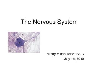
- 1. The Nervous System Mindy Milton, MPA, PA-C July 15, 2010
- 2. Neurological Anatomy • Neurons – Functional units of the nervous system • Fuel source is glucose • Does not require insulin for cellular uptake • Structure – Cell body: contains nucleus, neurofilaments, and neurotubules. Mitochondria active due to high energy needs, contain Nissl bodies that are responsible for the grey color of the neuron tissue – Dendrites: fine highly branched structures that are responsible for receiving information and transmitting to cell body – Axon: long projection that is capable of propagating an action potential
- 3. • Neurons – Neuronal structure • Anaxonic: small with no anatomical clues to identify dendrite from axon • Unipolar: single dendrite and axon that are essentially fused with cell body that lies off to the side • Bipolar: two distinct processes ie axon and dendrite • Multipolar: have two or more dendrites and a single axon
- 4. NEURON TYPES
- 5. Types of neuronal structure
- 7. Neuroglia Oligodendrocytes Astrocyte Ependymal Microglia Cells cells
- 8. Neurological Anatomy • Synapse – Definition: specialized site where the neuron communicates with another cell – Components • Pre-synaptic cell: neuron that sends the message • Post- synaptic cell: neuron or muscle cell that receives a message • Neurotransmitters: chemical substances that are released by the pre-synaptic cell and cause a change in electrical activity of the post-synaptic cell i.e stimulation of an impulse
- 11. NEUROMUSCULAR JUNCTION • Anatomy: – Axon terminal contains large numbers of mitochondria that are important in the synthesis of acetylcholine – Acetylcholine is stored in multiple vesicles – Synaptic cleft: area between the axon and the muscle fiber – Sub neural cleft: invagination of the muscle fibers that increases surface area
- 13. Neurotransmitters These have either excitatory functions, inhibitory or both • Acetylcholine • Norepinephrine • Serotonin • Dopamine • Histamine • Gamma aminobutyric acid (GABA) • Glycine • Glutamate and Aspartate • Endorphin • Substance P
- 14. Cerebral Cortex • General Principles – Each cerebral cortex receives information from and sends motor commands to the opposite side of the body – Two hemispheres have different functions – Assignment of specific functions to a specific region of the brain is imprecise at this time
- 17. Pre-Frontal Function • Alertness and orientation • Mood • Attention • Perception • Memory • Thought content • Thought process • Insight • Judgment • Language
- 18. Examination of pre-frontal function • Observation of alertness, dress, hygiene, manor. • Coma scale, e.g. Glascow • General conversation and history-taking • Mental status examinations • Special scales, e.g., depression, alcohol
- 19. CEREBRUM • FRONTAL LOBE • PARIETAL LOBE – Contains somatic motor – Contains somatic sensory areas areas • Voluntary • Primary • Premotor area • Association • Eye fields – Important in the interpretation of sensory – Important in the control data of body movements – Loss is associated with – Loss is associated with difficulty recognizing inability to direct and objects, forms, or having a program movements sense of body parts
- 20. Homunculus
- 22. CEREBRUM • TEMPORAL LOBE – Hearing and speech is located in the dominant hemisphere – Responsible for the interpretation and understanding of speech – Major area of long term memory storage – “General Interpretative or Wernicke’s area” – Vestibular sense
- 23. CEREBRUM • OCCIPITAL LOBE – Vision and the visual interpretative area • CORPUS CALLOSUM – Connects the two hemisphere’s and helps coordinate activities between them. Transfers learned descrimination, experiences, and memories
- 24. Neurological Anatomy • Cerebral Cortex – Association areas: storage, analysis and interpretation of sensory data • Somatic association area • Visual association area • Auditory association area
- 25. Neurological Anatomy • Integrative Areas – Integrate information from multiple association areas and direct complex motor activity – General Interpretive Area: (Wernike’s Area) • Present in the left hemisphere • Receives information from all of the sensory association areas • Provides the ability to interpret what is seen and heard, coordinates access to complex visual and auditory memories – Broca’s area: • Motor neurons in the general interpretative area that coordinate the activity of the respiratory, pharyngeal, and muscles of the tongue, cheeks, and jaws
- 26. Anatomy
- 27. CEREBRUM • BASAL GANGLION – Exerts regulating, controlling influences on motor integration – Numerous pathways from the motor cortex – Dopamine and GABA found here and are responsible for inhibition of tone
- 28. DIENCEPHLON • THALAMUS – Receives sensory information for somatic senses, taste, hearing, and relays to cerebral cortex – System of connecting nuclei • HYPOTHALAMUS – Temperature regulation, thirst center, ADH release – Behavior: affective nature of sensory stimuli – Hormonal control
- 29. BRAIN STEM • MIDBRAIN – Located between the Pons and diencephalon – Contains both motor and sensory pathways – Contains the nuclei for the third and fourth cranial nerves • MEDULLA – Located between the Pons and spinal cord – Center for vegetative functions – Cranial nerve tracts for 8,9,10,11,12
- 31. CEREBELLUM • CEREBELLUM – Important in synchronization of muscle movement – Monitors and makes corrective movements – Receives information from the motor and sensory cortex, and peripheral sensory receptors – Serves as feedback regulator – “Damping” of movement
- 32. Neurological Anatomy: CSF • Produced by the choroid plexus in the lateral, third and fourth ventricles • Flow pathway – Lateral ventricle – Foramen of Monroe – Third ventricle – Aqueduct of Sylvius – Fourth ventricle – Foramina of Luschka, Foramen of Magendi
- 33. CSF • Allows passage of nutrients between the blood and extracellular fluid of the brain • Reabsorbed by the arachnoid villi • Blood brain barrier: selective ability for substances to enter the brain through the capillaries of the choroid plexus.
- 34. CSF • Cerebral Spinal Fluid – Reabsorbed by the arachnoid villa secondary to pressure gradient – Arachnoid villa act as one way valve that moves CSF fluid out into blood – Obstruction of flow of CSF fluid will increase pressure within cranial cavity. – Hydrocephalus is the result of increased CSF
- 35. Hydrocephalus
- 36. MENINGES • Dura mater: tough outer meningeal membrane that lies directly below the skull. The epidural space lies between the dura and the skull • Arachnoid: middle meningeal membrane with tissue arranged in a web like fashion. Cross the subdural space and reabsorb CSF
- 37. MENINGES • PIA MATER: is a vascular thin membrane that covers the brain and the spinal cord – Vessels pass between the pia and the arachnoid through the subarachnoid space
- 39. Spinal Cord • Spinal Cord Nerves – Vertebrae • 8 cervical • 12 thoracic • 5 lumbar • 5 Sacral • 1 coccygeal – Divided into sensory and motor pathways
- 40. MOTOR SYSTEM • SPINAL CORD – Automatic reflex control – Thirty one pairs of spinal nerves with a sensory and motor root – Anterior Horn cells are large myelinated motor neurons that terminate on skeletal muscle
- 41. Spinal Cord View
- 42. SENSORY SYSTEM • Afferent transmission through posterior (dorsal) horns of the spinal cord • Sensory tracts – Spinothalamic • Small myelinated/non-myelinated fibers • Poorly localized sensation of crude touch, pain, temperature • Fibers synapse quickly on entering the spinal cord and cross over to the opposite side
- 43. SENSORY TRACTS • POSTERIOR COLUMN – Larger fibers that are myelinated with increased degree of spatialization – Carries fibers for touch requiring localization, vibratory sense, and position sense – Travels up same side of cord and synapses with second order neuron that travels to thalamus – Well spatialized: organized for increased ease of sensory interpretation and localization
- 44. • Sensory Pathways • Pain and temperature • Cross at Cord level. • Touch crosses at • Medulla
- 45. Neuro Anatomy: Sensory Tracts
- 46. Neuro Anatomy: Sensory Tracts
- 47. MOTOR SYSTEM • MOTOR CORTEX – Initiation of the pyramidal tract in the large Betz cells within the motor cortex – Impulses sent down to the motor tracts with collateral messages to the cerebellum, basal ganglia, and reticular activating system
- 48. Motor pathways • Lower motor • Upper motor – Pyramidal • Fine discrete – Extrapyramidal • Coordination- cerebellar • Tone- mid-brain
- 49. MOTOR TRACTS • CORTICOSPINAL - PYRAMIDAL – UMN Fibers pass from the cell body of the Betz cell in the motor cortex through the brain stem down the opposite side of the cord – Responsible for fine discrete motor movement – Synapse with inter-neurons or anterior horn cells in the spinal cord – Necessary for voluntary movement – Excitatory in nature
- 50. MOTOR TRACTS • BASAL GANGLIA - EXTRAPYRAMIDAL – All tracts outside the corticospinal system – Controls body tone and gross body movements • Cerebellum – Sensory and motor input – Controls posture and coordination
- 51. Motor Pathways • Most cross at Medulla in pyramid-shaped pathways. – Some don’t cross So whole tract is called Pyramidal tract.
- 52. Neuro Anatomy: Motor Tracts
- 54. Test Motor pathways • DTRs • Active range of motion (include CN III, IV, VI, VII, XI, XII) • Gait • Romberg • Coordination
- 55. Blood Supply: CNS • General Concepts – 20% of cardiac output per minute – Blood supply controlled largely by changes in the level of carbon dioxide • Arterial Blood Flow – Internal carotids enter through the skull, branch into the anterior and middle cerebral arteries – Vertebral arteries: originate from the subclavian, enter the skull through the foramen magnum, join to form the basilar artery, that will then divide and form the posterior cerebral arteries
- 56. Blood Supply: CNS • Circle of Willis – Formed by posterior cerebral arteries, posterior communicating arteries, internal carotid arteries, anterior cerebral arteries, and anterior communicating arteries – Ring shaped anastomosis allows for protection of blood flow interruption from the vertebral or carotid arteries
- 60. Peripheral Nervous System • Two Components – Spinal and cranial nerves – Autonomic Nervous system • Spinal Nerves – Mixed nerves as they contain both sensory and motor neurons – Divide and form posterior and anterior rami – Anterior rami then form plexuses • Brachial plexus: C 5-8 and T1 innervates the nerves of the arm, wrist, and hand • Lumbar plexus: L2-4 and sacral plexus: L5-S5 innervate the anterior and posterior portions of the lower body – Posterior rami form dermatomes that are distributed to specific areas of the body – Cranial Nerves: Most are mixed nerves that arrive from nuclei in the brain or brain stem
- 61. Neurological Anatomy • Peripheral Nervous System – All nervous tissue outside of the central nervous system – Important in the delivery of sensory data to the CNS and transmission of motor commands from the CNS – Two divisions • Afferent: brings sensory data into CNS • Efferent: transmits motor information from CNS – Somatic nervous system: skeletal muscles – Autonomic nervous system: visceral motor system
- 62. Sensory System • Smell • Vision • Hearing • Taste • Touch • Pain • Temperature • Pressure • Position • Vibration
- 63. Testing the Sensory System • Cranial nerves I, II, V, VII, VIII, IX, X • Body sensory touch, pain, temperature • DTRs • Vibratory • Position • Romberg
- 66. Optic Nerve • The visual signals of the right half of both eyes goes to left visual cortex
- 75. Lower Motor Neuron Pathway
- 76. Reflexes • Test sensory perception • Sensory pathway • Sensory-motor connection • Motor nuclei • Motor pathway • Muscle function • Neurotransmitters. • Flexible joint
- 77. Autonomic system • Sympathetic • Parasympathetic • Test largely by history • Note skin temp, moisture, • Vital signs • Pupillary response to light, accommodation
- 80. The End
Notas del editor
- \n
- \n
- \n
- \n
- \n
- \n
- \n
- \n
- \n
- \n
- \n
- \n
- \n
- \n
- \n
- \n
- \n
- \n
- \n
- \n
- \n
- \n
- \n
- \n
- \n
- \n
- \n
- \n
- \n
- \n
- \n
- \n
- \n
- \n
- \n
- \n
- \n
- \n
- \n
- \n
- \n
- \n
- \n
- \n
- \n
- \n
- \n
- \n
- \n
- \n
- \n
- \n
- \n
- \n
- \n
- \n
- \n
- \n
- \n
- \n
- \n
- \n
- \n
- \n
- \n
- \n
- \n
- \n
- \n
- \n
- \n
- \n
- \n
- \n
- \n
- \n
- \n
- \n
- \n
- \n