SlideShare una empresa de Scribd logo
1 de 26
.Geographic tongue
This condition is characterized by the disappearance of
the filiform papillae from irregular patches on the top surface of the
tongue. Then, the patches "heal" up and reoccur on another part of the tongue
at a later date. This process keeps going on and on over time, and one can see
lesions in varying stages of healing over large expanses of the tongue. No one
knows why some people get this condition. It is thought to be an oral form of
psoriasis (a common skin condition). Patients who live with this problem
frequently complain of pain on eating sharp foods. Serious outbreaks can be
treated with topical application of steroid gels. Otherwise it is not treated. It
is not a contagious condition. Recently, it has been noted that this
condition may be seen more frequently in AIDS patients, however the
presence of geographic tongue certainly does NOT mean that the patient has
AIDS. The reason that it may be more prevalent in persons with HIV is that
the immune system deficit seen in AIDS patients may lead to an increase in
dermatological abnormalities such as unusual forms of psoriasis. Click the
image on the right for a larger view.
Aphthous ulcer
Better known as Canker sores, these lesions are very painful (unlike most
oral cancers). The pain can be quite severe involving wide areas of the mouth
or head. (See discussion of referred pain.) Aphthous ulcers occur generally
on the soft unattached gingiva, in the vestibule or on the cheek mucosa,
on the floor of the mouth, or on the under-surface or lateral borders of the
tongue.
Upon rare occasion, they occur on the soft palate(see image on the left, and
click it for a larger view). They are characterized by a
white center surrounded by a thin red, inflamed border. No one knows
exactly why some people seem prone to these sores, or why they occur at all.
They are NOT due to a dietary deficiency! They are related to stress and
possibly food sensitivities. They generally disappear spontaneously within 10-
14 days.
Canker sores are not contagious. For more on aphthous, click on one of the
images above.
This condition happens in two varieties, each of which has its own
treatment protocol:
Minor aphthous is defined as the occasional, small ulcer that most persons
experience no more than once or twice a year. These lesions are generally
small (2-4 mm), and the ulcers are treated as isolated entities (one at a time).
Treatment for Minor Aphthous
Topical applications of steroids such as "Lidex gel" or "Kenalog in
Orabase" ® (Note: Lidex is approximately ten times stronger than Kenalog,
but Kenalog has the advantage of the Orabase which acts as a Band-Aid and
keeps the steroid in place longer). These drugs are applied after meals and
before bedtime, and both are prescription drugs. They generally reduce or
eliminate pain immediately and bring about resolution of the canker in two to
three days. I prefer Kenalog in Orabase in my own practice
Aphthasol paste ® is a prescription drug that is applied directly to the ulcer
four times a day (the same as Kenalog in Orabase).
Cautery using either chemical or laser treatment. This type of treatment is
palliative only, and does not treat the underlying condition. Cautery is done in
the dental office to relieve the pain caused by a specific aphthous ulcer.
Chemical cautery agents include silver nitrate(generally on a wooden
stick) or commercial agents such as Debacterol®, both of which are applied
by a dentist or physician and offer immediate pain relief.
Over-the-counter agents such as Zilactin®, Ora5® and Gly-Oxide® are mild
cautery agents that work more slowly.
Laser treatment is quick and painless and also offers immediate pain relief.
This is generally done only on small lesions in the dentist's office.
Major aphthous , also called Sutton's disease is defined as a chronic
condition in which patients are plagued with multiple aphthous lesions
occurring several times a month. These ulcers are generally quite large, on the
order of the size of a dime, and they often heal leaving scar tissue behind.
Major aphthous associated with HIV can cause much larger lesions. Major
aphthous has no known cure, but chronic aphthous lesions may be treated
using the following methods:
Treatment for Major Aphthous
If you suddenly develop chronic major aphthous lesions, It is wise to check
with your physician to see if there is an underlying cause such as an immune
problem, or an underlying chronic illness such as Bechet, Chrohn's or celiac
disease.
Vitamin B12 has been found to be effective in reducing the frequency and
severity of the lesions in patients suffering from major aphthous. 1 mg is
dissolved under the tongue every evening. Some formulations of vitamin B12
are manufactured specifically for this route of administration.
Other vitamin and mineral supplements such as iron,folic
acid and zinc have been known to reduce the number and frequency of
aphthous lesions.
Steroid mouth rinse-- Betamethasone sodium phosphate (Betnesol
mouthwash/Diprolene) one 0.5mg tablet dissolved in 5 to 10 ml of
water. Patients rinse using this solution four times a day (after meals and
before bed) whenever lesions are present. Another method is for the patient
to mix about 1/4 inch of Fluocinonide (Lidex) cream or gel in four ounces of
water. This mouth rinse is used the same way that betamethasone is used.
Remember--Never swallow a steroid mouth rinse! Steroids are powerful
drugs and mouth rinses made with them should be used sparingly since they
can have systemic effects, even when used topically.
Avoid oral products containing sodium lauryl sulfateas studies have
implicated this common ingredient as a causative factor in the formation of
aphthous.
More information on treatment modalities for major and minor aphthous can
be found by clicking here.
Liken Planus
Lichen Planus is actually a dermatological
autoimmune disease that is often first diagnosed by a dentist due to
its characteristic appearance in the mouth. In the mouth it appears
as a series of filamentous, white, lacy lines on the inside of the
cheeks or on nearly any other oral tissue. Lesions can occur on
other parts of the body as well, most notably on the skin of the
anticubital space (inside of the elbows). Most of these lesions are
painless, but sometimes they occur on attached tissue such as the
palate where they can be quite painful. They can also cause quite a
bit of burning in the mouth when eating sharp foods. The image on
the right is a fairly common presentation, and an obvious
diagnosis. the image on the left shows a
more subtle presentation under the tongue. Click on either image
to see it full size. This condition is thought to be an autoimmune
condition associated with exposure to drugs to which the patient
may be sensitive. It is especially associated with certain
antihypertensive drugs, NSAIDs, tetracycline and several
sulfonamides, as well as a number of "recreational" drugs. The
condition often improves with the cessation of the offending drug.
The condition is more of a nuisance than a disability. The oral
symptoms are often treated with steroid mouth rinses. If the
symptoms are not severe, it is not treated at all. Lichen planus is
not a contagious condition. Chronic lichen planus has been known
to (very rarely) morph into squamous cell oral cancer.
Fungal Infections (Candidiasis)
Oral Candidiasis, also called Candidosis is caused by a form of fungus
called Candida albicans. As many as 60 percent of healthy individuals may
unknowingly harbor this organism as a part of their normal oral floral pattern.
In healthy individuals, competing bacteria keep the fungus from overgrowing
and causing the infections that you will read about on this page.
The illnesses most commonly associated with the development of canidiasis
are systemic steroid therapy, and endocrine disturbances associated with
pregnancy, hypothyroidism, and diabetes. It is also associated with dry mouth
syndrome, Sjorgren's syndrome, cancer chemotherapy, chronic tissue
irritation (esp under dentures), smoking, antibiotic therapy and
immunosuppression disorders such as AIDS.
There are four forms of candidiasis
1. Pseudomembranous Candidiasis
Pseudomembranous candidiasis is characterized by the presence of a white
curd-like plaques that can easily be scraped off with a wooden tongue
depressor blade or wiped away with a 2 x 2 gauze sponge. The white curd
consists of the fungal organisms, including spores and mycelia. It happens in
healthy adults only rarely, and usually is an indication of a lowered immune
response. Often it is due to illnesses such as general viral infections or stress
related fatigue.
The term Thrush is a term for pseudomembranous candidiasis in infants. It
is a common problem for infants since their immune systems are not yet fully
developed.
2. Erythematous candidiasis
Erythematous candidiasis is characterized by red, inflamed tissue. This form
of candidiasis is especially prevalent in people who wear dentures and do not
take them out at night. It appears to be the result of decreased tissue
resistance because of the constant pressure from the prosthetic appliance.
Treatment for this condition is composed of an antifungal antibiotic combined
with the strong recommendation for the patient to remove his/her denture at
night!
In fact, pseudomembranous candidiasis, when scraped off the tissue reveals
red, inflamed tissues underneath. Thus erythematous candidiasis is probably
better characterized as the condition of the mucosa from which the white
plaque has been removed. The image above shows pharyngeal candidiasis in
an AIDS patient, and it demonstrates both pseudomembranous and
erythematous varieties of candidiasis, as well as antular cheilitis at the corners
of the lips.
3. Angular Cheilitis
Angular cheilitis is a very common infection of the corners of the lips. It
happens all the time to healthy people who tend to have moist lips, especially
in the cold winter months. This condition is caused by a persistent mixed
fungal/bacterial infection, and left untreated, may remain active for many
months. It generally looks like a reddened, dry area at the corners of the lips.
It is easily treated with Nystatin cream or nystatin/ triamcinolone ointment.
Nystatin is simply an antibiotic that kills the fungus. Triamcinolone is a light
steroid useful for reducing inflammatory responses to the fungus.(Mycolog®-
-nystatin and triamcinolone, or Vytone®--iodoquinol and hydrocortisone)
will work better to relieve the condition than an antifungal antibiotic alone.
Angular cheilosis is associated with chronic vitamin B deficiency, and ill fitting
dentures. Angular cheilits is not contagious.
4. Hyperplastic candidiasis
Hyperplastic candidiasis looks like pseudomembranous candidiasis, however
it cannot easily be wiped off with a gauze sponge. It is composed of elevated
white plaques, and may ealily be confused with leukoplakia. It is found most
often on the hard palate or the dorsal surface (top) of the tongue. It looks like
any other form of leukoplakik plaque, but it will respond to a therapeutic trial
of antifungal antibiotics. It can also be distinguished fron other keratotic
lesions by cytologic smear or biopsy.
Treatment of oral Candidiasis
1. Nystatin Ointment
Disp: 30 gm tube
Sig: Apply three times a day after meals or apply thin coat to inner surface of
denture three times a day after meals.
Safe: high compliance: inexpensive
2. Clortrimazole troches
Disp: #70
Sig: Disolve slowly five times a day until gone. do not chew
Safe: High compliance: pleasant tasting: High sugar (bad for teeth) There may
be side effects due to the large dose of medication.
3. Amphotericin oral suspension
Disp: 48 ml
Sig: Swish with 1 ml four times per day, and swallow until gone
Expensive: must remove dentures before using: there may be side effects.
4. Nystatin/triamcinolone ointment (Mycostatin®) (For
angular cheilitis)
Disp: 15 gm tube
Sig: Apply to corners of lips four times a day, after meals and before bedtime
Safe, inexpensive, high compliance
5. Ketaconazole 200 mg
disp: 14 tabs
Sig: One tab daily with meals
Used in cases which are refractory to treatment with topical antifgungal
infections. High compliance: Inexpensive: But there is a potential for liver
toxicity.
6. Fluconazole 100 mg (Diflucan®)
Disp: 15 tabs
Sig: take 2 tabs first day, then one tab every day til all are used.
Good compliance: expensive: There is a potential for liver toxicity.
Oral Bacterial Infections==>>
Bacterial diseases of the gums
Periodontal Disease
Periodontal disease is presented in this section as an aid to those who come
here looking for images of gum problems they may have noticed, and do not
know where to find information. This site contains two entire pages devoted
to periodontal disease, including one that explains thecauses of periodontal
disease and one on how it is treated. Periodontal disease is caused by poor
oral hygiene. This site includes a page on correct techniques of oral
hygiene that can prevent and even reverse periodontal disease.
Gingivitis
Gingivitis is caused by a chronic buildup of plaque around the teeth and is
characterized by a red, sometimes swollen appearance of the gums
immediately around the necks of the teeth. It is easily cured by good oral
hygiene, but left untreated, it generally leads to periodontal disease and
eventual loss of the teeth. Gingivitis is not contagious.
Periodontal disease
Outright periodontal disease may effect a single tooth, or any number of
teeth. It begins after about the age of 25, and becomes serious between the
ages of 35 and 50. Periodontal disease is painless in its early stages. It is the
outcome of a lifetime of poor oral hygiene, and begins with simple gingivitis.
In periodontal disease, the gums recede down the roots of the teeth, and the
teeth appear to be longer than normal (click on the image above to see more
about this image). Since the bone that maintains the teeth is effected by
periodontal disease, the teeth become loose, and eventually painful.
Periodontal disease is not contagious.
Halitosis
Bad breath is a chronic problem for persons with periodontal disease.
However, periodontal disease is not the only cause of bad breath, a I have
written an entire page devoted to the various causes and treatment of bad
breath.
Acute Necrotizing Ulcerative gingivitis (trench Mouth)
Acute Necrotizing UlcerativeGingivitis is
often called Trench Mouth. In ANUG, the gingiva immediately
surrounding the teeth becomes necrotic (dead). ANUG is often found in
people with poor oral hygiene who are either ill or under extreme physical
or emotional stress. (It was named "trench mouth" because it was common in
soldiers who fought in the trenches during world war I. These men were
certainly under extreme physical and emotional stress, and had little
opportunity to brush their teeth.)
ANUG, being a bacterial infection, is very
easily treated by gentle cleaning of the teeth and irrigation of the affected
gums with 3% hydrogen peroxide. The bacteria that take advantage of a
patient's run-down condition tend to be anaerobic which means that they die
in the presence of oxygen. Hydrogen peroxide liberates oxygen (hence the
bubbles) when it is exposed to blood, and the oxygen acts as an antiseptic and
speeds healing of the damaged gum tissue. The patient is sent home with a
prescription for Penicillin and instructions oncleaning the teeth to prevent
further problems. It is essential that the patient return to the dentist after the
initial infection for a professional cleaning to avoid a reoccurrence of the
disease. ANUG is not contagious.
Dentists today rarely see cases of ANUG, however the disease is making a
comeback in communities in which there is a lot of drug addiction. It is
especially prevalent in populations of methamphetamine addicts and is a part
of the syndrome now known as Meth Mouth.
Acute Necrotizing Oral Stomatitis
This is a sight we never see except in a
hospital setting. This man's mouth is being eaten alive by the same bacteria
that his immune system would ordinarily have no problem keeping at bay if it
were functioning normally. The difference between a dead body and a live one
from the point of view of everyday environmental bacteria is a functioning
immune system. AIDS attacks the immune system, and unless the disease and
the bacteria can be kept at bay by modern drug therapy, the human body has
no defense against parasitic bacteria and viruses.
Disorders caused by viruses
Hairy Leukoplakia
Hairy leukoplakia is a white,
corrugated or "hairy" "coating" on the lateral borders of the tongue. It is one
of the relatively few conditions seen in the oral cavity which is associated
almost exclusively with AIDS. Unlike Thrush, it is not easily scraped off. It is
painless, but patients occasionally complain of its appearance and texture. It
is caused by the body's reaction to the Epstein-Barr virus (responsible for
Mononucleosis), and can be eliminated with a viral antibiotic like acyclovir
(Zovirax®), famciclovir (Famvir®) or valacyclovir (Valtrex®).
This condition is rarely seen in patients not infected with HIV. However,
some healthy patients may develop a "callous" on the lateral borders of the
tongue due to the nervous habit of continually scraping the tongue over the
teeth. This can lead to embarrassment if the dentist suggests an AIDS test to a
person who believes such a suggestion is an insult! It is never meant as a
value judgment. Hairy Leukoplakia isnot contagious.
Herpes Zoster
Herpes Zoster (better known
as shingles) is caused by the same virus that causes Chicken Pox. Herpes
zoster "hides out" in a somatic nerve branch after the initial Chicken Pox
infection (which usually happens in childhood), and flares up again later in
life when the immune system begins to fail. Shingles is common in otherwise
healthy elderly persons. It generally does not occur in younger people unless
they are concurrently infected with the AIDS virus. The distribution of the
rash on the body is the key to the diagnosis of shingles, and distinguishes the
herpes zoster virus from other forms of herpes viruses. The distribution of the
rash caused by herpes zoster in shingles is almost always on one side of the
body, and is confined to the distribution of a single nerve root. The skin
surface distribution of each spinal or cranial nerve is called adermatome.
The image on the left above shows a rash which is confined to the dermatome
defined by the third branch of the trigeminal nerve. It is outlined in blue to
make it easier to see. Click the image to see larger images, as well as a great
deal more on the concept of somatic dermatomes. Shingles infections are
quite painful, and they generally go away after four or five weeks, but shingles
may reoccur again at a later date. It frequently leaves those so afflicted with
"postherpetic neuralgia" (PHN), which is severely sensitive skin, well
after the infection.
The image below shows the distribution of the herpes zoster rash from the
frontal aspect, this time confined to the distribution of the first and second
branches of the trigeminal nerve. Note the sharp delineation between the
affected side and the unaffected side in the forehead and nose regions, which
corresponds to the distribution of the first branch. The second branch,
corresponding to the region below the eye is billaterally (both sides) affected.
This image is presented compliments of Dr. Jonathan D. Trobe, MD at the
University of Michigan.
In the mouth, it looks very much like a
typical intraoral herpes simplex infection. It is, however, identified by its
distribution. It is limited to one side of the affected organ. The image to the
right shows the Herpes zoster virus infecting half of the upper posterior
palate. It is easy to confuse Herpes zoster with Herpes simplex which may
occur in the same distribution purely by chance. While the Herpes Zoster
virus is contagious, Shingles, surprisingly is not. Since a large percentage of
the population already has been exposed to Chicken pox, most people harbor
an immunity, and the probability that anyone will develop this disease
depends more on the state of their immune system than on recent exposure to
the virus.
Herpes Simplex(the "cold sore" or "fever blister" virus)
The Herpes simplex virus (HSV) is the most commonly occurring virus in the
oral cavity. There are two distinct subcategories of HSV; HSV-I is primarily
associated with infections in and around the mouth, and HSV-II is primarily
associated with genital lesions. However, recent studies have shown that this
type of site predilection is changing, probably due to changing sexual habits.
current estimates show that approximately 11% of genital herpes infections are
caused by HSV-I and about 2.5% of primary Herpes stomatitis infections
(mostly in immunocompromised adults) are caused by HSV-II.
Herpes Simplex (type I) is the virus that causes cold
sores (herpes labialis) in normal, healthy adults. The image at the right shows
a recurrent herpetic infection, in other words, a typical cold sore, sometimes
called a fever blister due to its propensity to appear when the patient has a cold or
other febrile (fever causing) illness. This is another bug that, likeShingles, tends to
"hang out" in a nerve root for the life of the patient after the initial infection, which
often occurs in childhood. Once infected, the patient remains infected for life. The
virus remains dormant inside the nerve root most of the time until the patient suffers
an illness or other problem which lowers his immune response. The virus takes
advantage of the drop in immune response to flare up in the typical cold sore seen in
this image. Click the image above for more information on recurrent
herpes labialis and many more images of herpes infections.
This image is what the initial infection
may look like when a child, or young adult is firstinfected with the Herpes
Simplex virus. While most young children and adolescents experience a
subclinical infection (no outward signs of the infection), a small percentage
will develop "Primary Herpes stomatitis". As you can see, it can look quite
severe with blisters both inside and outside the mouth. ("Stomatitis" means
inflammation of the entire mouth.) It starts out with "prodromal" symptoms
including fever, malaise, headache and irritability, and then progresses after
several days into severe gingival (gums) inflammation followed by the
outbreak of numerous small blisters both inside and around the mouth. The
blisters break leaving behind very painful sores with yellowish centers and red
borders. The sores in the mouth are generally accompanied by severe pain,
foul odor and increased salivation. Often the sores extend into the throat
(pharyngitis). On rare occasions, the primary herpes infection can be confined
to the throat. In any case, the patient is quite sick, but this primary infection
will disappear after 10-14 days with rest, Tylenol®l and lots of fluids. In
healthy people, this infection happens only once in a lifetime. Later in life, the
presence of the virus only becomes apparent whenever an "ordinary" cold sore
appears.
Approximately 30% - 40% of patients who have been exposed to HSV will
develop recurrent infections that will manifest as either recurrent herpes
labialis (cold sores on the lips) or recurrent intraoral herpes. The sores are
generally triggered by exposure to sunlight, fatigue, stress, hormonal changes
such as menstruation, gastrointestinal disturbances and oral trauma.
Whenever an adult appears in a clinic
with a case of Primary Herpes Stomatitis, this infers a severely depressed
immune response, and the dentist might consider referring the patient to a
physician for diagnosis of an underlying disorder. Adults presenting with
severe herpesstomatitis should consider being tested for HIV. It must be
remembered, however, that a primary herpes stomatitis can happen at any
time of life if the patient has never before had a cold sore. Click on the image
to see larger views of this condition.
For more basic information on the various forms herpes simplex takes,
visitHerpesEductaion.Org.
Intraoral Herpes
Herpes simplex blisters can sometimes occur in the oral cavity on tissues not
generally associated with cold sores. They always happen on tissue that is
firmly bound down to underlying bone, such as the gums immediately around
the teeth or on the roof of the mouth. As you can see, the appearance of this
infection in the mouth can easily be confused with Herpes Zoster (shingles),
especially if it occurs on only one side of the mouth. The viruses are closely
related, and the blisters in the oral cavity can look identical.
The presence of this type of infection in the mouth does not indicate the
presence of HIV, although it is more common in AIDS patients than in the
non-HIV population. This can happen to anyone who harbors the Herpes
Simplex virus. Left alone, provided the patient is not immunologically
compromised, it disappears in 10 to 14 days. The herpes simplex virus is very
contagious and if one person in a family develops a cold sore, then others in
the family may develop one as well.
Treatment or herpes infections
The treatment of herpes stomatitis is essentially palliative (treating the
symptoms only). In healthy individuals, bedrest, high fluid intake and Tylenol
work best to alleviate the symptoms. Aspirin and nonsteroidal anti-
inflammatories like Motrin® and Alive® are best avoided in viral infections.
New antibiotics like acyclovir (Zovirax®), famciclovir (Famvir®) or
valacyclovir (Valtrex®) are effective in suppressing the Herpes virus and will
generally alleviate the symptoms within a fairly short time. Systemic drugs
like these are generally used in very severe cases in immunocompromised
patients, or in herpes infections of the eye. In patients exposed to a lot of sun
who are prone to outbreaks of recurrent herpes labialis, a prophylactic
regimen of 400 mg of acyclovir twice a day may prevent the outbreaks
Unfortunately, the antibiotics do not often "cure" the disease since the virus
continues to remain in an inactive form inside a nerve root waiting for another
chance to cause an outbreak.
Recently, the Food and Drug Administration has approved penciclovir 1%
topical cream for the treatment of herpes labialis. This is applied every two
hours while awake, and will help to shorten the duration and severity of the
cold sore. Acyclovir cream (Zovirax®) works, but is less effective than
penciclovir.
Lysine (available at most drug stores) has been reported to reduce the severity
of recurrent outbreaks if taken in high enough doses (2-3 gm) at the first
prodromal signs (burning, tingling
For more information on HSV-II (genital herpes), please see my section on Oral
Cancer
Human Papillomavirus lesions (warts)
Warts are caused by a virus. In
the oral cavity, they tend to be somewhat flatter than the type occurring on
hands, but if they are dried with air, the tiny projections characteristic of
regular warts become evident. The causative agent is the Human
Papillomavirus (HPV). These growths generally are not painful. They may be
removed using lasers, cautery or cold steel blades. A majority of these lesions
have no consequences beyond the primary papilloma. Some, especially
toward the back of the mouth or throat may have more serious consequences
discussed below. HPV is contagious.
There are about 200 different strains of HPV. Most strains are relatively
harmless, or may cause papillomas like the one in the image above. Some
strains of HPV causegenital warts and are now known as the cause of cervical
cancer (cancer of the uterus) as well as anal, vulvar and penile cancers.
Although there are eighteen strains with serious pathological consequences,
one strain in particular, HPV-16 is quite dangerous in the oral cavity. It is a
major causative agent in oral cancers, and is transmitted to the oral cavity via
vigorous oral/sexual contact (oral sex). As a result of the stark increase in
promiscuous sexual activity since the late 1960's, this strain of HPV is
increasingly problematic, and seems to account for the serious rise in the
incidence of oral cancers in younger people, many of whom do not smoke or
drink regularly. For much more on this subject, please see my page on Oral
Cancer.
Five strains of HPV cause oral papillomas. These include 6, 7 11, 16 and 32.
Note that young persons who contract oral papillomas, even those
infected with HPV-16, will probably clear the infection eventually if
their immune systems are not adversely affected by other chronic
diseases or lifestyle issues like drug or alcohol addictions.
Because HPV-16 is associated with vigorous sexual oral/genital contact (oral
sex), most of the "warts" associated with this strain would most likely occur in
the back of the oral cavity, toward the back of the throat, around the tonsils,
on the soft palate, at the base and sides of the tongue, etc. They will rarely be
found toward the front of the mouth like the one pictured above. HPV-16 is
NOT transferred by "ordinary" kissing, but may be transmitted by vigorous
open mouth deep (French) kissing.
There is a saliva test that a dentist can perform that can diagnose hidden cases
of HPV. Dentists interested in offering this service to their patients can visit
thewebsite of OralDNA Labs to learn about this simple and accurate process.
In general, HPV is implicated as a causative agent only in squamous cell
carcinomas occurring at the base of the tongue (i.e. the non movable area in
the throat) the pharyngeal tonsils and the upper throat. It is in this subset of
oral squamous cell cancers that we find the stark increase in oral cancers in
young adults who neither drink nor smoke. An association between HPV 16
and squamous cell carcinomas of the anterior tongue and other more anterior
oral tissues has not yet been demonstrated.
Genital Herpes
Herpes Simplex type I (HSV-1) prefers to infect the face and oral cavity. It is
the virus most responsible for traditional cold sores and primary herpes
stomatitis. There is, however a second variety of Herpes that prefers to infect
the genital areas. Herpes Simplex Type II (HSV-2) is called "genital
Herpes" because of its venereal (sexually transmitted) qualities. Both
varieties produce similar lesions, the difference between them being their site
specific preferences. Both establish latency (take up permanent residence) in
nerve roots and once established, tend to cause occasional outbreaks with
active lesions (sores) in areas of the body serviced by that particular nerve
root. HSV-1 prefers to live in the trigeminal nerve root where it causes lesions
in the oral cavity and on the face. HSV-2 takes up residence in the sacral
ganglion at the base of the spine where it may cause genital lesions (see
thedermatome chart on the Herpes zoster page).
Even though each type has site specific preferences, the viruses are genetically
similar and can take up residence in nerve roots in other parts of the body,
including in each other's territory. Outside of their own home territories,
however, neither virus is especially virulent, and rarely cause recurrent
outbreaks.
HSV-2 causes approximately 90% of all cases of genital herpes. Genital
herpes caused by HSV-1 is generally much milder than that caused by HSV-2.
HSV-1 is usually transferred to the genital area by direct oral/genital contact,
although the virus is present in the saliva of infected individuals. Thus the use
of saliva as a lubricant can, in fact, transfer HSV-1 to the genital area. HSV-1
is found in only about 10% of all cases of genital herpes, however most people
infected with HSV-1 in the genital area have few, if any, outbreaks after the
initial episode. HSV-2 prefers to live in this area and causes a much more
virulent infection there.
On the other hand, HSV-1 causes almost all cases of oral and facial herpes.
Oral herpes caused by HSV-2 almost never reoccurs, except in
immunocompromised patients.
For more on this subject, visit this page.
Neoplasms (tumors, or "growths")
Kaposi's Sarcoma (KS) (pronounced "cap-o-zeez")
Kaposi's Sarcoma is a tumor composed of numerous tiny blood vessels. It
tends to be dark red or deep purple. It may be flat, or a swollen mass. These
growths are not generally painful unless secondarily infected by another type
of Herpes or bacteria. Thus good oral hygiene is important in the
management of these tumors if they occur in the mouth.
Kaposi's occurs most frequently on the
skin, although tumors can occur in the gastrointestinal tract and mouth. In
the oral cavity, the lesions occur mostly on the palate (the roof of the mouth).
Although they are technically a form of cancer, there is evidence that they are,
in fact the result of a secondary infection with Herpes virus type VIII. This
virus is found in high concentration in the saliva of infected individuals and
can cause Kaposi's Sarcoma only in patients with very compromised immune
systems. Some recent research has shown that this virus is transferred
through deep kissing.
K aposi's tumors are seen almost
exclusively in gay men with AIDS. The occurrence of one of these lesions
anywhere on the body of ayoung man is indicative of the presence of HIV.
Kaposi's is infrequent in women, even women with AIDS. It is also rare in
men who have contracted AIDS via intravenous drug use. It is not known why
women and heterosexual males with AIDS do not generally succumb to
Kaposi's sarcoma, although there is probably an association between the gay
male lifestyle and the transfer of the herpes type 8 virus. These lesions occur
as the initial manifestation of AIDS in approximately 11% of patients.
Prior to the AIDS epidemic, they were seen (rarely) only on the lower
extremities of elderly men. They probably occur in elderly men because of age
related immune depression. The reason for their appearance in elderly men
and not in women may also be associated with lifestyle issues.
For more information on HIV and AIDS, click hereto be directed to a page with
statistics, diagnostic criteria and the history of the epidemic. You will also find a
repeat of the images seen on this page with more specific information on how they
relate to AIDS patients.
Lymphoma (lymphatic cancer)
Non Hodgkin's Lymphoma (NHL) is a
cancer that starts in a lymph node and spreads to other areas of the body
through the lymphatic system and the blood vessels. Prior to the AIDS
epidemic, NHL generally effected older individuals (average age 67), however
the incidence of NHL has increased substantially in younger persons since the
beginning of the AIDS epidemic. Lesions (abnormalities) like those in the
image to the right, especially in a younger person, may be the first indication
that a patient has HIV, although it is usually accompanied by a
generalizedlymphadenopathy (swelling of lymph nodes all over the body).
A suppressed immune response is a strong factor in the development of NHL,
however persons with no history of immunosuppression (or HIV) may
contract the disease. There is some evidence that one or more secondary
viruses may bear the responsibility for the actual disease, the Epstein-Barr
(Mononucleosis) virus once again being a prime suspect. Treatment for this
condition usually involves chemotherapy and Radiation therapy. Oral
lymphoma can happen any place in the oral cavity. It generally begins as a
non specific swelling of the oral tissues in a localized area.
|
Geographic tongue
This condition is characterized by the disappearance of
the filiform papillae from irregular patches on the top surface of the
tongue. Then, the patches "heal" up and reoccur on another part of the tongue
at a later date. This process keeps going on and on over time, and one can see
lesions in varying stages of healing over large expanses of the tongue. No one
knows why some people get this condition. It is thought to be an oral form of
psoriasis (a common skin condition). Patients who live with this problem
frequently complain of pain on eating sharp foods. Serious outbreaks can be
treated with topical application of steroid gels. Otherwise it is not treated. It
is not a contagious condition. Recently, it has been noted that this
condition may be seen more frequently in AIDS patients, however the
presence of geographic tongue certainly does NOT mean that the patient has
AIDS. The reason that it may be more prevalent in persons with HIV is that
the immune system deficit seen in AIDS patients may lead to an increase in
dermatological abnormalities such as unusual forms of psoriasis. Click the
image on the right for a larger view.
Aphthous ulcer
Better known as Canker sores, these lesions are very painful (unlike most
oral cancers). The pain can be quite severe involving wide areas of the mouth
or head. (See discussion of referred pain.) Aphthous ulcers occur generally
on the soft unattached gingiva, in the vestibule or on the cheek mucosa,
on the floor of the mouth, or on the under-surface or lateral borders of the
tongue.
Upon rare occasion, they occur on the soft palate(see image on the left, and
click it for a larger view). They are characterized by a
white center surrounded by a thin red, inflamed border. No one knows
exactly why some people seem prone to these sores, or why they occur at all.
They are NOT due to a dietary deficiency! They are related to stress and
possibly food sensitivities. They generally disappear spontaneously within 10-
14 days.
Canker sores are not contagious. For more on aphthous, click on one of the
images above.
This condition happens in two varieties, each of which has its own
treatment protocol:
Minor aphthous is defined as the occasional, small ulcer that most persons
experience no more than once or twice a year. These lesions are generally
small (2-4 mm), and the ulcers are treated as isolated entities (one at a time).
Treatment for Minor Aphthous
Topical applications of steroids such as "Lidex gel" or "Kenalog in
Orabase" ® (Note: Lidex is approximately ten times stronger than Kenalog,
but Kenalog has the advantage of the Orabase which acts as a Band-Aid and
keeps the steroid in place longer). These drugs are applied after meals and
before bedtime, and both are prescription drugs. They generally reduce or
eliminate pain immediately and bring about resolution of the canker in two to
three days. I prefer Kenalog in Orabase in my own practice
Aphthasol paste ® is a prescription drug that is applied directly to the ulcer
four times a day (the same as Kenalog in Orabase).
Cautery using either chemical or laser treatment. This type of treatment is
palliative only, and does not treat the underlying condition. Cautery is done in
the dental office to relieve the pain caused by a specific aphthous ulcer.
Chemical cautery agents include silver nitrate(generally on a wooden
stick) or commercial agents such as Debacterol®, both of which are applied
by a dentist or physician and offer immediate pain relief.
Over-the-counter agents such as Zilactin®, Ora5® and Gly-Oxide® are mild
cautery agents that work more slowly.
Laser treatment is quick and painless and also offers immediate pain relief.
This is generally done only on small lesions in the dentist's office.
Major aphthous , also called Sutton's disease is defined as a chronic
condition in which patients are plagued with multiple aphthous lesions
occurring several times a month. These ulcers are generally quite large, on the
order of the size of a dime, and they often heal leaving scar tissue behind.
Major aphthous associated with HIV can cause much larger lesions. Major
aphthous has no known cure, but chronic aphthous lesions may be treated
using the following methods:
Treatment for Major Aphthous
If you suddenly develop chronic major aphthous lesions, It is wise to check
with your physician to see if there is an underlying cause such as an immune
problem, or an underlying chronic illness such as Bechet, Chrohn's or celiac
disease.
Vitamin B12 has been found to be effective in reducing the frequency and
severity of the lesions in patients suffering from major aphthous. 1 mg is
dissolved under the tongue every evening. Some formulations of vitamin B12
are manufactured specifically for this route of administration.
Other vitamin and mineral supplements such as iron,folic
acid and zinc have been known to reduce the number and frequency of
aphthous lesions.
Steroid mouth rinse-- Betamethasone sodium phosphate (Betnesol
mouthwash/Diprolene) one 0.5mg tablet dissolved in 5 to 10 ml of
water. Patients rinse using this solution four times a day (after meals and
before bed) whenever lesions are present. Another method is for the patient
to mix about 1/4 inch of Fluocinonide (Lidex) cream or gel in four ounces of
water. This mouth rinse is used the same way that betamethasone is used.
Remember--Never swallow a steroid mouth rinse! Steroids are powerful
drugs and mouth rinses made with them should be used sparingly since they
can have systemic effects, even when used topically.
Avoid oral products containing sodium lauryl sulfateas studies have
implicated this common ingredient as a causative factor in the formation of
aphthous.
More information on treatment modalities for major and minor aphthous can
be found by clicking here.
Liken Planus
Lichen Planus is actually a dermatological
autoimmune disease that is often first diagnosed by a dentist due to its
characteristic appearance in the mouth. In the mouth it appears as a series
of filamentous, white, lacy lines on the inside of the cheeks or on nearly
any other oral tissue. Lesions can occur on other parts of the body as well,
most notably on the skin of the anticubital space (inside of the elbows). Most
of these lesions are painless, but sometimes they occur on attached tissue such
as the palate where they can be quite painful. They can also cause quite a bit
of burning in the mouth when eating sharp foods. The image on the right is a
fairly common presentation, and an obvious
diagnosis. the image on the left shows a more subtle
presentation under the tongue. Click on either image to see it full size. This
condition is thought to be an autoimmune condition associated with exposure
to drugs to which the patient may be sensitive. It is especially associated with
certain antihypertensive drugs, NSAIDs, tetracycline and several
sulfonamides, as well as a number of "recreational" drugs. The condition
often improves with the cessation of the offending drug. The condition is
more of a nuisance than a disability. The oral symptoms are often treated
with steroid mouth rinses. If the symptoms are not severe, it is not treated at
all. Lichen planus is not a contagious condition. Chronic lichen planus has
been known to (very rarely) morph into squamous cell oral cancer.
<<==Fungal infections in the oral cavity

Más contenido relacionado

La actualidad más candente

La actualidad más candente (20)

Cheilitis
CheilitisCheilitis
Cheilitis
 
Disorders of lip
Disorders of lipDisorders of lip
Disorders of lip
 
Conjunctivities prevention-occupational health
Conjunctivities prevention-occupational healthConjunctivities prevention-occupational health
Conjunctivities prevention-occupational health
 
Cheilitis
CheilitisCheilitis
Cheilitis
 
Keratitis
KeratitisKeratitis
Keratitis
 
Red eye
Red eye Red eye
Red eye
 
How to get rid of pink eye (Conjunctivitis)
How to get rid of pink eye (Conjunctivitis)How to get rid of pink eye (Conjunctivitis)
How to get rid of pink eye (Conjunctivitis)
 
Dental care during covid 19
Dental care during covid 19Dental care during covid 19
Dental care during covid 19
 
Angular chilitis
Angular chilitisAngular chilitis
Angular chilitis
 
Diseases of lips & tongue
Diseases of lips & tongueDiseases of lips & tongue
Diseases of lips & tongue
 
Conjuctivitis
ConjuctivitisConjuctivitis
Conjuctivitis
 
Conjunctivitis
ConjunctivitisConjunctivitis
Conjunctivitis
 
3 disorders of the oral cavity
3 disorders of the oral cavity3 disorders of the oral cavity
3 disorders of the oral cavity
 
The RED EYE
The RED EYEThe RED EYE
The RED EYE
 
Thrush(2)
Thrush(2)Thrush(2)
Thrush(2)
 
Lip tongue lesions...quick summary
Lip tongue lesions...quick summaryLip tongue lesions...quick summary
Lip tongue lesions...quick summary
 
conjunctivitis *polet*
conjunctivitis *polet*conjunctivitis *polet*
conjunctivitis *polet*
 
Conjunctivitis for Nurses- Easy Explanation
Conjunctivitis for Nurses- Easy ExplanationConjunctivitis for Nurses- Easy Explanation
Conjunctivitis for Nurses- Easy Explanation
 
Conjunctivitis
ConjunctivitisConjunctivitis
Conjunctivitis
 
Conjunctivitis
ConjunctivitisConjunctivitis
Conjunctivitis
 

Similar a Geographic tongue: Causes, symptoms and treatment of this common oral condition

Acute gingival infrections
Acute gingival infrectionsAcute gingival infrections
Acute gingival infrectionsSaeed Bajafar
 
Inflammation milind
Inflammation milind Inflammation milind
Inflammation milind Milind Rajan
 
ORAL HEALTH CONCEPTS IN HIV PATIENTS
ORAL HEALTH CONCEPTS IN HIV PATIENTSORAL HEALTH CONCEPTS IN HIV PATIENTS
ORAL HEALTH CONCEPTS IN HIV PATIENTSIndian dental academy
 
5.GINGIVAL_INFECTIONS.ppt dental patholo
5.GINGIVAL_INFECTIONS.ppt dental patholo5.GINGIVAL_INFECTIONS.ppt dental patholo
5.GINGIVAL_INFECTIONS.ppt dental patholoMukelabaikatanekwa2
 
Salivary gland infections
Salivary gland infectionsSalivary gland infections
Salivary gland infectionsSaeed Bajafar
 
Conjunctivitis- Prevention Tips and Treatments
Conjunctivitis- Prevention Tips and TreatmentsConjunctivitis- Prevention Tips and Treatments
Conjunctivitis- Prevention Tips and TreatmentsMahavratPatel
 
How to soothe mouth ulcers - Karalee Family Dental
How to soothe mouth ulcers  - Karalee Family Dental How to soothe mouth ulcers  - Karalee Family Dental
How to soothe mouth ulcers - Karalee Family Dental Karalee Family Dental
 
11- gingival and periodontal diseses.pdf
11- gingival and periodontal diseses.pdf11- gingival and periodontal diseses.pdf
11- gingival and periodontal diseses.pdfEslam Elghazouly
 
DISORDERS OF THE ORAL CAVITY
DISORDERS OF THE ORAL CAVITYDISORDERS OF THE ORAL CAVITY
DISORDERS OF THE ORAL CAVITYRamya569989
 
K-oral.m_Infectious diseases-of-oral-mucosa
K-oral.m_Infectious diseases-of-oral-mucosaK-oral.m_Infectious diseases-of-oral-mucosa
K-oral.m_Infectious diseases-of-oral-mucosaYahya Almoussawy
 
Fungal Infections of Oral Mucosa
Fungal Infections of Oral MucosaFungal Infections of Oral Mucosa
Fungal Infections of Oral MucosaHadi Munib
 
Recurrent Aphthous Stomatitis
Recurrent Aphthous StomatitisRecurrent Aphthous Stomatitis
Recurrent Aphthous StomatitisSidra Fahim
 

Similar a Geographic tongue: Causes, symptoms and treatment of this common oral condition (20)

acanthameba keraitis.pptx
acanthameba keraitis.pptxacanthameba keraitis.pptx
acanthameba keraitis.pptx
 
Acute gingival infrections
Acute gingival infrectionsAcute gingival infrections
Acute gingival infrections
 
Inflammation milind
Inflammation milind Inflammation milind
Inflammation milind
 
Thrush
ThrushThrush
Thrush
 
ORAL HEALTH CONCEPTS IN HIV PATIENTS
ORAL HEALTH CONCEPTS IN HIV PATIENTSORAL HEALTH CONCEPTS IN HIV PATIENTS
ORAL HEALTH CONCEPTS IN HIV PATIENTS
 
Gingival disease in childhood
Gingival disease in childhoodGingival disease in childhood
Gingival disease in childhood
 
Oral Inflammation.docx
Oral Inflammation.docxOral Inflammation.docx
Oral Inflammation.docx
 
5.GINGIVAL_INFECTIONS.ppt dental patholo
5.GINGIVAL_INFECTIONS.ppt dental patholo5.GINGIVAL_INFECTIONS.ppt dental patholo
5.GINGIVAL_INFECTIONS.ppt dental patholo
 
Salivary gland infections
Salivary gland infectionsSalivary gland infections
Salivary gland infections
 
Conjunctivitis- Prevention Tips and Treatments
Conjunctivitis- Prevention Tips and TreatmentsConjunctivitis- Prevention Tips and Treatments
Conjunctivitis- Prevention Tips and Treatments
 
How to soothe mouth ulcers - Karalee Family Dental
How to soothe mouth ulcers  - Karalee Family Dental How to soothe mouth ulcers  - Karalee Family Dental
How to soothe mouth ulcers - Karalee Family Dental
 
Paronychia
ParonychiaParonychia
Paronychia
 
11- gingival and periodontal diseses.pdf
11- gingival and periodontal diseses.pdf11- gingival and periodontal diseses.pdf
11- gingival and periodontal diseses.pdf
 
DISORDERS OF THE ORAL CAVITY
DISORDERS OF THE ORAL CAVITYDISORDERS OF THE ORAL CAVITY
DISORDERS OF THE ORAL CAVITY
 
Orodental
OrodentalOrodental
Orodental
 
K-oral.m_Infectious diseases-of-oral-mucosa
K-oral.m_Infectious diseases-of-oral-mucosaK-oral.m_Infectious diseases-of-oral-mucosa
K-oral.m_Infectious diseases-of-oral-mucosa
 
Fungal Infections of Oral Mucosa
Fungal Infections of Oral MucosaFungal Infections of Oral Mucosa
Fungal Infections of Oral Mucosa
 
Recurrent Aphthous Stomatitis
Recurrent Aphthous StomatitisRecurrent Aphthous Stomatitis
Recurrent Aphthous Stomatitis
 
AKASH SINGH MONSOON.ppsx
AKASH SINGH MONSOON.ppsxAKASH SINGH MONSOON.ppsx
AKASH SINGH MONSOON.ppsx
 
Stomatitis
StomatitisStomatitis
Stomatitis
 

Geographic tongue: Causes, symptoms and treatment of this common oral condition

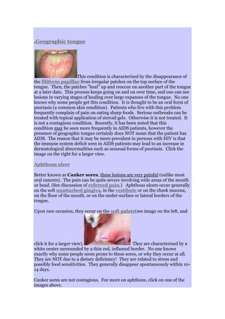
  • 1. .Geographic tongue This condition is characterized by the disappearance of the filiform papillae from irregular patches on the top surface of the tongue. Then, the patches "heal" up and reoccur on another part of the tongue at a later date. This process keeps going on and on over time, and one can see lesions in varying stages of healing over large expanses of the tongue. No one knows why some people get this condition. It is thought to be an oral form of psoriasis (a common skin condition). Patients who live with this problem frequently complain of pain on eating sharp foods. Serious outbreaks can be treated with topical application of steroid gels. Otherwise it is not treated. It is not a contagious condition. Recently, it has been noted that this condition may be seen more frequently in AIDS patients, however the presence of geographic tongue certainly does NOT mean that the patient has AIDS. The reason that it may be more prevalent in persons with HIV is that the immune system deficit seen in AIDS patients may lead to an increase in dermatological abnormalities such as unusual forms of psoriasis. Click the image on the right for a larger view. Aphthous ulcer Better known as Canker sores, these lesions are very painful (unlike most oral cancers). The pain can be quite severe involving wide areas of the mouth or head. (See discussion of referred pain.) Aphthous ulcers occur generally on the soft unattached gingiva, in the vestibule or on the cheek mucosa, on the floor of the mouth, or on the under-surface or lateral borders of the tongue. Upon rare occasion, they occur on the soft palate(see image on the left, and click it for a larger view). They are characterized by a white center surrounded by a thin red, inflamed border. No one knows exactly why some people seem prone to these sores, or why they occur at all. They are NOT due to a dietary deficiency! They are related to stress and possibly food sensitivities. They generally disappear spontaneously within 10- 14 days. Canker sores are not contagious. For more on aphthous, click on one of the images above.
  • 2. This condition happens in two varieties, each of which has its own treatment protocol: Minor aphthous is defined as the occasional, small ulcer that most persons experience no more than once or twice a year. These lesions are generally small (2-4 mm), and the ulcers are treated as isolated entities (one at a time). Treatment for Minor Aphthous Topical applications of steroids such as "Lidex gel" or "Kenalog in Orabase" ® (Note: Lidex is approximately ten times stronger than Kenalog, but Kenalog has the advantage of the Orabase which acts as a Band-Aid and keeps the steroid in place longer). These drugs are applied after meals and before bedtime, and both are prescription drugs. They generally reduce or eliminate pain immediately and bring about resolution of the canker in two to three days. I prefer Kenalog in Orabase in my own practice Aphthasol paste ® is a prescription drug that is applied directly to the ulcer four times a day (the same as Kenalog in Orabase). Cautery using either chemical or laser treatment. This type of treatment is palliative only, and does not treat the underlying condition. Cautery is done in the dental office to relieve the pain caused by a specific aphthous ulcer. Chemical cautery agents include silver nitrate(generally on a wooden stick) or commercial agents such as Debacterol®, both of which are applied by a dentist or physician and offer immediate pain relief. Over-the-counter agents such as Zilactin®, Ora5® and Gly-Oxide® are mild cautery agents that work more slowly. Laser treatment is quick and painless and also offers immediate pain relief. This is generally done only on small lesions in the dentist's office. Major aphthous , also called Sutton's disease is defined as a chronic condition in which patients are plagued with multiple aphthous lesions occurring several times a month. These ulcers are generally quite large, on the order of the size of a dime, and they often heal leaving scar tissue behind. Major aphthous associated with HIV can cause much larger lesions. Major aphthous has no known cure, but chronic aphthous lesions may be treated using the following methods: Treatment for Major Aphthous If you suddenly develop chronic major aphthous lesions, It is wise to check with your physician to see if there is an underlying cause such as an immune problem, or an underlying chronic illness such as Bechet, Chrohn's or celiac disease.
  • 3. Vitamin B12 has been found to be effective in reducing the frequency and severity of the lesions in patients suffering from major aphthous. 1 mg is dissolved under the tongue every evening. Some formulations of vitamin B12 are manufactured specifically for this route of administration. Other vitamin and mineral supplements such as iron,folic acid and zinc have been known to reduce the number and frequency of aphthous lesions. Steroid mouth rinse-- Betamethasone sodium phosphate (Betnesol mouthwash/Diprolene) one 0.5mg tablet dissolved in 5 to 10 ml of water. Patients rinse using this solution four times a day (after meals and before bed) whenever lesions are present. Another method is for the patient to mix about 1/4 inch of Fluocinonide (Lidex) cream or gel in four ounces of water. This mouth rinse is used the same way that betamethasone is used. Remember--Never swallow a steroid mouth rinse! Steroids are powerful drugs and mouth rinses made with them should be used sparingly since they can have systemic effects, even when used topically. Avoid oral products containing sodium lauryl sulfateas studies have implicated this common ingredient as a causative factor in the formation of aphthous. More information on treatment modalities for major and minor aphthous can be found by clicking here. Liken Planus
  • 4. Lichen Planus is actually a dermatological autoimmune disease that is often first diagnosed by a dentist due to its characteristic appearance in the mouth. In the mouth it appears as a series of filamentous, white, lacy lines on the inside of the cheeks or on nearly any other oral tissue. Lesions can occur on other parts of the body as well, most notably on the skin of the anticubital space (inside of the elbows). Most of these lesions are painless, but sometimes they occur on attached tissue such as the palate where they can be quite painful. They can also cause quite a bit of burning in the mouth when eating sharp foods. The image on the right is a fairly common presentation, and an obvious diagnosis. the image on the left shows a more subtle presentation under the tongue. Click on either image to see it full size. This condition is thought to be an autoimmune condition associated with exposure to drugs to which the patient may be sensitive. It is especially associated with certain antihypertensive drugs, NSAIDs, tetracycline and several sulfonamides, as well as a number of "recreational" drugs. The condition often improves with the cessation of the offending drug. The condition is more of a nuisance than a disability. The oral symptoms are often treated with steroid mouth rinses. If the symptoms are not severe, it is not treated at all. Lichen planus is not a contagious condition. Chronic lichen planus has been known to (very rarely) morph into squamous cell oral cancer.
  • 5. Fungal Infections (Candidiasis) Oral Candidiasis, also called Candidosis is caused by a form of fungus called Candida albicans. As many as 60 percent of healthy individuals may unknowingly harbor this organism as a part of their normal oral floral pattern. In healthy individuals, competing bacteria keep the fungus from overgrowing and causing the infections that you will read about on this page. The illnesses most commonly associated with the development of canidiasis are systemic steroid therapy, and endocrine disturbances associated with pregnancy, hypothyroidism, and diabetes. It is also associated with dry mouth syndrome, Sjorgren's syndrome, cancer chemotherapy, chronic tissue irritation (esp under dentures), smoking, antibiotic therapy and immunosuppression disorders such as AIDS. There are four forms of candidiasis 1. Pseudomembranous Candidiasis Pseudomembranous candidiasis is characterized by the presence of a white curd-like plaques that can easily be scraped off with a wooden tongue depressor blade or wiped away with a 2 x 2 gauze sponge. The white curd consists of the fungal organisms, including spores and mycelia. It happens in healthy adults only rarely, and usually is an indication of a lowered immune response. Often it is due to illnesses such as general viral infections or stress related fatigue.
  • 6. The term Thrush is a term for pseudomembranous candidiasis in infants. It is a common problem for infants since their immune systems are not yet fully developed. 2. Erythematous candidiasis Erythematous candidiasis is characterized by red, inflamed tissue. This form of candidiasis is especially prevalent in people who wear dentures and do not take them out at night. It appears to be the result of decreased tissue resistance because of the constant pressure from the prosthetic appliance. Treatment for this condition is composed of an antifungal antibiotic combined with the strong recommendation for the patient to remove his/her denture at night!
  • 7. In fact, pseudomembranous candidiasis, when scraped off the tissue reveals red, inflamed tissues underneath. Thus erythematous candidiasis is probably better characterized as the condition of the mucosa from which the white plaque has been removed. The image above shows pharyngeal candidiasis in an AIDS patient, and it demonstrates both pseudomembranous and erythematous varieties of candidiasis, as well as antular cheilitis at the corners of the lips. 3. Angular Cheilitis Angular cheilitis is a very common infection of the corners of the lips. It happens all the time to healthy people who tend to have moist lips, especially in the cold winter months. This condition is caused by a persistent mixed fungal/bacterial infection, and left untreated, may remain active for many months. It generally looks like a reddened, dry area at the corners of the lips. It is easily treated with Nystatin cream or nystatin/ triamcinolone ointment. Nystatin is simply an antibiotic that kills the fungus. Triamcinolone is a light steroid useful for reducing inflammatory responses to the fungus.(Mycolog®- -nystatin and triamcinolone, or Vytone®--iodoquinol and hydrocortisone) will work better to relieve the condition than an antifungal antibiotic alone. Angular cheilosis is associated with chronic vitamin B deficiency, and ill fitting dentures. Angular cheilits is not contagious. 4. Hyperplastic candidiasis
  • 8. Hyperplastic candidiasis looks like pseudomembranous candidiasis, however it cannot easily be wiped off with a gauze sponge. It is composed of elevated white plaques, and may ealily be confused with leukoplakia. It is found most often on the hard palate or the dorsal surface (top) of the tongue. It looks like any other form of leukoplakik plaque, but it will respond to a therapeutic trial of antifungal antibiotics. It can also be distinguished fron other keratotic lesions by cytologic smear or biopsy. Treatment of oral Candidiasis 1. Nystatin Ointment Disp: 30 gm tube Sig: Apply three times a day after meals or apply thin coat to inner surface of denture three times a day after meals. Safe: high compliance: inexpensive 2. Clortrimazole troches Disp: #70 Sig: Disolve slowly five times a day until gone. do not chew Safe: High compliance: pleasant tasting: High sugar (bad for teeth) There may be side effects due to the large dose of medication. 3. Amphotericin oral suspension Disp: 48 ml Sig: Swish with 1 ml four times per day, and swallow until gone Expensive: must remove dentures before using: there may be side effects. 4. Nystatin/triamcinolone ointment (Mycostatin®) (For angular cheilitis) Disp: 15 gm tube Sig: Apply to corners of lips four times a day, after meals and before bedtime Safe, inexpensive, high compliance 5. Ketaconazole 200 mg disp: 14 tabs Sig: One tab daily with meals Used in cases which are refractory to treatment with topical antifgungal infections. High compliance: Inexpensive: But there is a potential for liver toxicity.
  • 9. 6. Fluconazole 100 mg (Diflucan®) Disp: 15 tabs Sig: take 2 tabs first day, then one tab every day til all are used. Good compliance: expensive: There is a potential for liver toxicity. Oral Bacterial Infections==>> Bacterial diseases of the gums Periodontal Disease Periodontal disease is presented in this section as an aid to those who come here looking for images of gum problems they may have noticed, and do not know where to find information. This site contains two entire pages devoted to periodontal disease, including one that explains thecauses of periodontal disease and one on how it is treated. Periodontal disease is caused by poor
  • 10. oral hygiene. This site includes a page on correct techniques of oral hygiene that can prevent and even reverse periodontal disease. Gingivitis Gingivitis is caused by a chronic buildup of plaque around the teeth and is characterized by a red, sometimes swollen appearance of the gums immediately around the necks of the teeth. It is easily cured by good oral hygiene, but left untreated, it generally leads to periodontal disease and eventual loss of the teeth. Gingivitis is not contagious. Periodontal disease Outright periodontal disease may effect a single tooth, or any number of teeth. It begins after about the age of 25, and becomes serious between the ages of 35 and 50. Periodontal disease is painless in its early stages. It is the outcome of a lifetime of poor oral hygiene, and begins with simple gingivitis. In periodontal disease, the gums recede down the roots of the teeth, and the teeth appear to be longer than normal (click on the image above to see more about this image). Since the bone that maintains the teeth is effected by periodontal disease, the teeth become loose, and eventually painful. Periodontal disease is not contagious. Halitosis Bad breath is a chronic problem for persons with periodontal disease. However, periodontal disease is not the only cause of bad breath, a I have
  • 11. written an entire page devoted to the various causes and treatment of bad breath. Acute Necrotizing Ulcerative gingivitis (trench Mouth) Acute Necrotizing UlcerativeGingivitis is often called Trench Mouth. In ANUG, the gingiva immediately surrounding the teeth becomes necrotic (dead). ANUG is often found in people with poor oral hygiene who are either ill or under extreme physical or emotional stress. (It was named "trench mouth" because it was common in soldiers who fought in the trenches during world war I. These men were certainly under extreme physical and emotional stress, and had little opportunity to brush their teeth.) ANUG, being a bacterial infection, is very easily treated by gentle cleaning of the teeth and irrigation of the affected gums with 3% hydrogen peroxide. The bacteria that take advantage of a patient's run-down condition tend to be anaerobic which means that they die in the presence of oxygen. Hydrogen peroxide liberates oxygen (hence the bubbles) when it is exposed to blood, and the oxygen acts as an antiseptic and speeds healing of the damaged gum tissue. The patient is sent home with a prescription for Penicillin and instructions oncleaning the teeth to prevent further problems. It is essential that the patient return to the dentist after the initial infection for a professional cleaning to avoid a reoccurrence of the disease. ANUG is not contagious. Dentists today rarely see cases of ANUG, however the disease is making a comeback in communities in which there is a lot of drug addiction. It is especially prevalent in populations of methamphetamine addicts and is a part of the syndrome now known as Meth Mouth. Acute Necrotizing Oral Stomatitis
  • 12. This is a sight we never see except in a hospital setting. This man's mouth is being eaten alive by the same bacteria that his immune system would ordinarily have no problem keeping at bay if it were functioning normally. The difference between a dead body and a live one from the point of view of everyday environmental bacteria is a functioning immune system. AIDS attacks the immune system, and unless the disease and the bacteria can be kept at bay by modern drug therapy, the human body has no defense against parasitic bacteria and viruses.
  • 13. Disorders caused by viruses Hairy Leukoplakia
  • 14. Hairy leukoplakia is a white, corrugated or "hairy" "coating" on the lateral borders of the tongue. It is one of the relatively few conditions seen in the oral cavity which is associated almost exclusively with AIDS. Unlike Thrush, it is not easily scraped off. It is painless, but patients occasionally complain of its appearance and texture. It is caused by the body's reaction to the Epstein-Barr virus (responsible for Mononucleosis), and can be eliminated with a viral antibiotic like acyclovir (Zovirax®), famciclovir (Famvir®) or valacyclovir (Valtrex®). This condition is rarely seen in patients not infected with HIV. However, some healthy patients may develop a "callous" on the lateral borders of the tongue due to the nervous habit of continually scraping the tongue over the teeth. This can lead to embarrassment if the dentist suggests an AIDS test to a person who believes such a suggestion is an insult! It is never meant as a value judgment. Hairy Leukoplakia isnot contagious. Herpes Zoster Herpes Zoster (better known as shingles) is caused by the same virus that causes Chicken Pox. Herpes zoster "hides out" in a somatic nerve branch after the initial Chicken Pox infection (which usually happens in childhood), and flares up again later in life when the immune system begins to fail. Shingles is common in otherwise healthy elderly persons. It generally does not occur in younger people unless they are concurrently infected with the AIDS virus. The distribution of the rash on the body is the key to the diagnosis of shingles, and distinguishes the herpes zoster virus from other forms of herpes viruses. The distribution of the rash caused by herpes zoster in shingles is almost always on one side of the body, and is confined to the distribution of a single nerve root. The skin surface distribution of each spinal or cranial nerve is called adermatome. The image on the left above shows a rash which is confined to the dermatome
  • 15. defined by the third branch of the trigeminal nerve. It is outlined in blue to make it easier to see. Click the image to see larger images, as well as a great deal more on the concept of somatic dermatomes. Shingles infections are quite painful, and they generally go away after four or five weeks, but shingles may reoccur again at a later date. It frequently leaves those so afflicted with "postherpetic neuralgia" (PHN), which is severely sensitive skin, well after the infection. The image below shows the distribution of the herpes zoster rash from the frontal aspect, this time confined to the distribution of the first and second branches of the trigeminal nerve. Note the sharp delineation between the affected side and the unaffected side in the forehead and nose regions, which corresponds to the distribution of the first branch. The second branch, corresponding to the region below the eye is billaterally (both sides) affected. This image is presented compliments of Dr. Jonathan D. Trobe, MD at the University of Michigan. In the mouth, it looks very much like a typical intraoral herpes simplex infection. It is, however, identified by its distribution. It is limited to one side of the affected organ. The image to the right shows the Herpes zoster virus infecting half of the upper posterior palate. It is easy to confuse Herpes zoster with Herpes simplex which may occur in the same distribution purely by chance. While the Herpes Zoster virus is contagious, Shingles, surprisingly is not. Since a large percentage of the population already has been exposed to Chicken pox, most people harbor an immunity, and the probability that anyone will develop this disease depends more on the state of their immune system than on recent exposure to the virus. Herpes Simplex(the "cold sore" or "fever blister" virus)
  • 16. The Herpes simplex virus (HSV) is the most commonly occurring virus in the oral cavity. There are two distinct subcategories of HSV; HSV-I is primarily associated with infections in and around the mouth, and HSV-II is primarily associated with genital lesions. However, recent studies have shown that this type of site predilection is changing, probably due to changing sexual habits. current estimates show that approximately 11% of genital herpes infections are caused by HSV-I and about 2.5% of primary Herpes stomatitis infections (mostly in immunocompromised adults) are caused by HSV-II. Herpes Simplex (type I) is the virus that causes cold sores (herpes labialis) in normal, healthy adults. The image at the right shows a recurrent herpetic infection, in other words, a typical cold sore, sometimes called a fever blister due to its propensity to appear when the patient has a cold or other febrile (fever causing) illness. This is another bug that, likeShingles, tends to "hang out" in a nerve root for the life of the patient after the initial infection, which often occurs in childhood. Once infected, the patient remains infected for life. The virus remains dormant inside the nerve root most of the time until the patient suffers an illness or other problem which lowers his immune response. The virus takes advantage of the drop in immune response to flare up in the typical cold sore seen in this image. Click the image above for more information on recurrent herpes labialis and many more images of herpes infections. This image is what the initial infection may look like when a child, or young adult is firstinfected with the Herpes Simplex virus. While most young children and adolescents experience a subclinical infection (no outward signs of the infection), a small percentage will develop "Primary Herpes stomatitis". As you can see, it can look quite severe with blisters both inside and outside the mouth. ("Stomatitis" means inflammation of the entire mouth.) It starts out with "prodromal" symptoms including fever, malaise, headache and irritability, and then progresses after several days into severe gingival (gums) inflammation followed by the outbreak of numerous small blisters both inside and around the mouth. The blisters break leaving behind very painful sores with yellowish centers and red borders. The sores in the mouth are generally accompanied by severe pain, foul odor and increased salivation. Often the sores extend into the throat (pharyngitis). On rare occasions, the primary herpes infection can be confined
  • 17. to the throat. In any case, the patient is quite sick, but this primary infection will disappear after 10-14 days with rest, Tylenol®l and lots of fluids. In healthy people, this infection happens only once in a lifetime. Later in life, the presence of the virus only becomes apparent whenever an "ordinary" cold sore appears. Approximately 30% - 40% of patients who have been exposed to HSV will develop recurrent infections that will manifest as either recurrent herpes labialis (cold sores on the lips) or recurrent intraoral herpes. The sores are generally triggered by exposure to sunlight, fatigue, stress, hormonal changes such as menstruation, gastrointestinal disturbances and oral trauma. Whenever an adult appears in a clinic with a case of Primary Herpes Stomatitis, this infers a severely depressed immune response, and the dentist might consider referring the patient to a physician for diagnosis of an underlying disorder. Adults presenting with severe herpesstomatitis should consider being tested for HIV. It must be remembered, however, that a primary herpes stomatitis can happen at any time of life if the patient has never before had a cold sore. Click on the image to see larger views of this condition. For more basic information on the various forms herpes simplex takes, visitHerpesEductaion.Org. Intraoral Herpes Herpes simplex blisters can sometimes occur in the oral cavity on tissues not generally associated with cold sores. They always happen on tissue that is firmly bound down to underlying bone, such as the gums immediately around the teeth or on the roof of the mouth. As you can see, the appearance of this
  • 18. infection in the mouth can easily be confused with Herpes Zoster (shingles), especially if it occurs on only one side of the mouth. The viruses are closely related, and the blisters in the oral cavity can look identical. The presence of this type of infection in the mouth does not indicate the presence of HIV, although it is more common in AIDS patients than in the non-HIV population. This can happen to anyone who harbors the Herpes Simplex virus. Left alone, provided the patient is not immunologically compromised, it disappears in 10 to 14 days. The herpes simplex virus is very contagious and if one person in a family develops a cold sore, then others in the family may develop one as well. Treatment or herpes infections The treatment of herpes stomatitis is essentially palliative (treating the symptoms only). In healthy individuals, bedrest, high fluid intake and Tylenol work best to alleviate the symptoms. Aspirin and nonsteroidal anti- inflammatories like Motrin® and Alive® are best avoided in viral infections. New antibiotics like acyclovir (Zovirax®), famciclovir (Famvir®) or valacyclovir (Valtrex®) are effective in suppressing the Herpes virus and will generally alleviate the symptoms within a fairly short time. Systemic drugs like these are generally used in very severe cases in immunocompromised patients, or in herpes infections of the eye. In patients exposed to a lot of sun who are prone to outbreaks of recurrent herpes labialis, a prophylactic regimen of 400 mg of acyclovir twice a day may prevent the outbreaks Unfortunately, the antibiotics do not often "cure" the disease since the virus continues to remain in an inactive form inside a nerve root waiting for another chance to cause an outbreak. Recently, the Food and Drug Administration has approved penciclovir 1% topical cream for the treatment of herpes labialis. This is applied every two hours while awake, and will help to shorten the duration and severity of the cold sore. Acyclovir cream (Zovirax®) works, but is less effective than penciclovir. Lysine (available at most drug stores) has been reported to reduce the severity of recurrent outbreaks if taken in high enough doses (2-3 gm) at the first prodromal signs (burning, tingling For more information on HSV-II (genital herpes), please see my section on Oral Cancer Human Papillomavirus lesions (warts)
  • 19. Warts are caused by a virus. In the oral cavity, they tend to be somewhat flatter than the type occurring on hands, but if they are dried with air, the tiny projections characteristic of regular warts become evident. The causative agent is the Human Papillomavirus (HPV). These growths generally are not painful. They may be removed using lasers, cautery or cold steel blades. A majority of these lesions have no consequences beyond the primary papilloma. Some, especially toward the back of the mouth or throat may have more serious consequences discussed below. HPV is contagious. There are about 200 different strains of HPV. Most strains are relatively harmless, or may cause papillomas like the one in the image above. Some strains of HPV causegenital warts and are now known as the cause of cervical cancer (cancer of the uterus) as well as anal, vulvar and penile cancers. Although there are eighteen strains with serious pathological consequences, one strain in particular, HPV-16 is quite dangerous in the oral cavity. It is a major causative agent in oral cancers, and is transmitted to the oral cavity via vigorous oral/sexual contact (oral sex). As a result of the stark increase in promiscuous sexual activity since the late 1960's, this strain of HPV is increasingly problematic, and seems to account for the serious rise in the incidence of oral cancers in younger people, many of whom do not smoke or drink regularly. For much more on this subject, please see my page on Oral Cancer. Five strains of HPV cause oral papillomas. These include 6, 7 11, 16 and 32. Note that young persons who contract oral papillomas, even those infected with HPV-16, will probably clear the infection eventually if their immune systems are not adversely affected by other chronic diseases or lifestyle issues like drug or alcohol addictions. Because HPV-16 is associated with vigorous sexual oral/genital contact (oral sex), most of the "warts" associated with this strain would most likely occur in the back of the oral cavity, toward the back of the throat, around the tonsils, on the soft palate, at the base and sides of the tongue, etc. They will rarely be found toward the front of the mouth like the one pictured above. HPV-16 is NOT transferred by "ordinary" kissing, but may be transmitted by vigorous open mouth deep (French) kissing.
  • 20. There is a saliva test that a dentist can perform that can diagnose hidden cases of HPV. Dentists interested in offering this service to their patients can visit thewebsite of OralDNA Labs to learn about this simple and accurate process. In general, HPV is implicated as a causative agent only in squamous cell carcinomas occurring at the base of the tongue (i.e. the non movable area in the throat) the pharyngeal tonsils and the upper throat. It is in this subset of oral squamous cell cancers that we find the stark increase in oral cancers in young adults who neither drink nor smoke. An association between HPV 16 and squamous cell carcinomas of the anterior tongue and other more anterior oral tissues has not yet been demonstrated. Genital Herpes Herpes Simplex type I (HSV-1) prefers to infect the face and oral cavity. It is the virus most responsible for traditional cold sores and primary herpes stomatitis. There is, however a second variety of Herpes that prefers to infect the genital areas. Herpes Simplex Type II (HSV-2) is called "genital Herpes" because of its venereal (sexually transmitted) qualities. Both varieties produce similar lesions, the difference between them being their site specific preferences. Both establish latency (take up permanent residence) in nerve roots and once established, tend to cause occasional outbreaks with active lesions (sores) in areas of the body serviced by that particular nerve root. HSV-1 prefers to live in the trigeminal nerve root where it causes lesions in the oral cavity and on the face. HSV-2 takes up residence in the sacral ganglion at the base of the spine where it may cause genital lesions (see thedermatome chart on the Herpes zoster page). Even though each type has site specific preferences, the viruses are genetically similar and can take up residence in nerve roots in other parts of the body, including in each other's territory. Outside of their own home territories, however, neither virus is especially virulent, and rarely cause recurrent outbreaks. HSV-2 causes approximately 90% of all cases of genital herpes. Genital herpes caused by HSV-1 is generally much milder than that caused by HSV-2. HSV-1 is usually transferred to the genital area by direct oral/genital contact, although the virus is present in the saliva of infected individuals. Thus the use of saliva as a lubricant can, in fact, transfer HSV-1 to the genital area. HSV-1 is found in only about 10% of all cases of genital herpes, however most people infected with HSV-1 in the genital area have few, if any, outbreaks after the initial episode. HSV-2 prefers to live in this area and causes a much more virulent infection there. On the other hand, HSV-1 causes almost all cases of oral and facial herpes. Oral herpes caused by HSV-2 almost never reoccurs, except in immunocompromised patients. For more on this subject, visit this page.
  • 21. Neoplasms (tumors, or "growths") Kaposi's Sarcoma (KS) (pronounced "cap-o-zeez") Kaposi's Sarcoma is a tumor composed of numerous tiny blood vessels. It tends to be dark red or deep purple. It may be flat, or a swollen mass. These growths are not generally painful unless secondarily infected by another type of Herpes or bacteria. Thus good oral hygiene is important in the management of these tumors if they occur in the mouth. Kaposi's occurs most frequently on the skin, although tumors can occur in the gastrointestinal tract and mouth. In the oral cavity, the lesions occur mostly on the palate (the roof of the mouth). Although they are technically a form of cancer, there is evidence that they are, in fact the result of a secondary infection with Herpes virus type VIII. This virus is found in high concentration in the saliva of infected individuals and can cause Kaposi's Sarcoma only in patients with very compromised immune systems. Some recent research has shown that this virus is transferred through deep kissing. K aposi's tumors are seen almost exclusively in gay men with AIDS. The occurrence of one of these lesions anywhere on the body of ayoung man is indicative of the presence of HIV. Kaposi's is infrequent in women, even women with AIDS. It is also rare in men who have contracted AIDS via intravenous drug use. It is not known why women and heterosexual males with AIDS do not generally succumb to Kaposi's sarcoma, although there is probably an association between the gay male lifestyle and the transfer of the herpes type 8 virus. These lesions occur as the initial manifestation of AIDS in approximately 11% of patients. Prior to the AIDS epidemic, they were seen (rarely) only on the lower extremities of elderly men. They probably occur in elderly men because of age
  • 22. related immune depression. The reason for their appearance in elderly men and not in women may also be associated with lifestyle issues. For more information on HIV and AIDS, click hereto be directed to a page with statistics, diagnostic criteria and the history of the epidemic. You will also find a repeat of the images seen on this page with more specific information on how they relate to AIDS patients. Lymphoma (lymphatic cancer) Non Hodgkin's Lymphoma (NHL) is a cancer that starts in a lymph node and spreads to other areas of the body through the lymphatic system and the blood vessels. Prior to the AIDS epidemic, NHL generally effected older individuals (average age 67), however the incidence of NHL has increased substantially in younger persons since the beginning of the AIDS epidemic. Lesions (abnormalities) like those in the image to the right, especially in a younger person, may be the first indication that a patient has HIV, although it is usually accompanied by a generalizedlymphadenopathy (swelling of lymph nodes all over the body). A suppressed immune response is a strong factor in the development of NHL, however persons with no history of immunosuppression (or HIV) may contract the disease. There is some evidence that one or more secondary viruses may bear the responsibility for the actual disease, the Epstein-Barr (Mononucleosis) virus once again being a prime suspect. Treatment for this condition usually involves chemotherapy and Radiation therapy. Oral lymphoma can happen any place in the oral cavity. It generally begins as a non specific swelling of the oral tissues in a localized area. |
  • 23. Geographic tongue This condition is characterized by the disappearance of the filiform papillae from irregular patches on the top surface of the tongue. Then, the patches "heal" up and reoccur on another part of the tongue at a later date. This process keeps going on and on over time, and one can see lesions in varying stages of healing over large expanses of the tongue. No one knows why some people get this condition. It is thought to be an oral form of psoriasis (a common skin condition). Patients who live with this problem frequently complain of pain on eating sharp foods. Serious outbreaks can be treated with topical application of steroid gels. Otherwise it is not treated. It is not a contagious condition. Recently, it has been noted that this condition may be seen more frequently in AIDS patients, however the presence of geographic tongue certainly does NOT mean that the patient has AIDS. The reason that it may be more prevalent in persons with HIV is that the immune system deficit seen in AIDS patients may lead to an increase in dermatological abnormalities such as unusual forms of psoriasis. Click the image on the right for a larger view. Aphthous ulcer Better known as Canker sores, these lesions are very painful (unlike most oral cancers). The pain can be quite severe involving wide areas of the mouth or head. (See discussion of referred pain.) Aphthous ulcers occur generally on the soft unattached gingiva, in the vestibule or on the cheek mucosa, on the floor of the mouth, or on the under-surface or lateral borders of the tongue. Upon rare occasion, they occur on the soft palate(see image on the left, and click it for a larger view). They are characterized by a white center surrounded by a thin red, inflamed border. No one knows exactly why some people seem prone to these sores, or why they occur at all. They are NOT due to a dietary deficiency! They are related to stress and possibly food sensitivities. They generally disappear spontaneously within 10- 14 days. Canker sores are not contagious. For more on aphthous, click on one of the images above.
  • 24. This condition happens in two varieties, each of which has its own treatment protocol: Minor aphthous is defined as the occasional, small ulcer that most persons experience no more than once or twice a year. These lesions are generally small (2-4 mm), and the ulcers are treated as isolated entities (one at a time). Treatment for Minor Aphthous Topical applications of steroids such as "Lidex gel" or "Kenalog in Orabase" ® (Note: Lidex is approximately ten times stronger than Kenalog, but Kenalog has the advantage of the Orabase which acts as a Band-Aid and keeps the steroid in place longer). These drugs are applied after meals and before bedtime, and both are prescription drugs. They generally reduce or eliminate pain immediately and bring about resolution of the canker in two to three days. I prefer Kenalog in Orabase in my own practice Aphthasol paste ® is a prescription drug that is applied directly to the ulcer four times a day (the same as Kenalog in Orabase). Cautery using either chemical or laser treatment. This type of treatment is palliative only, and does not treat the underlying condition. Cautery is done in the dental office to relieve the pain caused by a specific aphthous ulcer. Chemical cautery agents include silver nitrate(generally on a wooden stick) or commercial agents such as Debacterol®, both of which are applied by a dentist or physician and offer immediate pain relief. Over-the-counter agents such as Zilactin®, Ora5® and Gly-Oxide® are mild cautery agents that work more slowly. Laser treatment is quick and painless and also offers immediate pain relief. This is generally done only on small lesions in the dentist's office. Major aphthous , also called Sutton's disease is defined as a chronic condition in which patients are plagued with multiple aphthous lesions occurring several times a month. These ulcers are generally quite large, on the order of the size of a dime, and they often heal leaving scar tissue behind. Major aphthous associated with HIV can cause much larger lesions. Major aphthous has no known cure, but chronic aphthous lesions may be treated using the following methods: Treatment for Major Aphthous If you suddenly develop chronic major aphthous lesions, It is wise to check with your physician to see if there is an underlying cause such as an immune problem, or an underlying chronic illness such as Bechet, Chrohn's or celiac disease.
  • 25. Vitamin B12 has been found to be effective in reducing the frequency and severity of the lesions in patients suffering from major aphthous. 1 mg is dissolved under the tongue every evening. Some formulations of vitamin B12 are manufactured specifically for this route of administration. Other vitamin and mineral supplements such as iron,folic acid and zinc have been known to reduce the number and frequency of aphthous lesions. Steroid mouth rinse-- Betamethasone sodium phosphate (Betnesol mouthwash/Diprolene) one 0.5mg tablet dissolved in 5 to 10 ml of water. Patients rinse using this solution four times a day (after meals and before bed) whenever lesions are present. Another method is for the patient to mix about 1/4 inch of Fluocinonide (Lidex) cream or gel in four ounces of water. This mouth rinse is used the same way that betamethasone is used. Remember--Never swallow a steroid mouth rinse! Steroids are powerful drugs and mouth rinses made with them should be used sparingly since they can have systemic effects, even when used topically. Avoid oral products containing sodium lauryl sulfateas studies have implicated this common ingredient as a causative factor in the formation of aphthous. More information on treatment modalities for major and minor aphthous can be found by clicking here. Liken Planus Lichen Planus is actually a dermatological autoimmune disease that is often first diagnosed by a dentist due to its characteristic appearance in the mouth. In the mouth it appears as a series of filamentous, white, lacy lines on the inside of the cheeks or on nearly any other oral tissue. Lesions can occur on other parts of the body as well, most notably on the skin of the anticubital space (inside of the elbows). Most of these lesions are painless, but sometimes they occur on attached tissue such as the palate where they can be quite painful. They can also cause quite a bit of burning in the mouth when eating sharp foods. The image on the right is a fairly common presentation, and an obvious
  • 26. diagnosis. the image on the left shows a more subtle presentation under the tongue. Click on either image to see it full size. This condition is thought to be an autoimmune condition associated with exposure to drugs to which the patient may be sensitive. It is especially associated with certain antihypertensive drugs, NSAIDs, tetracycline and several sulfonamides, as well as a number of "recreational" drugs. The condition often improves with the cessation of the offending drug. The condition is more of a nuisance than a disability. The oral symptoms are often treated with steroid mouth rinses. If the symptoms are not severe, it is not treated at all. Lichen planus is not a contagious condition. Chronic lichen planus has been known to (very rarely) morph into squamous cell oral cancer. <<==Fungal infections in the oral cavity