1. THE SPINAL CORD
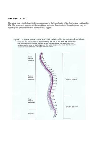
The spinal cord extends from the foramen magnum to the lower border of the first lumbar vertebra (Fig.
13). The nerve roots leave the cord at an oblique angle and thus the site of the cord damage may be
higher up the spine than the root number would suggest.
2. In the spinal cord, from dorsal to ventral, the inputs are somatic sensory and visceral sensory (Fig. 14,
right side in red), and the outputs are visceral motor and somatic motor (Fig. 14, left side in yellow).
3. Complete spinal cord transection (Fig. 15) causes:
• Loss of all sensory modalities below the lesion
• Complete flaccid paralysis below the lesion
• Inability to pass urine because of a flaccid bladder and constipation
• Lower motor neurone signs at the level of the lesion
• Sensory impairment in the area exclusively supplied at the level of the lesion (there is overlap)
• Reflexes reduced or absent at the level of the lesion (and possibly below this at the time of the
transection
• Upper motor neurone signs below the level of the lesion
4. Hemisection of the spinal cord (Fig. 16) produces:
• Loss of sensation and motor innervation and reflex activity at the level of the hemisection on the
same side
• Upper motor neurone type paralysis of muscles below the hemisection on the same side
• Loss of pain and temperature on the opposite side
• Loss of joint position and vibration sense on the same side
Because the spinal cord axons cannot usefully regenerate established changes are permanent.
5. Basic structure and function of the spinal cord
Figure 14 illustrates the anatomy of the spinal cord. In early quadrupeds impulses needed for fast
communication to coordinate movement, mostly of the tail (and thus assist balance), were notably:
• vestibulospinal (for equilibrium)
• reticulospinal, olivospinal (part of the extrapyramidal tract controlling movement)
• rubrospinal (basically reflecting influence of corpus striatum and perhaps cerebellum, mostly to
distal limb muscles)
• tectospinal (derived from visual information)
These pathways still persist in man but the anatomical tail has been replaced by the “physiological tail”
of the cerebellum. The final common pathway to striated muscle is provided by anterior horn cells and
their (lower) motor neurones which supply skeletal muscle fibres. The force of muscle contraction
depends on the number of muscle fibres activated and the frequency of nerve impulses received.
The corticospinal (pyramidal) tract mostly crosses the midline in the medullary decussation (Fig. 5)
developed later in mammals. It is the only uninterrupted descending pathway from the forebrain. Most
of the descending motor pathways have to cross superior to the cervical part of the spinal cord so that the
upper limbs receive appropriate motor instructions from the contralateral cortex. Some corticospinal
fibres do not decussate in the medulla but pass caudally as the anterior corticospinal tract and cross the
midline at the level of “their” anterior horn cells. The descending tracts (which are more medially
situated than most of the ascending tracts (Fig. 14) influence the final common pathway of output from
the anterior horn cells. Motor nerves which drive fine movements have fewer muscle fibres to innervate
(for example in the muscle that move they eye there is a one nerve to one muscle ratio). About half of
the descending fibres drive the arms and about one third drive the legs (reflecting the relative complexity
of actions). It must be stressed that neither structure nor function is as precisely localized as these
diagrams may suggest.
Sensory nerves feed into the dorsal columns of the spinal cord, the motor nerves leave from the ventral
columns and the visceral output leaves from the lateral horn of the spinal cord between these two. This
scheme persists in the medulla in a modified fashion. Somewhat similar nerve outgrowths occur in the
head segments to form trigeminal, facial, glossopharyngeal and vagus nerve sensory ganglia (link).
Joint position sense, vibration sense, and some (fine) touch ascend the posterior columns of the spinal
cord (Fig. 14) without crossing the midline to the medulla where they cross the midline (Fig. 5) in the
medial lemniscus. Some (crude) touch ascends in the anterior spinothalamic after crossing the midline.
Pain and temperature information crosses the midline shortly after entry into the spinal cord and ascend
in the lateral spinothalamic columns (Fig. 14). These spinothalamic ascending fibres rejoin their
initially non-crossing sensory partners in the thalamus.
Some diseases, such as syringomyelia, damage the center of the spinal cord by causing a cavity (syrinx)
which impairs or abolishes the crossing pain and temperature sensation but leaves the non-crossing
touch sensation intact (such patients usually unknowingly burn themselves by touching hot objects).
Somatic: bodily, usually referring to skeletal muscle
Visceral: refers to the inner organs
6. This dissociated sensory loss may be confined to the arms and hands because the crossing fibres from
the legs are situated more laterally than those from the hand and are thus away from the central
damaging syrinx. Syringomyelia usually occurs in the cervical cord producing wasting of the small
muscles of the hand (caused by involvement of the anterior horn cells) and loss of pain and temperature
in a “cape” distribution, often extending up the back of the head and often down to involve the hands
Spinal cord problems
• Problems caused by an inadequate blood supply usually cause death of the relevant part of the
cord. The anterior spinal artery supplies the anterior two thirds of the cord and the two posterior
spinal arteries supply the posterior cord. Haemorrhage in or around the cord is unusual
• Inherited degenerations of parts of the cord can occur, often affecting specific tracts and their
nucleiin the brain or brainstem
• Spina bifida, a condition in which the arches of the vertebrae fail to fuse, mayl cause spinal cord
signs
• Inflammation (myelitis) usually is caused by viruses although in the past tabes dorsalis (caused
by syphilis) used to affect the dorsal roots (to cause shooting ”lightning” pain) and posterior
columns (to cause loss of joint position sense, hence the characteristic stamping gait). Syphilis
could also cause masses, gummata, that presented as space occupying lesions, or
meningovascular damage, or meningoencephalomyelitis
• Deficiency of vitamin B12, usually associated with pernicious anaemia, produces subacute
combined degeneration of the cord with changes in the posterior and lateral columns and signs of
both peripheral nerve damage and spinal cord degeneration
• External or internal trauma may damage the cord directly or, by secondary pressure, by
interrupting blood flow
• Tumours in the cord itself are rare (problems are more likely to be caused by extrinsic
compression)