
Recomendados
Más contenido relacionado
La actualidad más candente
La actualidad más candente (20)
Destacado
Destacado (19)
Similar a The Veins
Similar a The Veins (20)
Más de meducationdotnet
Más de meducationdotnet (20)
The Veins
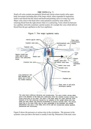
- 1. THE VEINS (Fig. 7) Nearly all veins contain non-pulsatile blood and venous return mostly relies upon body movement (including that of the lungs which, when expanded in inspiration, tend to suck blood into the chest) and heartward pointing valves in some leg veins. Major veins close to the heart show some pulsation caused by some reflux of returning blood from the right atrium when it is contracting. Veins which lie between two capillary networks constitute a portal system, for example the veins which deliver blood from the gut capillaries to the liver capillaries. Figure 8 shows the pressures at various sites in the circulation. The pressure in the systemic veins just above the heart is usually 0 mm Hg. Distension of the neck veins
- 2. does not usually occur because there is insufficient pressure inside the veins to produce distension. Veins can contract without affecting arterial peripheral resistance. The capacity of the blood vessels and the blood volume must be kept equal if there is to be sufficient venous blood to fill each ventricle for each contraction. The state of the venous system thus is an important determinant of heart function. For example after a brisk haemorrhage both veins and arterioles constrict (the veins to allow filling of ventricles and arterioles to maintain perfusion pressure). If this mechanism fails then shock results with an inadequate circulation and a low arterial blood pressure.
- 3. THE CAPILLARIES (=small hairs) By the time systemic arterial blood reaches the capillaries it is not pulsatile. Capillaries are permeable to most plasma constituents (except the plasma proteins) and most of the movement of fluids into and out of the vascular system occurs through the thin capillary walls. THE LYMPHATIC SYSTEM (Fig. 9) Exudation of extra tissue fluid occurs if arteriolar and capillary blood pressures tend to rise and the lymph collection system caters for this excess. The lymphatics, some of which possess valves, are a necessary alternative to the veins for returning fluid from the tissues back into the bloodstream. The final common pathway for lymph drainage is the thoracic duct which enters the systemic venous system at the junction of the left subclavian vein and the left internal jugular vein. If lymphatics are blocked then tissue swelling will be high in protein, relatively stiff, and less likely than venous oedema to pit when pressed with a finger.