SlideShare una empresa de Scribd logo
1 de 7
Descargar para leer sin conexión
ORIGINAL ARTICLES

   Adult-to-Adult Living Donor Liver Transplantation
  Using Right-Lobe Grafts: Results and Lessons Learned
            From a Single-Center Experience
            Thomas Bak,* Michael Wachs,* James Trotter,† Gregory Everson,†
         Thomas Trouillot,† Marcelo Kugelmas,† Tracy Steinberg,* and Igal Kam*

Living donor liver transplantation (LDLT) for adults is                   mulated to date a series of 41 adult-to-adult right-lobe
now a practical alternative to cadaveric liver transplanta-               LDLTs. The results of this series and the lessons learned
tion. Use of right-lobe grafts has become the preferred
donor procedure. Because of the complexity of this oper-
                                                                          from these cases are presented to show that this can be a
ation, a learning curve is to be expected. We report the                  safe and effective procedure.
outcome of our first 41 LDLTs at the University of Colo-                       Justification for the use of adult-to-adult liver trans-
rado Health Sciences Center (Denver, CO). We also dis-                    plantation is based on the critical shortage of adult
cuss the lessons learned and the resultant modifications in                organs available for cadaveric transplantation. A grow-
the procedure that evolved during our series. Patient
records were retrospectively reviewed between August
                                                                          ing number of people are being listed for transplanta-
1997 and February 2001 for the following end points:                      tion, whereas the availability of cadaveric donor organs
recipient survival, graft survival, and donor and recipient               is remaining fairly constant. The increasing use of mar-
complications. Thirty-eight of 41 living donor liver trans-               ginal donor organs may increase this number, but what
plant recipients (93%) are alive and well postoperatively                 cost this will have on short- and long-term survival of
with a mean follow-up of 9.6 months. Four patients
required retransplantation secondary to technical prob-                   the organs is yet to be determined. Waiting-list mortal-
lems (9.8%); all 4 patients were in our initial 11 cases.                 ity remains a problem and is approximately 10% per
Modification of the donor liver plane of transection                       year.7 Using LDLT has been shown to decrease waiting
resulted in venous outflow improvement. Also, biliary                      time on the list for the transplant recipients while free-
management was modified during the series. Donor com-                      ing organs for the remainder of the recipient pool who
plications are listed; all 41 donors have returned to normal
pretransplantation activity. Our results indicate that                    may not have potential living donors.8 When intro-
LDLT can be performed safely with excellent donor and                     duced for the pediatric population, LDLT reduced
recipient outcomes. Dissemination of our experience can                   mortality and waiting times for pediatric liver recipi-
help shorten the learning curve for other institutions.                   ents.9 Applying this concept to adults remained diffi-
(Liver Transpl 2001;7:680-686.)                                           cult because left lateral segments would not provide
                                                                          sufficient liver mass to meet an adult transplant recipi-

L    iving donor liver transplantation (LDLT) has
     undergone an evolution from its beginnings in
pediatric transplantation using left lateral segments to
                                                                          ent’s needs. Left-lobe grafting in the adult-to-adult set-
                                                                          ting was initiated with mixed results, with concerns of
                                                                          inadequate hepatic mass.10 The right-lobe graft has sev-
adult-to-adult left-lobe transplantation to the now                       eral advantages over the left lobe. These include
fairly well-accepted standard of adult-to-adult right-                    increased hepatic mass, better anatomic position for
lobe transplantation.1-5 We published a report of our                     anastomoses, and less concern for hepatic venous out-
first 2 cases using this technique and the rationale for                   flow obstruction.
our approach.6 Since these initial cases, we have accu-                       Of utmost importance in the use of this procedure is
                                                                          donor safety. Adequate liver volume must be left in
                                                                          place to avoid hepatic dysfunction. Technical resection
                                                                          of the right lobe of the liver has to be performed in a
    From the Divisions of *Transplant Surgery and †Gastroenterology/
Hepatology, University of Colorado Health Sciences Center, Denver, CO.
                                                                          setting in which donor morbidity and mortality are
    Address reprint requests to Thomas Bak, MD, Division of Transplant    minimal. Based on large surgical series of major hepatic
Surgery, University of Colorado Health Sciences Center, 4200 E 9th Ave,   resections, this donor operation should be able to be
C-318, Denver, CO 80262. Telephone: 303-372-8750; FAX: 303-               performed with a quoted mortality risk of less than 1%
372-8737; E-mail: thomas.bak@uchsc.edu                                    in experienced centers.11 Several reports document the
    Copyright © 2001 by the American Association for the Study of
Liver Diseases
                                                                          safety of living donor surgery in liver transplanta-
    1527-6465/01/0708-0011$35.00/0                                        tion.12,13 Realistic expectations for recovery include
    doi:10.1053/jlts.2001.26509                                           normal daily activity in 1 month, return to predonation

680                                  Liver Transplantation, Vol 7, No 8 (August), 2001: pp 680-686
Living Donor Liver Transplantation                                               681


work at 2 to 3 months, and a full recovery to normal at
3 to 4 months. This report describes our series of 41                                 Table 2. Recipient Demographics
patients undergoing this procedure and outcomes of
both donors and recipients. We also focus on the tech-                   Sex (M/F)                                      24/17
                                                                         Age (yr)                                       45.0 Ϯ 8.6
nical aspects of the surgery that have changed during
                                                                         Median waiting time (d)                        124
this period.                                                             Mean LOS (d)                                   21.5 Ϯ 19.5
                                                                         Median LOS (d)                                 12

Methods                                                                  NOTE. Values expressed as number or mean Ϯ SD.
                                                                         Abbreviation: LOS, length of stay.
Forty-one cases of adult-to-adult right-lobe LDLT were per-
formed at the University of Colorado Health Sciences Center
in Denver, CO, from August 1997 to March 2001. The
frequency of this operation has increased over time, with 12          Sharing (UNOS) status IIB at time of transplantation. Two
cases performed over the past 6 months. Donor demographics            recipients were status I with a diagnosis of fulminant hepatic
are listed in Table 1. Only one donor was not related to the          failure, and 4 transplant recipients were status IIA. Transplant
recipient. Donor age ranged from 19 to 54 years. Details of           recipients were evaluated and screened for listing similarly to
our donor evaluation are presented elsewhere.8 All donors             other liver transplant candidates. After being informed of the
presented on a strictly voluntary basis and were never solic-         option for LDLT and only after the recipient and/or donor
ited. Each donor underwent a thorough physical examination            presented voluntarily, further assessment was performed to
and laboratory evaluation. Imaging modalities of the donor            ensure good candidacy. This included the absence of morbid
included magnetic resonance angiography and cholangiogra-             obesity, limited previous major upper-abdominal surgery,
phy in all cases. Conventional angiography was used as a              good size matching between the pair, and stricter age criteria
confirmatory test in patients with arterial anomalies, espe-           (only 1 of 41 recipients was older than 60 years). Risks and
cially when there was a question of more than one artery              benefits of surgery for both donor and recipient were method-
supplying the right lobe. Absolute liver volume of the right          ically explained, including minimal risk for donor mortality.
lobe was calculated based on these scans, but the judgment                 Donor hepatectomy is performed under general anesthe-
and experience of the senior transplant physicians were used          sia, with a thoracic epidural catheter used for postoperative
to determine adequacy of mass, mainly based on donor and              pain management. A right subcostal incision with upper mid-
recipient body size, along with the imaging studies. Full psy-        line extension similar to that performed on the transplant
chological and social evaluations were performed on each              recipient is used. Mobilization of the right lobe of the liver
donor, and a stable support system was documented. Liver              proceeds, with care focused on leaving the attachments of the
biopsies were performed infrequently; only 3 of the 41 even-          left lobe intact to prevent future twisting or torsion. Accessory
tual donors underwent a biopsy. Biopsies were not deemed              hepatic veins are ligated and divided to free the vena cava.
necessary for patients considered to be at low risk for fatty liver   Branches greater than 1 cm are preserved. After cholecystec-
disease.                                                              tomy, an intraoperative cholangiogram is obtained. Vascular
    Recipient demographics are listed in Table 2. Of the 41           isolation of the right hepatic artery, right portal vein, and right
transplant recipients, 35 were United Network for Organ               hepatic vein is then completed. A transection line is marked
                                                                      on the liver with electrocautery. Using electrocautery and
                                                                      ligatures, parenchymal transection proceeds. As our series
                                                                      progressed, the line of transection shifted to run left of and
                 Table 1. Donor Demographics                          parallel to the middle hepatic vein branch draining segments 5
                                                                      and 8. This right branch of the middle hepatic vein is left
   No. of donors                                       41             intact on the edge of the graft and transected superiorly at its
   Sex (M/F)                                         26/15            junction with the middle hepatic vein. The right hepatic bile
   Mean age (yr)                                  30.8 Ϯ 11.2
                                                                      duct is transected, leaving a small cuff so as not to stricture the
   Mean LOS (d)                                    6.3 Ϯ 1.6
                                                                      main hepatic duct. Accessory posterior right-lobe bile ducts
   Relationship to recipient
     Son                                               12             draining to the left hepatic duct are identified on intraopera-
     Brother                                            7             tive cholangiography and also carefully identified at the tran-
     Sister                                             5             section plane, with the remaining left stumps oversewn.
     Daughter                                           6             Autologous blood transfusion systems were available for all
     Wife                                               2             donor operations. Parenchymal transection is completed,
     Father                                             1             leaving both lobes of the liver with intact blood supply. No
     Other                                              8             inflow occlusion is used. The raw surface of the liver is then
   NOTE. Values expressed as number or mean Ϯ SD.                     packed and re-evaluated 20 to 30 minutes after transection to
   Abbreviation: LOS, length of stay.                                 ensure no detectable bleeding or bile leak is present.
                                                                           The transplant recipient is taken to the operating room
682                                                           Bak et al


                              Figure 1. Donor hepatic vein anastomosis to recipient vena cava.


after the donor operation has started and shows no contrain-         fused at this point. The recipient right hepatic arterial branch
dications to proceeding. No donor operations were aborted in         is then spatulated and anastomosed in an end-to-end fashion
our series. Two transplant recipients with diagnoses of cancer       to the spatulated donor right hepatic artery using a running
underwent exploratory surgery to ensure that there was no            7-0 vasculature suture. Bile duct reconstruction then pro-
extrahepatic spread of disease before the donor operation            ceeds. Thirteen of 41 cases were reconstructed in a duct-to-
commenced. The recipient native hepatectomy proceeded in             duct fashion over an internal stent. The remainder of these
standard fashion, with extreme care used to ensure adequate          reconstructions were performed using a standard Roux-en-Y
hepatic artery and portal vein length. The piggyback tech-           small-bowel limb anastomosis to the right hepatic duct with
nique was also necessarily used in all cases.                        interrupted 6-0 sutures over an internal stent. Secondary and
    With the recipient team in the room, the vasculature to          occasional tertiary anastomoses were performed to the same
the right lobe of the donor liver is transected and the liver is     Roux limb with any significant (Ͼ3 mm) accessory bile ducts.
passed to the back table. The vascular stumps on the donor are       Small accessory bile ducts (Ͻ3 mm), particularly those in a
then closed with running monofilament vascular suture. The            very posterior position that would be technically difficult to
graft is immediately flushed through the portal vein with 1 L         reconstruct, are oversewn. Routine abdominal wall closure
of heparinized (10,000 U/L) iced saline followed by a second         follows in both the donor and recipient. One Jackson-Pratt
liter of iced saline. No systemic anticoagulation with heparin       drain is left in the donor between the hilum and the cut
is used in the donor.                                                surface of the liver. The recipient has 2 to 3 drains placed, with
    The liver is transported to the recipient room, where the        1 drain also along the cut liver surface. The donors recover in
native hepatectomy is completed. In most cases, we use a             the recovery room and are transferred to the ward. Twenty-
complete cross-clamp to allow easy access to the recipient vena      three of the recipients went to the surgical intensive care unit
cava. No venovenous bypass is used. The recipient middle and         postoperatively, and the remainder were transferred to the
left hepatic veins are oversewn, and the right hepatic vein cuff     ward from the recovery room.
is extended down the anterior surface of the vena cava, creat-
ing an orifice to match the right hepatic vein size on the donor
graft. The right hepatic vein is anastomosed to the caval open-      Results
ing with a running 5-0 vascular suture (Fig. 1). The recipient
portal vein is then anastomosed to the right portal vein orifice      All 41 donors are alive, well, and have returned to
using a running 6-0 vascular suture. The right lobe is reper-        normal predonation activity. Donor complications are
Living Donor Liver Transplantation                                     683


listed in Table 3. Of 41 donors, 2 required nonautolo-
gous blood transfusions (5%). One of these was for a
large hemothorax secondary to a central venous catheter
placed preoperatively by the anesthesia department.
The second was in a patient whose hematocrit de-
creased on postoperative day 2 and stabilized after 2
units of packed red blood cells were administered.
Three patients had postoperative bile leaks. Two of
these patients were returned to the operating room and
underwent direct repair of a leaking bile duct stump
within 3 days of the hepatectomy. One of these stumps
was the cystic duct, whereas the other was the right
hepatic duct stump. The other patient was treated con-
servatively with percutaneous drainage, resulting in res-
                                                                   Figure 2. Donor aspartate aminotransferase (AST) (s)
olution of the leak from the cut liver surface. One large
                                                                   and alanine aminotransferase (ALT) (}) levels versus
incisional hernia was repaired in a patient who returned           postoperative day.
to lifting concrete bags 2 weeks after her surgery. One
Jackson-Pratt drain was retrieved surgically after it
snapped off during its removal. One idiosyncratic med-             setting was consistent with poor graft function based on
ication reaction caused donor lethargy that resolved               worsening laboratory values and a deteriorating clinical
postoperative day 3. Also, a temporary neuropraxia                 picture. Ultrasound evaluation showed patent vessels
occurred in the dominant hand of one donor. All 41                 with no evidence of venous obstructions. Venous con-
donors are alive, well, and have returned to normal                gestion was confirmed on gross inspection and micro-
activity.                                                          scopic analysis of the explanted liver. One retransplan-
    Postoperative laboratory values are shown in Figure            tation was performed for hepatic artery thrombosis
2. Aspartate aminotransferase and alanine aminotrans-              postoperative day 12. The fourth retransplantation was
ferase levels peaked postoperative day 3, with a return to         performed for a persistent bile leak that 2 surgical
normal by day 7. Serum bilirubin levels peaked slightly            repairs failed to resolve. This patient underwent
later. Mean hospital stay for donors was 6.3 days.                 retransplantation postoperative day 50 and died of sep-
    Forty-one right-lobe adult-to-adult LDLTs have                 sis 4 weeks after retransplantation. The second of the 3
been completed using this technique. Thirty-eight of               deaths occurred 15 months posttransplantation because
41 recipients (93%) are alive and well, with a mean                of uncontrolled chronic rejection, whereas the third
follow-up of 9.6 months. Thirty-six of 41 grafts are               death was in a patient who developed multiple strokes
functioning, for a graft survival rate of 88%. Four                on postoperative day 1 and died of a cardiac arrest on
patients required retransplantation with cadaveric                 postoperative day 5.
grafts (9.8%). All 4 of these retransplantations occurred              Forty-four percent of transplant recipients under-
in our initial 11 cases, initiating several technical adjust-      went multiple bile duct reconstructions, with one
ments described next. Two retransplantations were per-             patient requiring 3 bile duct anastomoses. Overall bili-
formed for hepatic dysfunction resulting from hepatic              ary complication rates were 34%. This includes 3 anas-
venous outflow obstruction. These were performed                    tomotic leaks treated surgically (1 duct-to-duct, 2
postoperative days 14 and 5. In both cases, the clinical           Roux-en-Y). Three patients developed postoperative
                                                                   strictures. The first occurred in a duct-to-duct anasto-
                                                                   mosis, which was converted to a Roux. The other 2
                                                                   strictures were in patients with Roux limb anastomoses,
                 Table 3. Donor Complications                      and these were successfully treated with percutaneous
                                                                   transhepatic cholangioplasty. Nine patients developed
   Bile leak, reoperation                                2         cut surface leaks; 6 patients were treated with prolonged
   Bile leak, external drainage                          1         drainage and 3 patients underwent reoperation. Eight
   Incisional hernia, surgical repair                    1
   Neuropraxia, transient                                1
                                                                   of the 9 leaks resolved; the exception was the patient
   Drain retrieval, reoperation                          1         who died after retransplantation.
   Hemothorax from venous access                         1             Four patients required a second hepatic vein anasto-
                                                                   mosis because of an accessory hepatic vein greater than
684                                                        Bak et al


1 cm. One of these patients had 2 accessory hepatic               the right hepatic vein system.15 Since moving our tran-
veins. Three patients underwent 2 portal venous anas-             section plane in this manner, we have not experienced
tomoses without complications.                                    outflow problems causing graft dysfunction. Another
                                                                  venous drainage issue is that of accessory hepatic veins
                                                                  draining into the vena cava. Previous series of right-lobe
Discussion                                                        transplants have reported reanastomosis of any acces-
Our results suggest that adult-to-adult LDLT using a              sory vein greater than 5 mm.16 We have been more
right hepatic lobe graft is a safe and effective operation        selective in our series, and of the 41 grafts, have per-
for people requiring liver transplantation when careful           formed accessory venous anastomoses in only 4
donor and recipient screening is performed. Graft and             patients. We routinely reconstruct accessory veins
patient survival rates in our series are equal to our cur-        greater than 0.8 to 1 cm.
rent cadaveric transplantation results. Donor safety is of            Arterial anatomy is often quite variable in both
utmost importance for this procedure, and although                donor and recipient. Most commonly, the major right
there have been reported donor complications and                  hepatic artery of the donor graft can be directly anasto-
deaths,5,14 our series shows excellent donor outcomes             mosed to the recipient artery at either the bifurcation of
with 100% survival and return to normal activity.                 the right and left branches or a bifurcation patch created
   Despite these results, it should be stressed that a            at the branch of the cystic artery. We attempt to maxi-
learning curve exists with this procedure: as experience          mize right hepatic arterial length in the recipient but
and level of comfort increased, so did our overall success        stay extraparenchymal in this dissection. On donor
rates. The four retransplantations described during our           workup, a replaced right hepatic artery is not a contra-
early experience led us to 2 important technical                  indication; this frequently makes the operation techni-
changes. Specifically, our significant changes during               cally easier because of a longer length of graft artery. In
this procedure have been improvements in graft hepatic            this situation, an angiogram is often obtained to assess
venous outflow and the understanding of the need for               whether this right branch off the superior mesenteric
intraoperative cholangiography to help show and                   artery is completely replaced or whether it is an acces-
reconstruct accessory right bile ducts.                           sory branch. This allows us to conclude whether a sec-
   In our early experience, we transected the liver to the        ond arterial anastomosis is required. We have studied
right of the middle hepatic vein, even if the middle              arterial flow postoperatively with duplex ultrasonogra-
hepatic vein contributed drainage to segments 5 and 8.            phy on a daily basis through postoperative day 5.
Using this technique, 2 transplant recipients had severe          Patients are started on 81 mg of acetylsalicylic acid
graft congestion and poor function requiring retrans-             when a stable postoperative hematocrit is present. To
plantation. Their clinical picture was consistent with            date, we have had one hepatic artery thrombosis, which
poor graft function despite ultrasound evaluation show-           is similar in incidence to our cadaveric recipient popu-
ing patent vessels and no obstruction. We now believe             lation, and concerns of greater thrombosis rates caused
that primary nonfunction of these grafts is very                  by smaller vessels have not been realized. Arterial anas-
unlikely, and if the postoperative clinical course mimics         tomoses are performed with 3.5 ϫ loupe magnification
this picture, it is likely caused by a technical issue. These     with a running monofilament 7-0 vascular suture. In 2
cases prompted us to change our plane of transection to           cases, the donor right hepatic artery bifurcated into
the left. The right branch of the middle hepatic vein is          anterior and posterior branches that encircled the com-
preserved with the right-lobe graft and ligated superi-           mon bile duct. In the first instance, the anterior branch
orly at its junction with the main middle hepatic vein.           was small and therefore was ligated. In the second case,
We believe this prevents disruption of collateral drain-          both arteries were of equal size. The anterior artery was
age between the right hepatic vein and the right branch           transected at the time of graft removal to free it from the
of the middle hepatic vein. We do not believe it is               bile duct and was then repaired, with an end-to-end
necessary to connect this middle hepatic vein branch to           anastomosis performed on the back table.
the cava using a jump graft. When the graft is reper-                 Portal vein inflow has been adequate in all patients.
fused, this right branch of the middle hepatic vein is            Direct end-to-end anastomosis of the donor right por-
well decompressed, presumably by collaterals in the               tal vein to the right branch or common portal vein of
graft. It has been our experience and also recently               the recipient was performed in all but 3 cases. In these
reported that postoperative ultrasound examination of             cases, there were 2 separate major portal veins to the
the graft drainage shows reversal of flow in the right             right lobe. In 1 of these, 2 anastomoses were performed;
middle hepatic vein branch ultimately emptying into               one each to the right and left branches of the recipient
Living Donor Liver Transplantation                                          685


portal vein. In the other 2 cases, the graft veins were very    internal stent. In addition, Roux drainage is performed
close to each other, with liver parenchyma providing a          in cases of a single bile duct when there is concern about
common back wall, and the donor had a large native              tension or blood supply. We do not use t-tubes or other
portal system. A single anastomosis of the donor veins          external biliary drainage catheters. Biliary complica-
to the native portal vein branch was performed.                 tions have occurred in 34% of transplant recipients.
    A preoperative ultrasound showed one transplant             This is similar to rates previously reported in living
recipient to have a thrombosed portal vein, with patent         donor liver surgery.17-19 Most of our leaks (60%) have
splenic and superior mesenteric veins. This LDLT was            been raw surface leaks. These are usually managed con-
delayed until a cadaveric donor iliac vein of compatible        servatively with external drainage if the patient is clini-
blood type was available to use as a jump graft. At             cally stable. These usually occur while the surgical
surgery, a successful thrombectomy allowed for ade-             drains are still in place. Our experience has been that
quate inflow and no vascular graft was used. To date in          these will spontaneously seal, allowing for drain re-
our series, we have not had to use an arterial or venous        moval. If the patient shows signs of uncontrolled leak-
graft for reconstruction; however, we routinely repeat          age, such as pain or increasing bilirubin levels, they are
an ultrasound examination of the recipient 1 week               treated surgically. Biliary leak rates are slightly greater in
before surgery to rule out portal vein thrombosis. The          the Roux-drained patients, but this is likely influenced
use of a recipient saphenous vein graft may be required         by the fact that this group includes patients with mul-
in a setting in which the right lobe has 2 arterial inflows,     tiple duct drainage.
i.e., an accessory right branch as opposed to a totally             Postoperative management of living donor liver
replaced system.                                                transplant recipients has been similar to that of recipi-
    The second change in technique is in regard to our          ents of cadaveric grafts. Overall, 56% of recipients went
biliary management. Magnetic resonance cholangio-               to the intensive care unit postoperatively. This
graphy (MRC) imaging is used as preoperative screen-            decreased to 40% in the second half of our series as our
ing of donor biliary anatomy. This has proven to be a           level of comfort with these cases has increased. An
relatively accurate assessment of major biliary struc-          increase in frequency of surveillance ultrasonography is
tures. In our early cases, a transplant recipient experi-       performed, but this has not shown greater thrombosis
enced a bile leak from an accessory posterior duct that         rates. Hospital length of stay averaged 21.5 days for the
had not been recognized on the MRC or at the original           transplant recipients. We believe that this is lengthened
donor surgery. Despite reoperation and oversewing, the          somewhat because of the increase in biliary complica-
leak persisted, eventually leading to retransplantation,        tions compared with our cadaveric transplant recipients
sepsis, and death. We have since instituted routine             and a less predictable decrease in postoperative liver
intraoperative cholangiography and have identified and           function test results, leading to a heightened vigilance
reconstructed accessory bile ducts in 44% of our right-         for potential complications and rejection. However,
lobe grafts. On one occasion, a patient underwent 2             graft and patient survival rates ultimately are equal to
accessory biliary reconstructions. Identification and            cadaveric results.
drainage of this important posterior branch has pre-                These results must be viewed cautiously because
vented biliary complications in a significant number of          candidates are carefully selected for LDLT and in gen-
right-lobe grafts, and we have not performed a retrans-         eral are not the sickest liver transplant recipients. We
plantation for a biliary complication since. In 2               believe that the optimal candidates for this procedure
instances, we have oversewn a small (Ͻ3 mm) posterior           are patients who are UNOS status IIB and have a good
right duct, avoiding reconstruction. Neither of these 2         donor available. Status III patients should rarely
cases had a leak or signs of cholangitis in this small          undergo transplantation until more experience has been
undrained biliary section of hepatic parenchyma. These          gained to justify the risk to the donor. The use of LDLT
smaller ducts are usually located in a more posterior           is especially valuable in the patient population who has
location than the previously mentioned accessory duct,          a suspicion for or proven small hepatocellular carci-
which is significant and is reconstructed.                       noma (status IIB), but would unlikely be able to receive
    Biliary reconstruction is performed with chole-             a cadaveric graft before their cancer progressed to a
dochocholedochostomy when possible. The need for                nontransplantable stage. The role of this procedure as
Roux limb drainage is obvious in the case of primary            palliation for large hepatocellular cancers is still being
sclerosing cholangitis or when multiple anastomoses             debated. Our center has not performed this procedure
must be performed, and these are performed to separate          on a patient who would otherwise not be a candidate for
openings of the intestinal limb, each over an individual        a cadaveric graft.
686                                                                  Bak et al


    In conclusion, LDLT is a safe and effective proce-                       9. Sindhi R, Rosendale J, Mundy D, Taranto S, Baliga P, Reuben
dure for well-selected patients. We report a large series                       A, et al. Impact of segmental grafts on pediatric liver transplan-
                                                                                tation—A review of the United Network for Organ Sharing
of right-lobe grafts with similar graft and patient sur-
                                                                                Scientific Registry Data (1990-1996). J Pediatr Surg 1999;34:
vival statistics to our cadaveric transplant recipients.                        107-110.
Donor safety has been maintained throughout the                             10. Emond JC, Renz JF, Ferrell LD, Rosenthal P, Lim RC, Roberts
series. This procedure will continue to grow in impor-                          JP, et al. Functional analysis of grafts from living donors. Ann
tance in the national transplant community as a way to                          Surg 1996;224:544-552.
alleviate some of the pressure of growing waiting lists                     11. Belghiti J, Hiramatsu K, Benoist S, Massault P, Sauvanet A,
                                                                                Farges O. Seven hundred forty-seven hepatectomies in the
and stagnant cadaveric donor availability.                                      1990s: An update to evaluate the actual risk of liver resection.
                                                                                J Am Coll Surg 2000;191:38-46.
References                                                                  12. Fujita S, Kim ID, Uryuhara K, Asonuma K, Egawa H, Kiuchi T,
                                                                                et al. Hepatic grafts from live donors: Donor morbidity for 470
 1. Broelsch CE, Whitington PF, Emond JC, Heffron TG, This-
                                                                                cases of live donation. Transpl Int 2000;13:333-339.
    tlethwaite JR, Stevens L, et al. Liver transplantation in children
                                                                            13. Fan ST, Lo CM, Liu CL, Yong BH, Chan JK, Ng IO. Safety of
    from living related donors: Surgical techniques and results. Ann
                                                                                donors in live donor liver transplantation using right lobe grafts.
    Surg 1991;214:428-437.
                                                                                Arch Surg 2000;135:336-340.
 2. Broelsch CE, Burdelski M, Rogiers X, Gundlach M, Knoefel
                                                                            14. Grewal HP, Thistlewaite JR, Loss GE, Fisher JS, Cronin DC,
    WT, Langwieler T, et al. Living donor for liver transplantation.
                                                                                Siegal CT, et al. Complications in 100 living liver donors. Ann
    Hepatology 1994;20(suppl 1, part 2):49S-55S.
                                                                                Surg 1998;228:214-219.
 3. Kawasaki S, Makuuchi M, Matsunami H, Hashikura Y, Ikegami
                                                                            15. Kaneko T, Kaneko K, Sugimoto H, Inoue S, Hatsuno T, Sawada
    T, Nakazawa Y, et al. Living related liver transplantation in
    adults. Ann Surg 1998;227:269-274.                                          K, et al. Intrahepatic anastomosis formation between the hepatic
 4. Todo S, Furukawa H, Jin MB, Shimamura T. Living donor liver                 veins in the graft liver of the living related liver transplantation:
    transplantation in adults: Outcome in Japan. Liver Transpl                  Observation by Doppler ultrasonography. Transplantation
    2000;6(suppl 2):S66-S72.                                                    2000;70:982-985.
 5. Broelsch CE, Malago M, Testa G, Gamazo CU. Living donor                 16. Marcos A, Ham JM, Fisher RA, Olzinski AT, Posner MP.
    liver transplantation in adults: Outcome in Europe. Liver                   Single center analysis of the first 40 adult-to-adult living donor
    Transpl 2000;6(suppl 2):S64-S65.                                            liver transplants using the right lobe. Liver Transpl 2000;6:296-
 6. Wachs ME, Bak TE, Karrer FM, Everson GT, Shrestha R,                        301.
    Trouillot TE, et al. Adult living donor liver transplantation using     17. Marcos A, Fisher RA, Ham JM, Shiffman ML, Sanyal AJ, Luke-
    a right hepatic lobe. Transplantation 1998;66:1313-1316.                    tic VA, et al. Right lobe living donor liver transplantation. Trans-
 7. Annual Report of the US Scientific Registry for Organ Trans-                 plantation 1999;68:798-803.
    plantation and the Organ Procurement and Transplantation                18. Cronin D, Alonso E, Piper J, Newell K, Bruce D, Woodle P, et
    Network—Transplant Data 1988-2000. UNOS, Richmond,                          al. Biliary complications in living donor liver transplantation.
    VA, and the Division of Transplantation, Bureau of Health                   Transplant Proc 1997;29:419-420.
    Resources and Services Administration, US Department of                 19. Testa G, Malego M, Valentin-Gamazo C, Lindell G, Broelsch
    Health and Human Services, Rockville, MD, 2000.                             CE. Biliary anastomosis in living related liver transplantation
 8. Trotter JF. Selection of donors and recipients for living donor             using the right lobe: Techniques and complications. Liver
    liver transplantation. Liver Transpl 2000;6(suppl 2):S52-S58.               Transpl 2000;6:710-714.

Más contenido relacionado

La actualidad más candente

Transcaval Retrograde Transcatheter Aortic Valve Replacement for Patients Wit...
Transcaval Retrograde Transcatheter Aortic Valve Replacement for Patients Wit...Transcaval Retrograde Transcatheter Aortic Valve Replacement for Patients Wit...
Transcaval Retrograde Transcatheter Aortic Valve Replacement for Patients Wit...Pedro Martinez-Clark, M.D.
 
Marginal and extended criteria donors
Marginal and extended criteria donorsMarginal and extended criteria donors
Marginal and extended criteria donorsDr. Rohit Saini
 
British Columbia Medical Journal - May 2010: Cardiac transplantation in Briti...
British Columbia Medical Journal - May 2010: Cardiac transplantation in Briti...British Columbia Medical Journal - May 2010: Cardiac transplantation in Briti...
British Columbia Medical Journal - May 2010: Cardiac transplantation in Briti...British Columbia Medical Journal
 
Rui Maio - Portugal - Tuesday 29 - Organ Donor Care. New Alternatives
Rui Maio  - Portugal - Tuesday 29 - Organ Donor Care. New AlternativesRui Maio  - Portugal - Tuesday 29 - Organ Donor Care. New Alternatives
Rui Maio - Portugal - Tuesday 29 - Organ Donor Care. New Alternativesincucai_isodp
 
Bioartificial Kidney Project skt dr keniz
Bioartificial Kidney Project skt   dr kenizBioartificial Kidney Project skt   dr keniz
Bioartificial Kidney Project skt dr kenizMohamed Yassine Keniz
 
La mia esperienza innovativa è... la perfusione degli organi
La mia esperienza innovativa è... la perfusione degli organiLa mia esperienza innovativa è... la perfusione degli organi
La mia esperienza innovativa è... la perfusione degli organiNetwork Trapianti
 
Evaluation of kidney donor sudan 2017
Evaluation of kidney donor sudan 2017Evaluation of kidney donor sudan 2017
Evaluation of kidney donor sudan 2017FarragBahbah
 
Preparation for transplantation dr ahmed kamal
Preparation for transplantation dr ahmed kamalPreparation for transplantation dr ahmed kamal
Preparation for transplantation dr ahmed kamalFarragBahbah
 
immunosuppressive drugs
immunosuppressive drugsimmunosuppressive drugs
immunosuppressive drugsdrsalwa22000
 
How should recently symptomatic patients be treated urgent cea or cas
How should recently symptomatic patients be treated urgent cea or casHow should recently symptomatic patients be treated urgent cea or cas
How should recently symptomatic patients be treated urgent cea or casuvcd
 
Celulas tronco hematopoieticas
Celulas tronco hematopoieticasCelulas tronco hematopoieticas
Celulas tronco hematopoieticasdervoz2012
 
Impact Of Warm And Cold Ischemia Time On Kidney Transplantation
Impact Of Warm And Cold Ischemia Time On Kidney TransplantationImpact Of Warm And Cold Ischemia Time On Kidney Transplantation
Impact Of Warm And Cold Ischemia Time On Kidney TransplantationIvaylo Mitsiev
 
TCT 2010 research highlights: A slideshow presentation
TCT 2010 research highlights: A slideshow presentation TCT 2010 research highlights: A slideshow presentation
TCT 2010 research highlights: A slideshow presentation theheart.org
 

La actualidad más candente (20)

Biomaterials 2008
Biomaterials 2008Biomaterials 2008
Biomaterials 2008
 
Transcaval Retrograde Transcatheter Aortic Valve Replacement for Patients Wit...
Transcaval Retrograde Transcatheter Aortic Valve Replacement for Patients Wit...Transcaval Retrograde Transcatheter Aortic Valve Replacement for Patients Wit...
Transcaval Retrograde Transcatheter Aortic Valve Replacement for Patients Wit...
 
Marginal and extended criteria donors
Marginal and extended criteria donorsMarginal and extended criteria donors
Marginal and extended criteria donors
 
PD in AKI
PD in AKIPD in AKI
PD in AKI
 
British Columbia Medical Journal - May 2010: Cardiac transplantation in Briti...
British Columbia Medical Journal - May 2010: Cardiac transplantation in Briti...British Columbia Medical Journal - May 2010: Cardiac transplantation in Briti...
British Columbia Medical Journal - May 2010: Cardiac transplantation in Briti...
 
Rui Maio - Portugal - Tuesday 29 - Organ Donor Care. New Alternatives
Rui Maio  - Portugal - Tuesday 29 - Organ Donor Care. New AlternativesRui Maio  - Portugal - Tuesday 29 - Organ Donor Care. New Alternatives
Rui Maio - Portugal - Tuesday 29 - Organ Donor Care. New Alternatives
 
Bioartificial Kidney Project skt dr keniz
Bioartificial Kidney Project skt   dr kenizBioartificial Kidney Project skt   dr keniz
Bioartificial Kidney Project skt dr keniz
 
La mia esperienza innovativa è... la perfusione degli organi
La mia esperienza innovativa è... la perfusione degli organiLa mia esperienza innovativa è... la perfusione degli organi
La mia esperienza innovativa è... la perfusione degli organi
 
Evaluation of kidney donor sudan 2017
Evaluation of kidney donor sudan 2017Evaluation of kidney donor sudan 2017
Evaluation of kidney donor sudan 2017
 
Preparation for transplantation dr ahmed kamal
Preparation for transplantation dr ahmed kamalPreparation for transplantation dr ahmed kamal
Preparation for transplantation dr ahmed kamal
 
Kidney Donor evaluation
Kidney Donor evaluationKidney Donor evaluation
Kidney Donor evaluation
 
immunosuppressive drugs
immunosuppressive drugsimmunosuppressive drugs
immunosuppressive drugs
 
Application Brief: Abdominal Aortic Aneurysm
Application Brief: Abdominal Aortic AneurysmApplication Brief: Abdominal Aortic Aneurysm
Application Brief: Abdominal Aortic Aneurysm
 
How should recently symptomatic patients be treated urgent cea or cas
How should recently symptomatic patients be treated urgent cea or casHow should recently symptomatic patients be treated urgent cea or cas
How should recently symptomatic patients be treated urgent cea or cas
 
Celulas tronco hematopoieticas
Celulas tronco hematopoieticasCelulas tronco hematopoieticas
Celulas tronco hematopoieticas
 
Damage control resuscitation. history theory and technique
Damage control resuscitation.  history theory and techniqueDamage control resuscitation.  history theory and technique
Damage control resuscitation. history theory and technique
 
Impact Of Warm And Cold Ischemia Time On Kidney Transplantation
Impact Of Warm And Cold Ischemia Time On Kidney TransplantationImpact Of Warm And Cold Ischemia Time On Kidney Transplantation
Impact Of Warm And Cold Ischemia Time On Kidney Transplantation
 
TCT 2010 research highlights: A slideshow presentation
TCT 2010 research highlights: A slideshow presentation TCT 2010 research highlights: A slideshow presentation
TCT 2010 research highlights: A slideshow presentation
 
Cirurgia do cone
Cirurgia do coneCirurgia do cone
Cirurgia do cone
 
Tratamento IAo e IM - JACC
Tratamento IAo e IM -  JACCTratamento IAo e IM -  JACC
Tratamento IAo e IM - JACC
 

Similar a Adult to adult living donor liver transplantation

Bridge therapy in hepatocellular carcinoma before liver transplantation
Bridge therapy in hepatocellular carcinoma before liver  transplantationBridge therapy in hepatocellular carcinoma before liver  transplantation
Bridge therapy in hepatocellular carcinoma before liver transplantationRicardo Yanez
 
Future is in heart transplantation
Future is in heart transplantationFuture is in heart transplantation
Future is in heart transplantationdrucsamal
 
Oscar Imventarza - Argentina - Tuesday 29 - Organ Allocation Optimizing dono...
Oscar Imventarza  - Argentina - Tuesday 29 - Organ Allocation Optimizing dono...Oscar Imventarza  - Argentina - Tuesday 29 - Organ Allocation Optimizing dono...
Oscar Imventarza - Argentina - Tuesday 29 - Organ Allocation Optimizing dono...incucai_isodp
 
Early liver transplantation after resection for hcc
Early liver transplantation after resection for hccEarly liver transplantation after resection for hcc
Early liver transplantation after resection for hcchr77
 
liver transplantation.pptx
liver transplantation.pptxliver transplantation.pptx
liver transplantation.pptxVigneshSNair3
 
2. continuous renal replacement therapy recent advances and future research
2. continuous renal replacement therapy recent advances and future research2. continuous renal replacement therapy recent advances and future research
2. continuous renal replacement therapy recent advances and future researchEdleo13
 
Selection and maintenance of a cadaver donor for liver transplantation
Selection and maintenance of a cadaver donor for liver transplantationSelection and maintenance of a cadaver donor for liver transplantation
Selection and maintenance of a cadaver donor for liver transplantationApollo Hospitals
 
Intestinal transplant registry report global activity and trends
Intestinal transplant registry report  global activity and trendsIntestinal transplant registry report  global activity and trends
Intestinal transplant registry report global activity and trendsSilvio Dantas
 
Sheet1Sleep with No SMSleep withusing social media Paticipants 18.docx
Sheet1Sleep with No SMSleep withusing social media Paticipants 18.docxSheet1Sleep with No SMSleep withusing social media Paticipants 18.docx
Sheet1Sleep with No SMSleep withusing social media Paticipants 18.docxbjohn46
 
Kidney transplantation1408
Kidney transplantation1408Kidney transplantation1408
Kidney transplantation1408bondafairieal
 
recent advances in hepatobiliary and GI surgery
recent advances in hepatobiliary and GI surgeryrecent advances in hepatobiliary and GI surgery
recent advances in hepatobiliary and GI surgeryhr77
 
Stem Cell Therapy in Liver Cirrhosis
Stem Cell Therapy in Liver CirrhosisStem Cell Therapy in Liver Cirrhosis
Stem Cell Therapy in Liver Cirrhosisdbpublications
 
Provision of ideal transfusion support – The essence of thalassemia care
Provision of ideal transfusion support – The essence of thalassemia careProvision of ideal transfusion support – The essence of thalassemia care
Provision of ideal transfusion support – The essence of thalassemia careApollo Hospitals
 
Chapter 21 Organ Transplantation
Chapter 21 Organ Transplantation Chapter 21 Organ Transplantation
Chapter 21 Organ Transplantation MeganSimpson27
 
Hepatoblastoma and hepatocellular carcinoma in children
Hepatoblastoma and hepatocellular carcinoma in children Hepatoblastoma and hepatocellular carcinoma in children
Hepatoblastoma and hepatocellular carcinoma in children Fanny Yeh
 
Jose Maria Morales - Spain - Tuesday 29 - HLA for Renal Allocation
Jose Maria Morales - Spain - Tuesday 29 - HLA for Renal AllocationJose Maria Morales - Spain - Tuesday 29 - HLA for Renal Allocation
Jose Maria Morales - Spain - Tuesday 29 - HLA for Renal Allocationincucai_isodp
 

Similar a Adult to adult living donor liver transplantation (20)

Donor hepatectomy
Donor hepatectomyDonor hepatectomy
Donor hepatectomy
 
Bridge therapy in hepatocellular carcinoma before liver transplantation
Bridge therapy in hepatocellular carcinoma before liver  transplantationBridge therapy in hepatocellular carcinoma before liver  transplantation
Bridge therapy in hepatocellular carcinoma before liver transplantation
 
Future is in heart transplantation
Future is in heart transplantationFuture is in heart transplantation
Future is in heart transplantation
 
Oscar Imventarza - Argentina - Tuesday 29 - Organ Allocation Optimizing dono...
Oscar Imventarza  - Argentina - Tuesday 29 - Organ Allocation Optimizing dono...Oscar Imventarza  - Argentina - Tuesday 29 - Organ Allocation Optimizing dono...
Oscar Imventarza - Argentina - Tuesday 29 - Organ Allocation Optimizing dono...
 
Early liver transplantation after resection for hcc
Early liver transplantation after resection for hccEarly liver transplantation after resection for hcc
Early liver transplantation after resection for hcc
 
liver transplantation.pptx
liver transplantation.pptxliver transplantation.pptx
liver transplantation.pptx
 
2. continuous renal replacement therapy recent advances and future research
2. continuous renal replacement therapy recent advances and future research2. continuous renal replacement therapy recent advances and future research
2. continuous renal replacement therapy recent advances and future research
 
Selection and maintenance of a cadaver donor for liver transplantation
Selection and maintenance of a cadaver donor for liver transplantationSelection and maintenance of a cadaver donor for liver transplantation
Selection and maintenance of a cadaver donor for liver transplantation
 
Intestinal transplant registry report global activity and trends
Intestinal transplant registry report  global activity and trendsIntestinal transplant registry report  global activity and trends
Intestinal transplant registry report global activity and trends
 
Sheet1Sleep with No SMSleep withusing social media Paticipants 18.docx
Sheet1Sleep with No SMSleep withusing social media Paticipants 18.docxSheet1Sleep with No SMSleep withusing social media Paticipants 18.docx
Sheet1Sleep with No SMSleep withusing social media Paticipants 18.docx
 
Kidney transplantation1408
Kidney transplantation1408Kidney transplantation1408
Kidney transplantation1408
 
recent advances in hepatobiliary and GI surgery
recent advances in hepatobiliary and GI surgeryrecent advances in hepatobiliary and GI surgery
recent advances in hepatobiliary and GI surgery
 
JCO study_Wang_Nov 2011
JCO study_Wang_Nov 2011JCO study_Wang_Nov 2011
JCO study_Wang_Nov 2011
 
Stem Cell Therapy in Liver Cirrhosis
Stem Cell Therapy in Liver CirrhosisStem Cell Therapy in Liver Cirrhosis
Stem Cell Therapy in Liver Cirrhosis
 
Provision of ideal transfusion support – The essence of thalassemia care
Provision of ideal transfusion support – The essence of thalassemia careProvision of ideal transfusion support – The essence of thalassemia care
Provision of ideal transfusion support – The essence of thalassemia care
 
2098
20982098
2098
 
Chapter 21 Organ Transplantation
Chapter 21 Organ Transplantation Chapter 21 Organ Transplantation
Chapter 21 Organ Transplantation
 
Hepatoblastoma and hepatocellular carcinoma in children
Hepatoblastoma and hepatocellular carcinoma in children Hepatoblastoma and hepatocellular carcinoma in children
Hepatoblastoma and hepatocellular carcinoma in children
 
Jose Maria Morales - Spain - Tuesday 29 - HLA for Renal Allocation
Jose Maria Morales - Spain - Tuesday 29 - HLA for Renal AllocationJose Maria Morales - Spain - Tuesday 29 - HLA for Renal Allocation
Jose Maria Morales - Spain - Tuesday 29 - HLA for Renal Allocation
 
Elliott bennett guerrero - Recess trial NEJM 2015
Elliott bennett guerrero - Recess trial NEJM 2015Elliott bennett guerrero - Recess trial NEJM 2015
Elliott bennett guerrero - Recess trial NEJM 2015
 

Adult to adult living donor liver transplantation

  • 1. ORIGINAL ARTICLES Adult-to-Adult Living Donor Liver Transplantation Using Right-Lobe Grafts: Results and Lessons Learned From a Single-Center Experience Thomas Bak,* Michael Wachs,* James Trotter,† Gregory Everson,† Thomas Trouillot,† Marcelo Kugelmas,† Tracy Steinberg,* and Igal Kam* Living donor liver transplantation (LDLT) for adults is mulated to date a series of 41 adult-to-adult right-lobe now a practical alternative to cadaveric liver transplanta- LDLTs. The results of this series and the lessons learned tion. Use of right-lobe grafts has become the preferred donor procedure. Because of the complexity of this oper- from these cases are presented to show that this can be a ation, a learning curve is to be expected. We report the safe and effective procedure. outcome of our first 41 LDLTs at the University of Colo- Justification for the use of adult-to-adult liver trans- rado Health Sciences Center (Denver, CO). We also dis- plantation is based on the critical shortage of adult cuss the lessons learned and the resultant modifications in organs available for cadaveric transplantation. A grow- the procedure that evolved during our series. Patient records were retrospectively reviewed between August ing number of people are being listed for transplanta- 1997 and February 2001 for the following end points: tion, whereas the availability of cadaveric donor organs recipient survival, graft survival, and donor and recipient is remaining fairly constant. The increasing use of mar- complications. Thirty-eight of 41 living donor liver trans- ginal donor organs may increase this number, but what plant recipients (93%) are alive and well postoperatively cost this will have on short- and long-term survival of with a mean follow-up of 9.6 months. Four patients required retransplantation secondary to technical prob- the organs is yet to be determined. Waiting-list mortal- lems (9.8%); all 4 patients were in our initial 11 cases. ity remains a problem and is approximately 10% per Modification of the donor liver plane of transection year.7 Using LDLT has been shown to decrease waiting resulted in venous outflow improvement. Also, biliary time on the list for the transplant recipients while free- management was modified during the series. Donor com- ing organs for the remainder of the recipient pool who plications are listed; all 41 donors have returned to normal pretransplantation activity. Our results indicate that may not have potential living donors.8 When intro- LDLT can be performed safely with excellent donor and duced for the pediatric population, LDLT reduced recipient outcomes. Dissemination of our experience can mortality and waiting times for pediatric liver recipi- help shorten the learning curve for other institutions. ents.9 Applying this concept to adults remained diffi- (Liver Transpl 2001;7:680-686.) cult because left lateral segments would not provide sufficient liver mass to meet an adult transplant recipi- L iving donor liver transplantation (LDLT) has undergone an evolution from its beginnings in pediatric transplantation using left lateral segments to ent’s needs. Left-lobe grafting in the adult-to-adult set- ting was initiated with mixed results, with concerns of inadequate hepatic mass.10 The right-lobe graft has sev- adult-to-adult left-lobe transplantation to the now eral advantages over the left lobe. These include fairly well-accepted standard of adult-to-adult right- increased hepatic mass, better anatomic position for lobe transplantation.1-5 We published a report of our anastomoses, and less concern for hepatic venous out- first 2 cases using this technique and the rationale for flow obstruction. our approach.6 Since these initial cases, we have accu- Of utmost importance in the use of this procedure is donor safety. Adequate liver volume must be left in place to avoid hepatic dysfunction. Technical resection of the right lobe of the liver has to be performed in a From the Divisions of *Transplant Surgery and †Gastroenterology/ Hepatology, University of Colorado Health Sciences Center, Denver, CO. setting in which donor morbidity and mortality are Address reprint requests to Thomas Bak, MD, Division of Transplant minimal. Based on large surgical series of major hepatic Surgery, University of Colorado Health Sciences Center, 4200 E 9th Ave, resections, this donor operation should be able to be C-318, Denver, CO 80262. Telephone: 303-372-8750; FAX: 303- performed with a quoted mortality risk of less than 1% 372-8737; E-mail: thomas.bak@uchsc.edu in experienced centers.11 Several reports document the Copyright © 2001 by the American Association for the Study of Liver Diseases safety of living donor surgery in liver transplanta- 1527-6465/01/0708-0011$35.00/0 tion.12,13 Realistic expectations for recovery include doi:10.1053/jlts.2001.26509 normal daily activity in 1 month, return to predonation 680 Liver Transplantation, Vol 7, No 8 (August), 2001: pp 680-686
  • 2. Living Donor Liver Transplantation 681 work at 2 to 3 months, and a full recovery to normal at 3 to 4 months. This report describes our series of 41 Table 2. Recipient Demographics patients undergoing this procedure and outcomes of both donors and recipients. We also focus on the tech- Sex (M/F) 24/17 Age (yr) 45.0 Ϯ 8.6 nical aspects of the surgery that have changed during Median waiting time (d) 124 this period. Mean LOS (d) 21.5 Ϯ 19.5 Median LOS (d) 12 Methods NOTE. Values expressed as number or mean Ϯ SD. Abbreviation: LOS, length of stay. Forty-one cases of adult-to-adult right-lobe LDLT were per- formed at the University of Colorado Health Sciences Center in Denver, CO, from August 1997 to March 2001. The frequency of this operation has increased over time, with 12 Sharing (UNOS) status IIB at time of transplantation. Two cases performed over the past 6 months. Donor demographics recipients were status I with a diagnosis of fulminant hepatic are listed in Table 1. Only one donor was not related to the failure, and 4 transplant recipients were status IIA. Transplant recipient. Donor age ranged from 19 to 54 years. Details of recipients were evaluated and screened for listing similarly to our donor evaluation are presented elsewhere.8 All donors other liver transplant candidates. After being informed of the presented on a strictly voluntary basis and were never solic- option for LDLT and only after the recipient and/or donor ited. Each donor underwent a thorough physical examination presented voluntarily, further assessment was performed to and laboratory evaluation. Imaging modalities of the donor ensure good candidacy. This included the absence of morbid included magnetic resonance angiography and cholangiogra- obesity, limited previous major upper-abdominal surgery, phy in all cases. Conventional angiography was used as a good size matching between the pair, and stricter age criteria confirmatory test in patients with arterial anomalies, espe- (only 1 of 41 recipients was older than 60 years). Risks and cially when there was a question of more than one artery benefits of surgery for both donor and recipient were method- supplying the right lobe. Absolute liver volume of the right ically explained, including minimal risk for donor mortality. lobe was calculated based on these scans, but the judgment Donor hepatectomy is performed under general anesthe- and experience of the senior transplant physicians were used sia, with a thoracic epidural catheter used for postoperative to determine adequacy of mass, mainly based on donor and pain management. A right subcostal incision with upper mid- recipient body size, along with the imaging studies. Full psy- line extension similar to that performed on the transplant chological and social evaluations were performed on each recipient is used. Mobilization of the right lobe of the liver donor, and a stable support system was documented. Liver proceeds, with care focused on leaving the attachments of the biopsies were performed infrequently; only 3 of the 41 even- left lobe intact to prevent future twisting or torsion. Accessory tual donors underwent a biopsy. Biopsies were not deemed hepatic veins are ligated and divided to free the vena cava. necessary for patients considered to be at low risk for fatty liver Branches greater than 1 cm are preserved. After cholecystec- disease. tomy, an intraoperative cholangiogram is obtained. Vascular Recipient demographics are listed in Table 2. Of the 41 isolation of the right hepatic artery, right portal vein, and right transplant recipients, 35 were United Network for Organ hepatic vein is then completed. A transection line is marked on the liver with electrocautery. Using electrocautery and ligatures, parenchymal transection proceeds. As our series progressed, the line of transection shifted to run left of and Table 1. Donor Demographics parallel to the middle hepatic vein branch draining segments 5 and 8. This right branch of the middle hepatic vein is left No. of donors 41 intact on the edge of the graft and transected superiorly at its Sex (M/F) 26/15 junction with the middle hepatic vein. The right hepatic bile Mean age (yr) 30.8 Ϯ 11.2 duct is transected, leaving a small cuff so as not to stricture the Mean LOS (d) 6.3 Ϯ 1.6 main hepatic duct. Accessory posterior right-lobe bile ducts Relationship to recipient Son 12 draining to the left hepatic duct are identified on intraopera- Brother 7 tive cholangiography and also carefully identified at the tran- Sister 5 section plane, with the remaining left stumps oversewn. Daughter 6 Autologous blood transfusion systems were available for all Wife 2 donor operations. Parenchymal transection is completed, Father 1 leaving both lobes of the liver with intact blood supply. No Other 8 inflow occlusion is used. The raw surface of the liver is then NOTE. Values expressed as number or mean Ϯ SD. packed and re-evaluated 20 to 30 minutes after transection to Abbreviation: LOS, length of stay. ensure no detectable bleeding or bile leak is present. The transplant recipient is taken to the operating room
  • 3. 682 Bak et al Figure 1. Donor hepatic vein anastomosis to recipient vena cava. after the donor operation has started and shows no contrain- fused at this point. The recipient right hepatic arterial branch dications to proceeding. No donor operations were aborted in is then spatulated and anastomosed in an end-to-end fashion our series. Two transplant recipients with diagnoses of cancer to the spatulated donor right hepatic artery using a running underwent exploratory surgery to ensure that there was no 7-0 vasculature suture. Bile duct reconstruction then pro- extrahepatic spread of disease before the donor operation ceeds. Thirteen of 41 cases were reconstructed in a duct-to- commenced. The recipient native hepatectomy proceeded in duct fashion over an internal stent. The remainder of these standard fashion, with extreme care used to ensure adequate reconstructions were performed using a standard Roux-en-Y hepatic artery and portal vein length. The piggyback tech- small-bowel limb anastomosis to the right hepatic duct with nique was also necessarily used in all cases. interrupted 6-0 sutures over an internal stent. Secondary and With the recipient team in the room, the vasculature to occasional tertiary anastomoses were performed to the same the right lobe of the donor liver is transected and the liver is Roux limb with any significant (Ͼ3 mm) accessory bile ducts. passed to the back table. The vascular stumps on the donor are Small accessory bile ducts (Ͻ3 mm), particularly those in a then closed with running monofilament vascular suture. The very posterior position that would be technically difficult to graft is immediately flushed through the portal vein with 1 L reconstruct, are oversewn. Routine abdominal wall closure of heparinized (10,000 U/L) iced saline followed by a second follows in both the donor and recipient. One Jackson-Pratt liter of iced saline. No systemic anticoagulation with heparin drain is left in the donor between the hilum and the cut is used in the donor. surface of the liver. The recipient has 2 to 3 drains placed, with The liver is transported to the recipient room, where the 1 drain also along the cut liver surface. The donors recover in native hepatectomy is completed. In most cases, we use a the recovery room and are transferred to the ward. Twenty- complete cross-clamp to allow easy access to the recipient vena three of the recipients went to the surgical intensive care unit cava. No venovenous bypass is used. The recipient middle and postoperatively, and the remainder were transferred to the left hepatic veins are oversewn, and the right hepatic vein cuff ward from the recovery room. is extended down the anterior surface of the vena cava, creat- ing an orifice to match the right hepatic vein size on the donor graft. The right hepatic vein is anastomosed to the caval open- Results ing with a running 5-0 vascular suture (Fig. 1). The recipient portal vein is then anastomosed to the right portal vein orifice All 41 donors are alive, well, and have returned to using a running 6-0 vascular suture. The right lobe is reper- normal predonation activity. Donor complications are
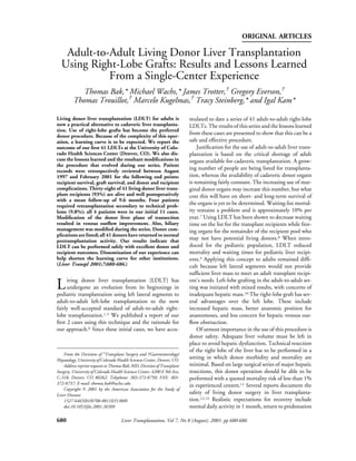
  • 4. Living Donor Liver Transplantation 683 listed in Table 3. Of 41 donors, 2 required nonautolo- gous blood transfusions (5%). One of these was for a large hemothorax secondary to a central venous catheter placed preoperatively by the anesthesia department. The second was in a patient whose hematocrit de- creased on postoperative day 2 and stabilized after 2 units of packed red blood cells were administered. Three patients had postoperative bile leaks. Two of these patients were returned to the operating room and underwent direct repair of a leaking bile duct stump within 3 days of the hepatectomy. One of these stumps was the cystic duct, whereas the other was the right hepatic duct stump. The other patient was treated con- servatively with percutaneous drainage, resulting in res- Figure 2. Donor aspartate aminotransferase (AST) (s) olution of the leak from the cut liver surface. One large and alanine aminotransferase (ALT) (}) levels versus incisional hernia was repaired in a patient who returned postoperative day. to lifting concrete bags 2 weeks after her surgery. One Jackson-Pratt drain was retrieved surgically after it snapped off during its removal. One idiosyncratic med- setting was consistent with poor graft function based on ication reaction caused donor lethargy that resolved worsening laboratory values and a deteriorating clinical postoperative day 3. Also, a temporary neuropraxia picture. Ultrasound evaluation showed patent vessels occurred in the dominant hand of one donor. All 41 with no evidence of venous obstructions. Venous con- donors are alive, well, and have returned to normal gestion was confirmed on gross inspection and micro- activity. scopic analysis of the explanted liver. One retransplan- Postoperative laboratory values are shown in Figure tation was performed for hepatic artery thrombosis 2. Aspartate aminotransferase and alanine aminotrans- postoperative day 12. The fourth retransplantation was ferase levels peaked postoperative day 3, with a return to performed for a persistent bile leak that 2 surgical normal by day 7. Serum bilirubin levels peaked slightly repairs failed to resolve. This patient underwent later. Mean hospital stay for donors was 6.3 days. retransplantation postoperative day 50 and died of sep- Forty-one right-lobe adult-to-adult LDLTs have sis 4 weeks after retransplantation. The second of the 3 been completed using this technique. Thirty-eight of deaths occurred 15 months posttransplantation because 41 recipients (93%) are alive and well, with a mean of uncontrolled chronic rejection, whereas the third follow-up of 9.6 months. Thirty-six of 41 grafts are death was in a patient who developed multiple strokes functioning, for a graft survival rate of 88%. Four on postoperative day 1 and died of a cardiac arrest on patients required retransplantation with cadaveric postoperative day 5. grafts (9.8%). All 4 of these retransplantations occurred Forty-four percent of transplant recipients under- in our initial 11 cases, initiating several technical adjust- went multiple bile duct reconstructions, with one ments described next. Two retransplantations were per- patient requiring 3 bile duct anastomoses. Overall bili- formed for hepatic dysfunction resulting from hepatic ary complication rates were 34%. This includes 3 anas- venous outflow obstruction. These were performed tomotic leaks treated surgically (1 duct-to-duct, 2 postoperative days 14 and 5. In both cases, the clinical Roux-en-Y). Three patients developed postoperative strictures. The first occurred in a duct-to-duct anasto- mosis, which was converted to a Roux. The other 2 strictures were in patients with Roux limb anastomoses, Table 3. Donor Complications and these were successfully treated with percutaneous transhepatic cholangioplasty. Nine patients developed Bile leak, reoperation 2 cut surface leaks; 6 patients were treated with prolonged Bile leak, external drainage 1 drainage and 3 patients underwent reoperation. Eight Incisional hernia, surgical repair 1 Neuropraxia, transient 1 of the 9 leaks resolved; the exception was the patient Drain retrieval, reoperation 1 who died after retransplantation. Hemothorax from venous access 1 Four patients required a second hepatic vein anasto- mosis because of an accessory hepatic vein greater than
  • 5. 684 Bak et al 1 cm. One of these patients had 2 accessory hepatic the right hepatic vein system.15 Since moving our tran- veins. Three patients underwent 2 portal venous anas- section plane in this manner, we have not experienced tomoses without complications. outflow problems causing graft dysfunction. Another venous drainage issue is that of accessory hepatic veins draining into the vena cava. Previous series of right-lobe Discussion transplants have reported reanastomosis of any acces- Our results suggest that adult-to-adult LDLT using a sory vein greater than 5 mm.16 We have been more right hepatic lobe graft is a safe and effective operation selective in our series, and of the 41 grafts, have per- for people requiring liver transplantation when careful formed accessory venous anastomoses in only 4 donor and recipient screening is performed. Graft and patients. We routinely reconstruct accessory veins patient survival rates in our series are equal to our cur- greater than 0.8 to 1 cm. rent cadaveric transplantation results. Donor safety is of Arterial anatomy is often quite variable in both utmost importance for this procedure, and although donor and recipient. Most commonly, the major right there have been reported donor complications and hepatic artery of the donor graft can be directly anasto- deaths,5,14 our series shows excellent donor outcomes mosed to the recipient artery at either the bifurcation of with 100% survival and return to normal activity. the right and left branches or a bifurcation patch created Despite these results, it should be stressed that a at the branch of the cystic artery. We attempt to maxi- learning curve exists with this procedure: as experience mize right hepatic arterial length in the recipient but and level of comfort increased, so did our overall success stay extraparenchymal in this dissection. On donor rates. The four retransplantations described during our workup, a replaced right hepatic artery is not a contra- early experience led us to 2 important technical indication; this frequently makes the operation techni- changes. Specifically, our significant changes during cally easier because of a longer length of graft artery. In this procedure have been improvements in graft hepatic this situation, an angiogram is often obtained to assess venous outflow and the understanding of the need for whether this right branch off the superior mesenteric intraoperative cholangiography to help show and artery is completely replaced or whether it is an acces- reconstruct accessory right bile ducts. sory branch. This allows us to conclude whether a sec- In our early experience, we transected the liver to the ond arterial anastomosis is required. We have studied right of the middle hepatic vein, even if the middle arterial flow postoperatively with duplex ultrasonogra- hepatic vein contributed drainage to segments 5 and 8. phy on a daily basis through postoperative day 5. Using this technique, 2 transplant recipients had severe Patients are started on 81 mg of acetylsalicylic acid graft congestion and poor function requiring retrans- when a stable postoperative hematocrit is present. To plantation. Their clinical picture was consistent with date, we have had one hepatic artery thrombosis, which poor graft function despite ultrasound evaluation show- is similar in incidence to our cadaveric recipient popu- ing patent vessels and no obstruction. We now believe lation, and concerns of greater thrombosis rates caused that primary nonfunction of these grafts is very by smaller vessels have not been realized. Arterial anas- unlikely, and if the postoperative clinical course mimics tomoses are performed with 3.5 ϫ loupe magnification this picture, it is likely caused by a technical issue. These with a running monofilament 7-0 vascular suture. In 2 cases prompted us to change our plane of transection to cases, the donor right hepatic artery bifurcated into the left. The right branch of the middle hepatic vein is anterior and posterior branches that encircled the com- preserved with the right-lobe graft and ligated superi- mon bile duct. In the first instance, the anterior branch orly at its junction with the main middle hepatic vein. was small and therefore was ligated. In the second case, We believe this prevents disruption of collateral drain- both arteries were of equal size. The anterior artery was age between the right hepatic vein and the right branch transected at the time of graft removal to free it from the of the middle hepatic vein. We do not believe it is bile duct and was then repaired, with an end-to-end necessary to connect this middle hepatic vein branch to anastomosis performed on the back table. the cava using a jump graft. When the graft is reper- Portal vein inflow has been adequate in all patients. fused, this right branch of the middle hepatic vein is Direct end-to-end anastomosis of the donor right por- well decompressed, presumably by collaterals in the tal vein to the right branch or common portal vein of graft. It has been our experience and also recently the recipient was performed in all but 3 cases. In these reported that postoperative ultrasound examination of cases, there were 2 separate major portal veins to the the graft drainage shows reversal of flow in the right right lobe. In 1 of these, 2 anastomoses were performed; middle hepatic vein branch ultimately emptying into one each to the right and left branches of the recipient
  • 6. Living Donor Liver Transplantation 685 portal vein. In the other 2 cases, the graft veins were very internal stent. In addition, Roux drainage is performed close to each other, with liver parenchyma providing a in cases of a single bile duct when there is concern about common back wall, and the donor had a large native tension or blood supply. We do not use t-tubes or other portal system. A single anastomosis of the donor veins external biliary drainage catheters. Biliary complica- to the native portal vein branch was performed. tions have occurred in 34% of transplant recipients. A preoperative ultrasound showed one transplant This is similar to rates previously reported in living recipient to have a thrombosed portal vein, with patent donor liver surgery.17-19 Most of our leaks (60%) have splenic and superior mesenteric veins. This LDLT was been raw surface leaks. These are usually managed con- delayed until a cadaveric donor iliac vein of compatible servatively with external drainage if the patient is clini- blood type was available to use as a jump graft. At cally stable. These usually occur while the surgical surgery, a successful thrombectomy allowed for ade- drains are still in place. Our experience has been that quate inflow and no vascular graft was used. To date in these will spontaneously seal, allowing for drain re- our series, we have not had to use an arterial or venous moval. If the patient shows signs of uncontrolled leak- graft for reconstruction; however, we routinely repeat age, such as pain or increasing bilirubin levels, they are an ultrasound examination of the recipient 1 week treated surgically. Biliary leak rates are slightly greater in before surgery to rule out portal vein thrombosis. The the Roux-drained patients, but this is likely influenced use of a recipient saphenous vein graft may be required by the fact that this group includes patients with mul- in a setting in which the right lobe has 2 arterial inflows, tiple duct drainage. i.e., an accessory right branch as opposed to a totally Postoperative management of living donor liver replaced system. transplant recipients has been similar to that of recipi- The second change in technique is in regard to our ents of cadaveric grafts. Overall, 56% of recipients went biliary management. Magnetic resonance cholangio- to the intensive care unit postoperatively. This graphy (MRC) imaging is used as preoperative screen- decreased to 40% in the second half of our series as our ing of donor biliary anatomy. This has proven to be a level of comfort with these cases has increased. An relatively accurate assessment of major biliary struc- increase in frequency of surveillance ultrasonography is tures. In our early cases, a transplant recipient experi- performed, but this has not shown greater thrombosis enced a bile leak from an accessory posterior duct that rates. Hospital length of stay averaged 21.5 days for the had not been recognized on the MRC or at the original transplant recipients. We believe that this is lengthened donor surgery. Despite reoperation and oversewing, the somewhat because of the increase in biliary complica- leak persisted, eventually leading to retransplantation, tions compared with our cadaveric transplant recipients sepsis, and death. We have since instituted routine and a less predictable decrease in postoperative liver intraoperative cholangiography and have identified and function test results, leading to a heightened vigilance reconstructed accessory bile ducts in 44% of our right- for potential complications and rejection. However, lobe grafts. On one occasion, a patient underwent 2 graft and patient survival rates ultimately are equal to accessory biliary reconstructions. Identification and cadaveric results. drainage of this important posterior branch has pre- These results must be viewed cautiously because vented biliary complications in a significant number of candidates are carefully selected for LDLT and in gen- right-lobe grafts, and we have not performed a retrans- eral are not the sickest liver transplant recipients. We plantation for a biliary complication since. In 2 believe that the optimal candidates for this procedure instances, we have oversewn a small (Ͻ3 mm) posterior are patients who are UNOS status IIB and have a good right duct, avoiding reconstruction. Neither of these 2 donor available. Status III patients should rarely cases had a leak or signs of cholangitis in this small undergo transplantation until more experience has been undrained biliary section of hepatic parenchyma. These gained to justify the risk to the donor. The use of LDLT smaller ducts are usually located in a more posterior is especially valuable in the patient population who has location than the previously mentioned accessory duct, a suspicion for or proven small hepatocellular carci- which is significant and is reconstructed. noma (status IIB), but would unlikely be able to receive Biliary reconstruction is performed with chole- a cadaveric graft before their cancer progressed to a dochocholedochostomy when possible. The need for nontransplantable stage. The role of this procedure as Roux limb drainage is obvious in the case of primary palliation for large hepatocellular cancers is still being sclerosing cholangitis or when multiple anastomoses debated. Our center has not performed this procedure must be performed, and these are performed to separate on a patient who would otherwise not be a candidate for openings of the intestinal limb, each over an individual a cadaveric graft.
  • 7. 686 Bak et al In conclusion, LDLT is a safe and effective proce- 9. Sindhi R, Rosendale J, Mundy D, Taranto S, Baliga P, Reuben dure for well-selected patients. We report a large series A, et al. Impact of segmental grafts on pediatric liver transplan- tation—A review of the United Network for Organ Sharing of right-lobe grafts with similar graft and patient sur- Scientific Registry Data (1990-1996). J Pediatr Surg 1999;34: vival statistics to our cadaveric transplant recipients. 107-110. Donor safety has been maintained throughout the 10. Emond JC, Renz JF, Ferrell LD, Rosenthal P, Lim RC, Roberts series. This procedure will continue to grow in impor- JP, et al. Functional analysis of grafts from living donors. Ann tance in the national transplant community as a way to Surg 1996;224:544-552. alleviate some of the pressure of growing waiting lists 11. Belghiti J, Hiramatsu K, Benoist S, Massault P, Sauvanet A, Farges O. Seven hundred forty-seven hepatectomies in the and stagnant cadaveric donor availability. 1990s: An update to evaluate the actual risk of liver resection. J Am Coll Surg 2000;191:38-46. References 12. Fujita S, Kim ID, Uryuhara K, Asonuma K, Egawa H, Kiuchi T, et al. Hepatic grafts from live donors: Donor morbidity for 470 1. Broelsch CE, Whitington PF, Emond JC, Heffron TG, This- cases of live donation. Transpl Int 2000;13:333-339. tlethwaite JR, Stevens L, et al. Liver transplantation in children 13. Fan ST, Lo CM, Liu CL, Yong BH, Chan JK, Ng IO. Safety of from living related donors: Surgical techniques and results. Ann donors in live donor liver transplantation using right lobe grafts. Surg 1991;214:428-437. Arch Surg 2000;135:336-340. 2. Broelsch CE, Burdelski M, Rogiers X, Gundlach M, Knoefel 14. Grewal HP, Thistlewaite JR, Loss GE, Fisher JS, Cronin DC, WT, Langwieler T, et al. Living donor for liver transplantation. Siegal CT, et al. Complications in 100 living liver donors. Ann Hepatology 1994;20(suppl 1, part 2):49S-55S. Surg 1998;228:214-219. 3. Kawasaki S, Makuuchi M, Matsunami H, Hashikura Y, Ikegami 15. Kaneko T, Kaneko K, Sugimoto H, Inoue S, Hatsuno T, Sawada T, Nakazawa Y, et al. Living related liver transplantation in adults. Ann Surg 1998;227:269-274. K, et al. Intrahepatic anastomosis formation between the hepatic 4. Todo S, Furukawa H, Jin MB, Shimamura T. Living donor liver veins in the graft liver of the living related liver transplantation: transplantation in adults: Outcome in Japan. Liver Transpl Observation by Doppler ultrasonography. Transplantation 2000;6(suppl 2):S66-S72. 2000;70:982-985. 5. Broelsch CE, Malago M, Testa G, Gamazo CU. Living donor 16. Marcos A, Ham JM, Fisher RA, Olzinski AT, Posner MP. liver transplantation in adults: Outcome in Europe. Liver Single center analysis of the first 40 adult-to-adult living donor Transpl 2000;6(suppl 2):S64-S65. liver transplants using the right lobe. Liver Transpl 2000;6:296- 6. Wachs ME, Bak TE, Karrer FM, Everson GT, Shrestha R, 301. Trouillot TE, et al. Adult living donor liver transplantation using 17. Marcos A, Fisher RA, Ham JM, Shiffman ML, Sanyal AJ, Luke- a right hepatic lobe. Transplantation 1998;66:1313-1316. tic VA, et al. Right lobe living donor liver transplantation. Trans- 7. Annual Report of the US Scientific Registry for Organ Trans- plantation 1999;68:798-803. plantation and the Organ Procurement and Transplantation 18. Cronin D, Alonso E, Piper J, Newell K, Bruce D, Woodle P, et Network—Transplant Data 1988-2000. UNOS, Richmond, al. Biliary complications in living donor liver transplantation. VA, and the Division of Transplantation, Bureau of Health Transplant Proc 1997;29:419-420. Resources and Services Administration, US Department of 19. Testa G, Malego M, Valentin-Gamazo C, Lindell G, Broelsch Health and Human Services, Rockville, MD, 2000. CE. Biliary anastomosis in living related liver transplantation 8. Trotter JF. Selection of donors and recipients for living donor using the right lobe: Techniques and complications. Liver liver transplantation. Liver Transpl 2000;6(suppl 2):S52-S58. Transpl 2000;6:710-714.