
Recomendados
Más contenido relacionado
La actualidad más candente
La actualidad más candente (20)
Similar a Chapter 6 - The Muscular System
Similar a Chapter 6 - The Muscular System (20)
Más de mpattani
Más de mpattani (20)
Último
Último (20)
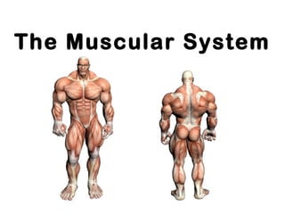
Chapter 6 - The Muscular System
- 2. Did you know that ? - more than 50% of body weight is muscle ! - And muscle is made up of proteins and water
- 4. The Muscular System • Muscles are responsible for all movement of the body • There are three basic types of muscle – Skeletal – Cardiac – Smooth
- 5. Info About Muscles • Only body tissue able to contract • create movement by flexing and extending joints • Body energy converters (many muscle cells contain many mitochondria)
- 6. 3 Types of Muscles
- 7. Three types of muscle Skeletal Cardiac Smooth
- 8. Classification of Muscle Skeletal- found in limbs Cardiac- found in heart Smooth- Found in viscera Striated, multi- nucleated Striated, 1 nucleus Not striated, 1 nucleus voluntary involuntary involuntary
- 9. Characteristics of Muscle • Skeletal and smooth muscle are elongated • Muscle cell = muscle fiber • Contraction of a muscle is due to movement of microfilaments (protein fibers) • All muscles share some terminology – Prefixes myo and mys refer to muscle – Prefix sarco refers to flesh
- 10. Shapes of Muscles • Triangular- shoulder, neck • Spindle- arms, legs • Flat- diaphragm, forehead • Circular- mouth, anus
- 12. Skeletal Muscle • Most are attached by tendons to bones • Cells have more than one nucleus (multinucleated) • Striated- have stripes, banding • Voluntary- subject to conscious control • Tendons are mostly made of collagen fibers • Found in the limbs • Produce movement, maintain posture, generate heat, stabilize joints
- 13. Structure of skeletal muscle • Each cell (fibre) is long and cylindrical • Muscle fibres are multi-nucleated • Typically 50-60mm in diameter, and up to 10cm long • The contractile elements of skeletal muscle cells are myofibrils
- 14. Skeletal muscle - Summary • Voluntary movement of skeletal parts • Spans joints and attached to skeleton • Multi-nucleated, striated, cylindrical fibres
- 15. Smooth Muscle • No striations • Spindle shaped • Single nucleus • Involuntary- no conscious control • Found mainly in the walls of hollow organs
- 16. Smooth muscle • Lines walls of viscera • Found in longitudinal or circular arrangement • Alternate contraction of circular & longitudinal muscle in the intestine leads to peristalsis
- 17. Smooth Muscle
- 18. Structure of smooth muscle • Spindle shaped uni-nucleated cells • Striations not observed • Actin and myosin filaments are present( protein fibers)
- 19. Smooth muscle - Summary • Found in walls of hollow internal organs • Involuntary movement of internal organs • Elongated, spindle shaped fibre with single nucleus
- 20. Cardiac Muscle • Striations • Branching cells • Involuntary • Found only in the heart • Usually has a single nucleus, but can have more than one
- 21. Cardiac muscle • Main muscle of heart • Pumping mass of heart • Critical in humans • Heart muscle cells behave as one unit • Heart always contracts to it’s full extent
- 22. Structure of cardiac muscle • Cardiac muscle cells (fibres) are short, branched and interconnected • Cells are striated & usually have 1 nucleus • Adjacent cardiac cells are joined via electrical synapses (gap junctions) • These gap junctions appear as dark lines and are called
- 23. Cardiac muscle - Summary • Found in the heart • Involuntary rhythmic contraction • Branched, striated fibre with single nucleus and intercalated discs
- 24. Muscle Control Type of muscle Nervous control Type of control Example SkeletalSkeletal Controlled by CNS Voluntary Lifting a glass Cardiac Regulated by ANS Involuntary Heart beating Smooth Controlled by ANS Involuntary Peristalsis
- 25. Types of Responses • Twitch- – A single brief contraction – Not a normal muscle function • Tetanus – One contraction immediately followed by another – Muscle never completely returns to a relaxed state – Effects are compounded
- 26. Where Does the Energy Come From? • Energy is stored in the muscles in the form of ATP • ATP comes from the breakdown of glucose during Cellular Respiration • This all happens in the Mitochondria of the cell • When a muscle is fatigued (tired) it is unable to contract because of lack of Oxygen
- 27. Fast Twitch and Slow Twitch Fibers Fast Twitch vs Slow Twitch
- 28. Exercise and Muscles • Isotonic- muscles shorten and movement occurs ( most normal exercise) • Isometric- tension in muscles increases, no movement occurs (pushing one hand against the other)
- 29. How are Muscles Attached to Bone? • Origin- attachment to immovable bone • Insertion- attachment to a movable bone • Muscles are always attached to at least 2 points • Movement is attained due to a muscle moving an attached bone
- 31. Muscle Attachments • The origin is on the clavicle and sternum. • The insertion is on the skull. • When the muscle contracts it will shorten the distance between the origin and insertion. • The head will move when this muscle contracts.
- 32. Flexion Types of Musculo-Skeletal Movement
- 33. Extension
- 34. Hyperextension
- 36. Rotation
- 37. More Types of Movement…… • Inversion- turn sole of foot medially • Eversion- turn sole of foot laterally • Pronation- palm facing down • Supination- palm facing up • Opposition- thumb touches tips of fingers on the same hand
- 38. The Skeletal Muscles There are about 650 muscles in the human body. They enable us to move, maintain posture and generate heat. In this section we will only study a sample of the major muscles.
- 40. Sternocleidomastoideus • Sometimes called the sternocleitomastoid. • It is the same neck muscle shown on the previous slide. • This muscle has two origins. – The first origin is on the sternum manubrium. – The second origin is on the clavicle. • The insertion is on the mastoid process of the skull. • Contraction of both sternocleidomastoideus muscles will flex the head. If just one of the muscles contracts, the head will rotate.
- 42. Masseter • The masseter is one of major chewing muscles. • The origin of the masseter is on the zygomatic arch. • The insertion is on the mandible. • Contraction of the masseter will elevate the jaw.
- 43. Temporalis Elevate & Retract Mandible
- 44. Temporalis • The temporalis is another chewing muscle. – Note how it attaches on the side of skull. • It also elevates the mandible. • You do not need to know the insertions and origins for this muscle
- 45. Trapezius Extend Head, Adduct, Elevate or Depress Scapula
- 46. Trapezius • The trapezius is a large muscle in the upper back. • It attaches to the skull, shoulder and vertebrae of the back. • When this muscle contracts it will cause the head to extend. • It will also move the scapula. • The direction the scapula moves depends on which part of the trapezius contracts. • The trapezius may elevate or depress the scapula.
- 47. Latissimus Dorsi Extend, Adduct & Rotate Arm Medially
- 48. Latissimus Dorsi • The latissimus dorsi is a large muscle in the back. – It is often referred to as a lat. • It has origins on the vertebrae, ilium ribs and scapula. • The insertion is on the humerus. – When it contracts it moves the humerus. • It can extend, adduct and rotate the arm medially. • This is the main muscle used in movement such as pounding a nail with a hammer.
- 49. Deltoid Abduct, Flex & Extend Arm
- 50. Deltoid • The deltoid covers the shoulder and has the shape of a delta. • It has origins on the scapula and clavicle. – The deltoid inserts on the deltoid tuberosity of the humerus. • Contraction of the deltoid will adduct the arm. • If only the anterior fibers of the muscle contract it will flex the arm. • Contraction of the posterior fibers will extend the arm.
- 51. Pectoralis Major Flexes, adducts & rotates arm medially
- 52. Pectoralis Major • The pectoralis major is a large muscle in the pectoral region of the body. • It has origins on the clavicle and sternum. – The insertion is on the greater tubercle of the humerus. • Contraction of the pectoralis major will flex the arm. • It will also adduct and rotate the arm medially. • The pectoralis major is used in movements such a climbing, throwing and doing pushups.
- 53. Biceps Brachii Flexes Elbow Joint
- 54. Biceps Brachii • The biceps brachii is located on the anterior side of the upper arm. • It is often just called the biceps. – There is a biceps femoris in the leg we will study shortly. • The biceps has two origins. One origin is on the corocoid process and the other on the Glenoid cavity of the scapula. • The “bi” in biceps refers to the two origins. – It inserts on the radial tuberosity. • Contraction of the biceps will cause flexing at the elbow joint.
- 55. Triceps Brachii Extend Elbow Joint
- 56. Triceps Brachii • The triceps is on the back of the upper arm. • It has three origins. • Two origins are on the back of the humerus and one on the scapula. • The triceps inserts on the olecranon. • Movement of the triceps will extend the elbow joint.
- 58. Rectus Abdominus • Rectus abdominus is a long muscle in the abdomen. • The muscle originates on the pubis. • It inserts on the xiphoid process of the sternum and also on cartilage of the ribs. • When rectus abdominus contracts it will flex the abdomen.
- 60. External Oblique • Another muscle in the abdomen is the external oblique. • It has muscle fibers that run in an oblique direction across the abdomen. • Contraction of the external oblique will compress the abdomen.
- 62. External Intercostals • There are two groups of muscles that run between the ribs. • The first are the external intercostals. • They will elevate the ribs.
- 64. Internal Intercostals • The internal intercostals are also located between the ribs. • They will depress the ribs.
- 66. Diaphragm • This is an inferior view of the diaphragm. • This muscle separates the abdominal cavity from the thoracic cavity. • When it contracts it will cause inspiration.
- 67. Forearm Muscles
- 68. Forearm Muscles • Flexor carpi—Flexes wrist • Extensor carpi—Extends wrist • Flexor digitorum—Flexes fingers • Extensor digitorum—Extends fingers • Pronator—Pronates • Supinator—Supinates
- 69. Gluteus Maximus Extends & Rotates Thigh Laterally
- 70. Gluteus Maximus • The large muscle on the posterior side of the body at the top of each leg is the gluteus maximus. • The gluteus maximus originates on the ilium, sacrum and coccyx. • It inserts on the gluteal tuberosity of the femur. • This muscle will extend and rotate the thigh laterally.
- 71. Rectus Femoris Flexes Thigh, Extends Lower Leg
- 72. Rectus Femoris • Rectus femoris is located on the anterior side of the thigh. • It originates on the ilium. • The insertion is on the patella and the tibial tuberosity. • When rectus femoris contracts it will flex the thigh and extend the lower leg.
- 73. Gracilis Adducts and Flexes Thigh
- 74. Gracilis • The gracilis is on the medial side of the thigh. • It adducts and flexes the thigh.
- 75. Sartorius Flexes Thigh, & Rotates Thigh Laterally
- 76. Sartorius • Sartorius is a long, strap like muscle. • It originates on the anterior superior iliac spine of the ilium. • The insertion is on the medial side of the tibia. • Contraction of the sartorius flexes the thigh and rotates the thigh laterally. • This is the muscle used when crossing the legs to sit on the floor.
- 77. Biceps Femoris Extends Thigh & Flexes Lower Leg
- 78. Biceps Femoris • Biceps femoris is one of the hamstring muscles. • The origin is on the ischial tuberosity. • Biceps femoris inserts on the tibia and fibula. • This muscle extends the thigh and flexes the lower leg.
- 79. Gastrocnemius Plantar Flexes Foot & Flex Lower Leg
- 80. Gastrocnemius • Gastrocnemius is commonly called the calf muscle. • It originates on the distal end of the femur. • The insertion is on the calcaneus bone of the foot. • It will cause plantar flexion of the foot and also flex the lower leg.
- 81. Tibialis Anterior Dorsiflexes and Inverts Foot
- 82. Tibialis Anterior • Tibialis anterior is located on the anterior side of the tibia. • It will dorsiflex and invert the foot.
Notas del editor
- Muscles attach in at least two places in the body. *The origin is the attachment that moves the least. *The insertion is the attachment that moves the most. This diagram illustrates the origin and insertion of one of the neck muscles. Note the origin is on the clavicle and sternum. The insertion is on the skull. When the muscle contracts it will shorten the distance between the origin and insertion. The head will move when this muscle contracts. Remember the insertion is the end of the muscle that moves the most. Since the head moves the attachment on the head is called the insertion. The origin is generally on a larger body part will move the least. The chest does not move when this muscle contract. The bones in the chest are therefore the origin.
- You will need to know the action or movement performed for each of the muscles we study. The first type of movement is called flexion. Note the lower leg is being flexed in this diagram. During flexion the angle of joint is decreased. As the knee is flexed, the angle between the lower leg and the thigh is decreased. Flexion of the upper arm is also illustrated in this diagram. Here the angle between the arm and the frontal plane is decreased.
- Extension is the opposite of flexion. In extension the angle of a joint is increased. Extension of the lower leg causes an increased angle between the lower leg and the thigh.
- Flexion and extension also apply to the neck. When a joint is extended past the anatomical position the movement is called hyperextension.
- Abduction refers to moving away from the median plane of the body. Adduction is the opposite movement to abduction. It is moving toward the medial plane. Circumduction refers to inscribing a circle while moving a limb.
- Rotation is turning a bone on its own axis. Moving the head back and forth to indicate “no” is an example of rotation. Note the difference between medial and lateral rotation. In lateral rotation the limb is rotated the lateral side of the body. Medial rotation rotates the limb toward the medial side of the body.
- There are about 650 muscles in the human body. They enable us to move, maintain posture and generate heat. In this unit will only study a sample of the major muscles.
- The first muscle we will learn is the sternocleidomastoideus. It is sometime called the sternocleitomastoid. It is the same neck muscle shown on the previous slide. * This muscle has two origins. The first origin is on the sternum manubrium. The second origin is on the clavicle. *The insertion is on the mastoid process of the skull. *Contraction of both sternocleidomastoideus muscles will flex the head. If just on of the muscles contracts, the head will rotate.
- The masseter is one of major chewing muscles. *The origin of the masseter is on the zygomatic arch. *The insertion is on the mandible. *Contraction of the masseter will elevate the jaw.
- The temporalis is another chewing muscle. Note how it attaches on the side of skull. *It also elevates the mandible. Also note I did not give the origin and insertion of this muscle. You will only need to know the origin and insertion of a few of the muscles in this unit.
- The trapezius is a large muscle in the upper back. It attaches to the skull, shoulder and vertebrae of the back. *When this muscle contracts it will cause the head to extend. It will also move the scapula. The direction the scapula moves depends on which part of the trapezius contracts. The trapezius may adduct elevate or depress the scapula.
- The latissimus dorsi is a large muscle in the back. It is often referred to as a lat. *It has origins on the vertebrae, ilium ribs and scapula. *The insertion is on the humerus. When it contracts it moves the humerus. *It can extend, adduct and rotate the arm medially. This is the main muscle used in movement such as pounding a nail with a hammer.
- The deltoid covers the shoulder and has the shape of a delta. *It has origins on the scapula and clavicle. *The deltoid inserts on the deltoid tuberosity of the humerus. *Contraction of the deltoid will adduct the arm. If only the anterior fibers of the muscle contract it will flex the arm. Contraction of the posterior fibers will extend the arm.
- The pectoralis major is a large muscle in the pectoral region of the body. *It has origins on the clavicle and sternum. *The insertion is on the greater tubercle of the humerus. *Contraction of the pectoralis major will flex the arm. It will also adduct and rotate the arm medially. The pectoralis major is used in movements such a climbing, throwing and doing pushups.
- The biceps brachii is located on the anterior side of the upper arm. It is often just called the biceps. There is a biceps femoris in the leg we will study shortly. *The biceps has two origins. One origin is on the corocoid process and the other on the Glenoid cavity of the scapula. The “bi” in biceps refers to the two origins. *It inserts on the radial tuberosity. *Contraction of the biceps will cause flexing at the elbow joint.
- The triceps is on the back of the upper arm. *It has three origins. Two origins are on the back of the humerus and one on the scapula. *The triceps inserts on the olecranon. *Movement of the triceps will extend the elbow joint.
- Rectus abdominus is a long muscle in the abdomen. *The muscle originates on the pubis. *It inserts on the xiphoid process of the sternum and also on cartilage of the ribs. *When rectus abdominus contracts it will flex the abdomen.
- Another muscle in the abdomen is the external oblique. It has muscle fibers that run in an oblique direction across the abdomen. *Contraction of the external oblique will compress the abdomen.
- There are two groups of muscles that run between the ribs. The first are the external intercostals. *They will elevate the ribs.
- The internal intercostals are also located between the ribs. *They will depress the ribs.
- This is an inferior view of the diaphragm. This muscle separates the abdominal cavity from the thoracic cavity. *When it contracts it will cause inspiration.
- There are several muscles in the forearm that have important movements. *The flexor carpi are used to flex the wrist. *Extensor carpi extend the wrist. *Flexor digitorum flexes the fingers. *Extensor digitorum extends the fingers. *The pronator pronates. *A supinator muscle will supinate.
- The large muscle on the posterior side of the body at the top of each leg is the gluteus maximus. *The gluteus maximus originates on the ilium, sacrum and coccyx. *It inserts on the gluteal tuberosity of the femur. *This muscle will extend and rotate the thigh laterally.
- Rectus femoris is located on the anterior side of the thigh. *It originates on the ilium. *The insertion is on the patella and the tibial tuberosity. *When rectus femoris contracts it will flex the thigh and extend the lower leg.
- The gracilis is on the medial side of the thigh. *It adducts and flexes the thigh.
- Sartorius is a long, strap like muscle. *It originates on the anterior superior iliac spine of the ilium. *The insertion is on the medial side of the tibia. * Contraction of the sartorius flexes the thigh and rotates the thigh laterally. This is the muscle used when crossing the legs to sit on the floor.
- Biceps femoris is one of the hamstring muscles. *The origin is on the ischial tuberosity. *Biceps femoris inserts on the tibia and fibula. This muscle extends the thigh and flexes the lower leg.
- Gastrocnemius is commonly called the calf muscle. *It originates on the distal end of the femur. *The insertion is on the calcaneus bone of the foot. *It will cause plantar flexion of the foot and also flex the lower leg.
- Tibialis anterior is located on the anterior side of the tibia. *It will dorsiflex and invert the foot.