SlideShare una empresa de Scribd logo
1 de 11
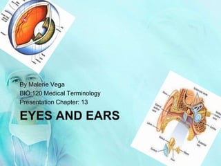
EYES AND EARS
By Malerie Vega
BIO:120 Medical Terminology
Presentation Chapter: 13
The Eye
The Eyeball
The Anatomy of the Eyeball
• Anterior Chamber: The space in the eye that is
behind the cornea and in front of the iris.
• Upper & lower lid: a thin fold of skin that covers and
protects the eye.
• Conjunctiva: a clear mucuse membrane consisting of
cells and underlying basement membrane
that covers the sclera (white part of the
eye) and lines the inside part of the eyelid.
• Pupil: a hole located in the center of the iris of the eye
that allows light to enter the eye
• Cornea: a transparent front part of the eye that covers the
iris and the pupil.
• Aqueous humor: The liquid between the lens and the
cornea.
• Posterior chamber: A narrow crack behind the
perpherial part of the iris
• Fovea centralis: The part of the eye located in the center
of the Macula region of the retina, this part is
responsible for the sharp central vision.
• Optic nerve: Transmits visual information from the
retina to the Brain.
• Central retinal artery: It pierces the optic nerve close to
the eyeball, sending branches over the internal
surface of the retina, and these terminal
branches are the only blood supply to the larger
part of the eye
• Sclera: fibrous, protective, outer layer of the eye
containing collagen and elastic fiber.
• Choroid: The vascular layer containing connective
tissue, of the eye lying between the retina and
the sclera. In humans its thickness is about 0.5 mm.
The choroid provides oxygen and nourishment to the
outer layers of the retina
• Retina: A light-sensitive tissue lining the inner
surface of the eye
Muscles of the eye
The extraocular
muscles are the six
muscles that control
the movements of the
(human) eye. The
actions of the
extraocular muscles
depend on the position
of the eye at the time
of muscle contraction.
Lacriminal apparatuse
The lacrimal apparatus is the
physiologic system containing the
orbital structures for tear production
and drainage[1]. It consists of
(a) the lacrimal gland, which
secretes the tears, and its
excretory ducts, which convey
the fluid to the surface of the
eye
(b) the lacrimal canaliculi, the
lacrimal sac, and the
nasolacrimal duct, by which the
fluid is conveyed into the cavity
of the nose, emptying
anterioinferiorly to the inferior
nasal conchae at the
nasolacrimal duct.
(c) the nerve supply of lacrimal
apparatus done by carotid
plexuse of nerves along artery
internal and external
sympathetically but
parasympathetic from lacrimal
nucleus of the facial nerve
• Vision begins when light rays are reflected off an
object and enter the eyes through the cornea, the
transparent outer covering of the eye. The cornea
bends or refracts the rays that pass through a round
hole called the pupil. The iris, or colored portion of the
eye that surrounds the pupil, opens and closes
(making the pupil bigger or smaller) to regulate the
amount of light passing through. The light rays then
pass through the lens, which actually changes shape
so it can further bend the rays and focus them on the
retina at the back of the eye. The retina is a thin layer
of tissue at the back of the eye that contains millions of
tiny light-sensing nerve cells called rods and cones,
which are named for their distinct shapes. Cones are
concentrated in the center of the retina, in an area
called the macula. In bright light conditions, cones
provide clear, sharp central vision and detect colors
and fine details. Rods are located outside the macula
and extend all the way to the outer edge of the retina.
They provide peripheral or side vision. Rods also allow
the eyes to detect motion and help us see in dim light
and at night. These cells in the retina convert the light
into electrical impulses. The optic nerve sends these
impulses to the brain where an image is produced.
How we see
The Ear
External ear
The outer ear has no
bones. It is the
external portion of the
ear, which consists of
the pinna, concha, and
auditory meatus. It
gathers sound energy
and focuses it on the
eardrum (tympanic
membrane).
Middle ear
The middle ear is the portion
of the ear internal to the
eardrum, and external to the
oval window of the cochlea.
The mammalian middle ear
contains three ossicles, which
couple vibration of the
eardrum into waves in the
fluid and membranes of the
inner ear. The hollow space of
the middle ear has also been
called the tympanic cavity, or
cavum tympani. The
eustachian tube joins the
tympanic cavity with the nasal
cavity (nasopharynx),
allowing pressure to equalize
between the middle ear and
throat.
Inner ear
The inner ear is the
innermost part of the
vertebrate ear. It consists of
the bony labyrinth, a
system of passages
comprising two main
functional parts:The
cochlea is dedicated to
hearingThe vestibular
system is dedicated to
balanceThe inner ear is
found in all vertebrates,
with substantial variations
in form and function. The
inner ear is innervated by
the eight cranial nerve in all
vertebrates.
How we hear
When something makes a noise, it sends
vibrations, or sound waves, through the
air. The human eardrum is a stretched
membrane, like the skin of a drum.
When the sound waves hit your eardrum,
it vibrates and the brain interprets these
vibrations as. After the vibrations hit
your eardrum, a chain reaction is set off.
Your eardrum, which is smaller and
thinner than the nail on your pinky
finger, sends the vibrations to the three
smallest bones in your body. First the
hammer, then the anvil, and finally, the
stirrup. The stirrup passes those
vibrations along a coiled tub in the inner
ear called the cochlea. Inside the cochlea
there are thousands of hair-like nerve
endings, cilia. When the Cochlea
vibrates, the cilia move. Your brain is
sent these messages (translated from
vibrations by the cilia) through the
auditory nerve. Your brain then
translates all that and tells you what you
are hearing. Neurologists don't yet fully
understand how we process raw sound
data once it enters the cerebral cortex in
the brain.

Más contenido relacionado

La actualidad más candente

La actualidad más candente (20)

Anatomy of the eye
Anatomy of the eyeAnatomy of the eye
Anatomy of the eye
 
Eye
EyeEye
Eye
 
The human eye
The human eyeThe human eye
The human eye
 
Human eye
Human eyeHuman eye
Human eye
 
Human eye
Human eyeHuman eye
Human eye
 
diagram of eye
diagram of eyediagram of eye
diagram of eye
 
The eye
The eyeThe eye
The eye
 
Mammalian eye
Mammalian eyeMammalian eye
Mammalian eye
 
The human eye
The human eyeThe human eye
The human eye
 
Human eye
Human eyeHuman eye
Human eye
 
ppt on EYE
ppt on EYEppt on EYE
ppt on EYE
 
Eye
EyeEye
Eye
 
Human eye and working of human eye
Human eye and working of human eyeHuman eye and working of human eye
Human eye and working of human eye
 
Parts of eyes
Parts of eyesParts of eyes
Parts of eyes
 
Human psychology assignment
Human psychology assignment Human psychology assignment
Human psychology assignment
 
Human eye
Human eyeHuman eye
Human eye
 
Special Senses
Special SensesSpecial Senses
Special Senses
 
Special Senses For Review
Special Senses For ReviewSpecial Senses For Review
Special Senses For Review
 
Structure of Eyes - Special Senses_ Human Anatomy & Physiology 1st
Structure of Eyes - Special Senses_ Human Anatomy & Physiology 1stStructure of Eyes - Special Senses_ Human Anatomy & Physiology 1st
Structure of Eyes - Special Senses_ Human Anatomy & Physiology 1st
 
Structure and function of eye
Structure and function of eyeStructure and function of eye
Structure and function of eye
 

Destacado

Malerie's presentation#13
Malerie's presentation#13Malerie's presentation#13
Malerie's presentation#13mvega0722
 
Malerie's presentation#13
Malerie's presentation#13Malerie's presentation#13
Malerie's presentation#13mvega0722
 
Malerie's presentation#13
Malerie's presentation#13Malerie's presentation#13
Malerie's presentation#13mvega0722
 
Malerie's Presentation
Malerie's PresentationMalerie's Presentation
Malerie's Presentationmvega0722
 
Biology presentation12
Biology presentation12Biology presentation12
Biology presentation12mvega0722
 
Ian Johnson Cv2012 D Luxury
Ian Johnson Cv2012 D LuxuryIan Johnson Cv2012 D Luxury
Ian Johnson Cv2012 D LuxuryBigfaluda
 
Biology presentation12
Biology presentation12Biology presentation12
Biology presentation12mvega0722
 
How to stop panic attacks fast and for good
How to stop panic attacks fast and for goodHow to stop panic attacks fast and for good
How to stop panic attacks fast and for goodnaztez
 
Biology presentation12
Biology presentation12Biology presentation12
Biology presentation12mvega0722
 
Desert
DesertDesert
Desertnlttms
 
Diversidade biologica
Diversidade biologicaDiversidade biologica
Diversidade biologicaJosenildocte
 
Biology presentation12
Biology presentation12Biology presentation12
Biology presentation12mvega0722
 
Presentation
Presentation Presentation
Presentation mvega0722
 
Desert
DesertDesert
Desertnlttms
 
Ian johnson CV 2012
Ian johnson CV 2012Ian johnson CV 2012
Ian johnson CV 2012Bigfaluda
 
Teaching Students with Emojis, Emoticons, & Textspeak
Teaching Students with Emojis, Emoticons, & TextspeakTeaching Students with Emojis, Emoticons, & Textspeak
Teaching Students with Emojis, Emoticons, & TextspeakShelly Sanchez Terrell
 

Destacado (20)

Malerie's presentation#13
Malerie's presentation#13Malerie's presentation#13
Malerie's presentation#13
 
Malerie's presentation#13
Malerie's presentation#13Malerie's presentation#13
Malerie's presentation#13
 
Malerie's presentation#13
Malerie's presentation#13Malerie's presentation#13
Malerie's presentation#13
 
Malerie's Presentation
Malerie's PresentationMalerie's Presentation
Malerie's Presentation
 
Biology presentation12
Biology presentation12Biology presentation12
Biology presentation12
 
Ian Johnson Cv2012 D Luxury
Ian Johnson Cv2012 D LuxuryIan Johnson Cv2012 D Luxury
Ian Johnson Cv2012 D Luxury
 
Biology presentation12
Biology presentation12Biology presentation12
Biology presentation12
 
How to stop panic attacks fast and for good
How to stop panic attacks fast and for goodHow to stop panic attacks fast and for good
How to stop panic attacks fast and for good
 
Biology presentation12
Biology presentation12Biology presentation12
Biology presentation12
 
Gestion de la seguridad
Gestion de la seguridadGestion de la seguridad
Gestion de la seguridad
 
Desert
DesertDesert
Desert
 
Diversidade biologica
Diversidade biologicaDiversidade biologica
Diversidade biologica
 
Biology presentation12
Biology presentation12Biology presentation12
Biology presentation12
 
Presentation
Presentation Presentation
Presentation
 
Tecnicas de Busqueda de Informacion en Google
Tecnicas de Busqueda de Informacion en GoogleTecnicas de Busqueda de Informacion en Google
Tecnicas de Busqueda de Informacion en Google
 
Desert
DesertDesert
Desert
 
Ian johnson CV 2012
Ian johnson CV 2012Ian johnson CV 2012
Ian johnson CV 2012
 
Guia adopción internacional
Guia adopción internacionalGuia adopción internacional
Guia adopción internacional
 
Inaugural Addresses
Inaugural AddressesInaugural Addresses
Inaugural Addresses
 
Teaching Students with Emojis, Emoticons, & Textspeak
Teaching Students with Emojis, Emoticons, & TextspeakTeaching Students with Emojis, Emoticons, & Textspeak
Teaching Students with Emojis, Emoticons, & Textspeak
 

Similar a Malerie's presentation#13

Med Term Presentation #3
Med Term Presentation #3Med Term Presentation #3
Med Term Presentation #3nhalm
 
Structure and Functions of the Eye and the Ear
Structure and Functions of the Eye and the EarStructure and Functions of the Eye and the Ear
Structure and Functions of the Eye and the EarGOPIKA NAIR
 
Med Term Presentation #3
Med Term Presentation #3Med Term Presentation #3
Med Term Presentation #3nhalm
 
Unit 4; Special Senses, Educational Platform.pptx
Unit 4; Special Senses, Educational Platform.pptxUnit 4; Special Senses, Educational Platform.pptx
Unit 4; Special Senses, Educational Platform.pptxZiaUddin5613
 
HEALTH ASSESSMENT AND DIAGNOSTIC TESTS OF EYE AND ENT DISORDERSDIAGNOSTIC TES...
HEALTH ASSESSMENT AND DIAGNOSTIC TESTS OF EYE AND ENT DISORDERSDIAGNOSTIC TES...HEALTH ASSESSMENT AND DIAGNOSTIC TESTS OF EYE AND ENT DISORDERSDIAGNOSTIC TES...
HEALTH ASSESSMENT AND DIAGNOSTIC TESTS OF EYE AND ENT DISORDERSDIAGNOSTIC TES...JishaSrivastava
 
the Sensory system Eye and Ear
the Sensory system Eye and Earthe Sensory system Eye and Ear
the Sensory system Eye and EarMawuogo
 
Chapter 13 Special Senses
Chapter 13 Special SensesChapter 13 Special Senses
Chapter 13 Special SensesPrincessQiana
 
14th aSpecial Senses of sight.pptx
14th aSpecial Senses of sight.pptx14th aSpecial Senses of sight.pptx
14th aSpecial Senses of sight.pptxChangezKhan33
 
181-Anatomy-Eyes-and-Ears.ppt
181-Anatomy-Eyes-and-Ears.ppt181-Anatomy-Eyes-and-Ears.ppt
181-Anatomy-Eyes-and-Ears.pptssuser0e32dd
 
A&P Lab 2 midterm review
A&P Lab 2 midterm review A&P Lab 2 midterm review
A&P Lab 2 midterm review guest6e42be7
 
A&P Chapter 24 Special Senses
A&P Chapter 24 Special SensesA&P Chapter 24 Special Senses
A&P Chapter 24 Special Sensesmarydelaney
 
Histology of Sensory organs
Histology of Sensory organsHistology of Sensory organs
Histology of Sensory organsEneutron
 
Special Senses eye, ear , nose, tongue ppt
Special Senses eye, ear , nose, tongue pptSpecial Senses eye, ear , nose, tongue ppt
Special Senses eye, ear , nose, tongue pptRupaSingh83
 

Similar a Malerie's presentation#13 (20)

Med Term Presentation #3
Med Term Presentation #3Med Term Presentation #3
Med Term Presentation #3
 
Structure and Functions of the Eye and the Ear
Structure and Functions of the Eye and the EarStructure and Functions of the Eye and the Ear
Structure and Functions of the Eye and the Ear
 
Med Term Presentation #3
Med Term Presentation #3Med Term Presentation #3
Med Term Presentation #3
 
Unit 4; Special Senses, Educational Platform.pptx
Unit 4; Special Senses, Educational Platform.pptxUnit 4; Special Senses, Educational Platform.pptx
Unit 4; Special Senses, Educational Platform.pptx
 
SENSE ORGAN
SENSE ORGANSENSE ORGAN
SENSE ORGAN
 
HEALTH ASSESSMENT AND DIAGNOSTIC TESTS OF EYE AND ENT DISORDERSDIAGNOSTIC TES...
HEALTH ASSESSMENT AND DIAGNOSTIC TESTS OF EYE AND ENT DISORDERSDIAGNOSTIC TES...HEALTH ASSESSMENT AND DIAGNOSTIC TESTS OF EYE AND ENT DISORDERSDIAGNOSTIC TES...
HEALTH ASSESSMENT AND DIAGNOSTIC TESTS OF EYE AND ENT DISORDERSDIAGNOSTIC TES...
 
the Sensory system Eye and Ear
the Sensory system Eye and Earthe Sensory system Eye and Ear
the Sensory system Eye and Ear
 
Chapter 13 Special Senses
Chapter 13 Special SensesChapter 13 Special Senses
Chapter 13 Special Senses
 
Eye + ear
Eye + earEye + ear
Eye + ear
 
Senses ppy
Senses ppySenses ppy
Senses ppy
 
14th aSpecial Senses of sight.pptx
14th aSpecial Senses of sight.pptx14th aSpecial Senses of sight.pptx
14th aSpecial Senses of sight.pptx
 
181-Anatomy-Eyes-and-Ears.ppt
181-Anatomy-Eyes-and-Ears.ppt181-Anatomy-Eyes-and-Ears.ppt
181-Anatomy-Eyes-and-Ears.ppt
 
Chapter 13
Chapter 13Chapter 13
Chapter 13
 
Special senses
Special sensesSpecial senses
Special senses
 
A&P Lab 2 midterm review
A&P Lab 2 midterm review A&P Lab 2 midterm review
A&P Lab 2 midterm review
 
A&P Chapter 24 Special Senses
A&P Chapter 24 Special SensesA&P Chapter 24 Special Senses
A&P Chapter 24 Special Senses
 
Histology of Sensory organs
Histology of Sensory organsHistology of Sensory organs
Histology of Sensory organs
 
Pns
PnsPns
Pns
 
Special Senses eye, ear , nose, tongue ppt
Special Senses eye, ear , nose, tongue pptSpecial Senses eye, ear , nose, tongue ppt
Special Senses eye, ear , nose, tongue ppt
 
Sensory organs
Sensory organsSensory organs
Sensory organs
 

Malerie's presentation#13

  • 1. EYES AND EARS By Malerie Vega BIO:120 Medical Terminology Presentation Chapter: 13
  • 3. The Eyeball The Anatomy of the Eyeball • Anterior Chamber: The space in the eye that is behind the cornea and in front of the iris. • Upper & lower lid: a thin fold of skin that covers and protects the eye. • Conjunctiva: a clear mucuse membrane consisting of cells and underlying basement membrane that covers the sclera (white part of the eye) and lines the inside part of the eyelid. • Pupil: a hole located in the center of the iris of the eye that allows light to enter the eye • Cornea: a transparent front part of the eye that covers the iris and the pupil. • Aqueous humor: The liquid between the lens and the cornea. • Posterior chamber: A narrow crack behind the perpherial part of the iris • Fovea centralis: The part of the eye located in the center of the Macula region of the retina, this part is responsible for the sharp central vision. • Optic nerve: Transmits visual information from the retina to the Brain. • Central retinal artery: It pierces the optic nerve close to the eyeball, sending branches over the internal surface of the retina, and these terminal branches are the only blood supply to the larger part of the eye • Sclera: fibrous, protective, outer layer of the eye containing collagen and elastic fiber. • Choroid: The vascular layer containing connective tissue, of the eye lying between the retina and the sclera. In humans its thickness is about 0.5 mm. The choroid provides oxygen and nourishment to the outer layers of the retina • Retina: A light-sensitive tissue lining the inner surface of the eye
  • 4. Muscles of the eye The extraocular muscles are the six muscles that control the movements of the (human) eye. The actions of the extraocular muscles depend on the position of the eye at the time of muscle contraction.
  • 5. Lacriminal apparatuse The lacrimal apparatus is the physiologic system containing the orbital structures for tear production and drainage[1]. It consists of (a) the lacrimal gland, which secretes the tears, and its excretory ducts, which convey the fluid to the surface of the eye (b) the lacrimal canaliculi, the lacrimal sac, and the nasolacrimal duct, by which the fluid is conveyed into the cavity of the nose, emptying anterioinferiorly to the inferior nasal conchae at the nasolacrimal duct. (c) the nerve supply of lacrimal apparatus done by carotid plexuse of nerves along artery internal and external sympathetically but parasympathetic from lacrimal nucleus of the facial nerve
  • 6. • Vision begins when light rays are reflected off an object and enter the eyes through the cornea, the transparent outer covering of the eye. The cornea bends or refracts the rays that pass through a round hole called the pupil. The iris, or colored portion of the eye that surrounds the pupil, opens and closes (making the pupil bigger or smaller) to regulate the amount of light passing through. The light rays then pass through the lens, which actually changes shape so it can further bend the rays and focus them on the retina at the back of the eye. The retina is a thin layer of tissue at the back of the eye that contains millions of tiny light-sensing nerve cells called rods and cones, which are named for their distinct shapes. Cones are concentrated in the center of the retina, in an area called the macula. In bright light conditions, cones provide clear, sharp central vision and detect colors and fine details. Rods are located outside the macula and extend all the way to the outer edge of the retina. They provide peripheral or side vision. Rods also allow the eyes to detect motion and help us see in dim light and at night. These cells in the retina convert the light into electrical impulses. The optic nerve sends these impulses to the brain where an image is produced. How we see
  • 8. External ear The outer ear has no bones. It is the external portion of the ear, which consists of the pinna, concha, and auditory meatus. It gathers sound energy and focuses it on the eardrum (tympanic membrane).
  • 9. Middle ear The middle ear is the portion of the ear internal to the eardrum, and external to the oval window of the cochlea. The mammalian middle ear contains three ossicles, which couple vibration of the eardrum into waves in the fluid and membranes of the inner ear. The hollow space of the middle ear has also been called the tympanic cavity, or cavum tympani. The eustachian tube joins the tympanic cavity with the nasal cavity (nasopharynx), allowing pressure to equalize between the middle ear and throat.
  • 10. Inner ear The inner ear is the innermost part of the vertebrate ear. It consists of the bony labyrinth, a system of passages comprising two main functional parts:The cochlea is dedicated to hearingThe vestibular system is dedicated to balanceThe inner ear is found in all vertebrates, with substantial variations in form and function. The inner ear is innervated by the eight cranial nerve in all vertebrates.
  • 11. How we hear When something makes a noise, it sends vibrations, or sound waves, through the air. The human eardrum is a stretched membrane, like the skin of a drum. When the sound waves hit your eardrum, it vibrates and the brain interprets these vibrations as. After the vibrations hit your eardrum, a chain reaction is set off. Your eardrum, which is smaller and thinner than the nail on your pinky finger, sends the vibrations to the three smallest bones in your body. First the hammer, then the anvil, and finally, the stirrup. The stirrup passes those vibrations along a coiled tub in the inner ear called the cochlea. Inside the cochlea there are thousands of hair-like nerve endings, cilia. When the Cochlea vibrates, the cilia move. Your brain is sent these messages (translated from vibrations by the cilia) through the auditory nerve. Your brain then translates all that and tells you what you are hearing. Neurologists don't yet fully understand how we process raw sound data once it enters the cerebral cortex in the brain.