1. Retrospective Study of the Utility of 18F Sodium Fluoride PET Bone Scans in Patients with
Differentiated Thyroid Cancer with Suspected Bone Metastases
Orquiza M1, Schneider M1, Garcia1, O’Neil J1, Van Nostrand D1, Wexler J2, Burman K2, Wartofsky L3
Div of Nuclear Medicine1, Div of Endocrinology2, Dept of Medicine3, MedStar Washington Hospital Center
All patients were given thyroid hormone suppression with either L- Table 1
thyroxine or liothyronine. Of the 16 pts, noninvasive diagnostic Patient Demographics Discussion
Mean Age (range) 71 yr (32-87)
tests and invasive procedures were eliminated in 13 (81%) and 11
Gender F/M (%) 9 (56%) / 7 ( 44%)
In this review, we evaluated both pre and post 18F
(69%) pts, respectively. The management plan was altered in 10
ABSTRACT (63%) pts, including a decision to (i) treat with 131I in 1 (6%) (ii) Histology (%)
NaF PET bone scan NOPR forms for the scan’s
-Papillary 9 (53%) utility in assessing the following three items:
The use of 18F sodium fluoride (18F NaF) was first administer external radiation therapy and/or bisphosphonates in 7 -Hürthle cell 4 (23%) elimination of noninvasive diagnostic
used in the 1960’s for skeletal scintigraphy, but was (41%), (iii) perform a tissue biopsy in 1 (6%), and (iv) cancel a -Follicular 2 (12%)
-Papilliary-follicular 1 (6%) testing, elimination of invasive procedures, and
rapidly replaced by 99mTc-phosphate agents to detect tissue biopsy in 2 (12%).
Anti-thyroglobulin Ab (%) alteration of management as a result of the 18F NaF
bone metastasis because the latter were less CONCLUSION: The preliminary data suggest that 18F NaF PET
- Not Detected 16 (81%) PET bone scan.
expensive and allowed for better images. bone scans may eliminate additional noninvasive diagnostic - Detected 3 (19%)
The study has strengths and limitations. The
However, with the availability of PET testing in 81% of patients, eliminate invasive procedures in 69% of Median (range) strengths of this study are the evaluation of 18F NaF
scanners, better skeletal images have been obtained patients, and alter management in 63% of selected patients with -TSH 0.42 (0.012-162.4) uIU/mL
-TG 63.3 (<0.2->35000) ng/mL PET in a specific cancer type as well as the
with 18F NaF. thyroid cancer.
evaluation of the alteration of management. The
OBJECTIVES: The objective of this study was to Graph 1 limitations of the study include a small number of
evaluate the utility of 18F sodium fluoride (18F NaF) Methods Utility of 18FNa
patients, no data available on alteration of
positron emission tomography (PET) bone scans in 90%
81%
outcomes, and the potential for bias of the referring
altering the management of patients (pts) suspected A retrospective review was performed of all pts with DTC and an 80%
18F- NaF PET bone scan performed at MedStar Washington
69%
endocrinologist completing the NOPR forms.
of having bone metastases from well-differentiated 70% 63%
thyroid cancer. Hospital Center from July 2011 to July 2012. Whole body 18F NaF
METHODS: A retrospective review was performed of PET scans were performed 1 h after the injection of 444-555 MBq
60%
50%
Summary
all pts with differentiated thyroid cancer (DTC) who (12-15 mCi) of 18F NaF. The average number of bed positions was 40%
20, and the time/bed position was 1.5 mins (legs) and 2.0 mins The preliminary data suggest that 18F NaF PET bone
had an 18F NaF PET bone scan performed at 30%
(trunk) with an average total scan time of 45 mins. Data submitted scans may eliminate additional noninvasive
MedStar Washington Hospital Center from July 2011
by referring physicians on the National Oncology PET Registry
20%
diagnostic testing and invasive procedures in as
to July 2012. Whole body 18F NaF PET scans were
(NOPR) pre and post 18F NaF PET bone scan forms were used, and
10%
many as 81% and 69% of patients, respectively, and
performed 1 h after the injection of 444-555 MBq
three parameters were assessed: elimination of noninvasive 0%
alter management in 63% of patients with
(12-15 mCi) of 18F NaF. The average number of bed Avoided noninvasive diagnostic tests Avoided invasive procedures Altered management plan
diagnostic testing, elimination of invasive procedures, and alteration differentiated thyroid cancer suspected of skeletal
positions was 20, and the time/bed position was 1.5 Figure 1
of management as a result of the 18F NaF PET bone scan. metastases. Further studies with a larger number of
mins (legs) and 2.0 mins (trunk) with an average
patients is warranted.
total scan time of 45 mins. Data submitted by
referring physicians on the National Oncology PET Results
Registry pre- and post-18F NaF PET bone scan References
forms were used, and three parameters were Sixteen 18F NaF PET bone scans were performed in 16 pts with
assessed: elimination of noninvasive diagnostic DTC. The demographics of the pts are shown in Table 1. Mean age 1. Even-Sapir E, Metser U, Mishani E. The Detection of Bone Metastases in Patients with High-Risk Prostate Cancer:
99mTc-MDP Planar Bone Scintigraphy, Single- and Multi-Field-of-View SPECT, 18F-Fluoride PET, and 18F-Fluoride
PET/CT. J Nucl Med. 2006;47:287–297.
testing, elimination of invasive procedures, and was 71 yr (range, 32-87). Gender: 9 women, 7 men. All patients 2. Even-Sapir E, Metser U, Flusser G. Assessment of Malignant Skeletal Disease: Initial Experience with 18F-Fluoride
PET/CT and Comparison Between18F-Fluoride PET and 18F-Fluoride PET/CT. J Nucl Med. 2004;45:272–278.
alteration of management as a result of the 18F NaF were given thyroid hormone with either L-thyroxine or liothyronine in 2. Grant FD, Frederic FH, Packard AB, et al. Skeletal PET with 18F-Fluoride: Applying new technology to an Old tracer.
J Nucl Med. 2008;49:68-78.
PET bone scan. order to achieve thyroid suppression. Of the 16 pts, noninvasive 3. Iagaru A, Mittra E, Dick DW, et al. Prospective Evaluation fo 99mTc MDP Scintigraphy, 18F NaF PET/CT, and 18F
FDG PET/CT for Detection of Skeletal Metastases. Mol Imaging Biol. 2012;14:252-259.
4. Kruger S, Buck A, Mottaghy F, et al. Detection of Bone Metastases in Patients with Lung Cancer: 99mTc-MDP Planar
RESULTS: Sixteen 18F NaF PET bone scans were
diagnostic tests and invasive procedures were avoided in 13 (81%) Bone Scintigraphy, 18F-Fluoride PET or 18F-FDG PET/CT. Eur J Nucl Med Mol Imaging. 2009;36:1807-1812.
5. Lin JD, Huang MJ, Juang JH, et al. Factors Related to the Survival of Papillary and Follicular Thyroid Carcinoma
performed in 16 pts with DTC. Mean age was 71 yr and 11 (69%) pts, respectively. The management plan was altered in 6.
Patients with Distant Metastases. Thyroid. 2009; 227-1235.
Muresan M, Olivier P, Leclere J, et al. Bone Metastases from Differentiated Thyroid Carcinoma. Endocr Relat Cancer.
(range, 32-87). Gender: 9 women, 7 men. Histology: 10 (63%) pts, including a decision to (i) treat with 131I in 1 (6%) (ii) 7.
2008;15:37-49.
National Oncology PET Registry (http://www.cancerpetregistry.org)
8. Schneider M, Garcia C, O'Neil J, et al. Retrospective study of the utility of 18F sodium fluoride PET bone scans in
9 papillary, 4 Hürthle cell, 2 follicular, and 1 papillary- administer external radiation therapy and/or bisphosphonates in 7 patients with thyroid cancer. J Nucl Med. Med. (Meeting Abstracts), 2012;53:2067.
9. Schirrmeister H, Buck A, Guhlmann A, et al. Anatomical distribution and sclerotic activity of bone metastasis from
follicular variant. Anti-thyroglobulin antibodies were (41%), (iii) perform a tissue biopsy in 1 (6%), and/or (iv) cancel a 10.
thyroid cancer assessed with F-18 sodium fluoride positron emission tomography. Thyroid. 2001;11:677-683.
Schirrmeister H, Guhlmann A, Elsner K, et al. Sensitivity in Detecting Osseous Lesions Depends on Anatomic
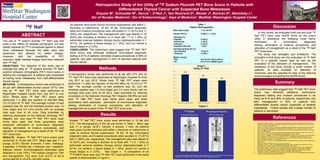
detected in 3 pts (19%). The median (range) for TSH tissue biopsy in 2 (12%). See Graph 1. A comparison of an 11.
Localization: Planar Bone Scintigraphy Versus 18F PET. J Nucl Med .1999;40:1623-1629.
Tickoo SK, Pittas AG, Adler M, et al. Bone Metastases from Thyroid Carcinoma: A Histopathologic Study with Clinical
99mTCMDP bone scan and 18F-NaF PET scan performed in the same Correlates. Arch Pathol Lab Med. 2000;124:1440-1447.
and thyroglobulin (Tg) were 0.42 (0.012 to 62.4)
uIU/ml and 63.3 (<0.2 to >35,000) ng/mL. patient is demonstrated in Figure 1.