

Recomendados
Recomendados
Más contenido relacionado
La actualidad más candente
La actualidad más candente (20)
Similar a Murray2005
Similar a Murray2005 (20)
Último
Último (20)
Murray2005
- 1. Review Synthèse E ctopic pregnancy, in which the gestational sac is outside the uterus, is the most common life- threatening emergency in early pregnancy. The in- cidence in the United States has increased greatly in the last few decades, from 4.5 per 1000 pregnancies in 1970 to an estimated 19.7 per 1000 pregnancies in 1992.1,2 Al- though spontaneous resolution of ectopic pregnancy can occur, patients are at risk of tubal rupture and catastrophic hemorrhage.3,4 Ectopic pregnancy remains an important cause of maternal death, accounting for about 4% of the approximately 20 annual pregnancy-related deaths in Canada.5 Despite the relatively high frequency of this seri- ous condition, early detection can be challenging. In up to half of all women with ectopic pregnancy presenting to an emergency department, the condition is not identified at the initial medical assessment.6 Although the incidence of ectopic pregnancy in the general population is about 2%, the prevalence among pregnant patients presenting to an emergency department with first-trimester bleeding or pain, or both, is 6% to 16%.7–14 Thus, greater suspicion and a lower threshold for investigation are justified. The availability of newer hormonal markers and ultra- sound imaging has increased the complexity of the diagnos- tic workup in patients suspected of having an ectopic preg- nancy, and the evolution of less invasive surgical techniques and noninvasive medical management has altered the treat- ment landscape. In this review we summarize the current literature examining the impact of recent advances in the diagnosis and treatment of ectopic pregnancy. Articles cited were identified with use of the keywords “ectopic preg- nancy,” “epidemiology,” “diagnosis,” “radiography,” “ultrasonography,” “therapy,” “surgery,” “methotrexate,” “emergency department” and “emergency” in searches of MEDLINE, EMBASE and the Cochrane Database of Sys- tematic Reviews. Diagnosis Historical features and physical findings Ectopic pregnancy is usually diagnosed in the first trimester of pregnancy. The most common gestational age at diagnosis is 6 to 10 weeks, but fetal viability can be dis- covered until the time of delivery.15,16 Ectopic pregnancy has about the same frequency across a wide range of mater- nal ages and ethnic origins. Documentation of risk factors (Table 19,17,18 ) is an essential part of history-taking, and asymptomatic clinic patients with risk factors may benefit from routine early imaging.19 However, more than half of identified ectopic pregnancies are in women without known risk factors.8,9 The physical findings depend on whether tubal rupture has occurred. Women with intraperitoneal hemorrhage Diagnosis and treatment of ectopic pregnancy Heather Murray, Hanadi Baakdah, Trevor Bardell, Togas Tulandi Abstract ECTOPIC PREGNANCY IS A LIFE- AND FERTILITY-threatening condition that is commonly seen in Canadian emergency departments. In- creases in the availability and use of hormonal markers, coupled with advances in formal and emergency ultrasonography have changed the diagnostic approach to the patient in the emergency department with first-trimester bleeding or pain. Ultrasonography should be the initial investigation for symptomatic women in their first trimester; when the results are indeterminate, the serum β hu- man chorionic gonadotropin (β-hCG) concentration should be measured. Serial measurement of β-hCG and progesterone con- centrations may be useful when the diagnosis remains unclear. Advances in surgical and medical therapy for ectopic pregnancy have allowed the proliferation of minimally invasive or noninva- sive treatment. Guidelines for laparoscopy and for methotrexate therapy are provided. CMAJ 2005;173(8):905-12 DOI:10.1503/cmaj.050222 CMAJ • OCT. 11, 2005; 173 (8) 905 © 2005 CMA Media Inc. or its licensors Table 1: Risk factors for ectopic pregnancy 9,17,18 OR (and 95% CI) Factor Ankum et al17 Mol et al18 Dart et al9 Previous tubal surgery 21 (9.3–47) – – Previous ectopic pregnancy 8.3 (6.0–11.5) – – In utero DES exposure 5.6 (2.4–13) – – History of PID 2.5 (2.1–3.0) – – History of infertility 2.5–21* – 5.0 (1.1–28) History of chlamydial or gonococcal cervicitis 2.8–3.7* – – Documented tubal abnormality 3.5–25* – – Tubal ligation – 9.3 (4.9–18) 18 (3.0–139) Current IUD use – 4.2–45* 5.0 (1.1–28) Note: OR = odds ratio, CI = confidence interval, DES = diethylstilbestrol, PID = pelvic inflammatory disease, IUD = intrauterine device. *Range; summary OR not calculated owing to significant heterogeneity between studies.
- 2. present with significant abdominal pain and tenderness, along with various degrees of hemodynamic instability. However, women without rupture may also present with pelvic pain or vaginal bleeding, or both.8,9,20,21 Several inves- tigators have measured the predictive value of specific risk factors and physical findings alone or in combination: no combination correctly and consistently ruled out ectopic pregnancy.8,9,21 Given the high prevalence of ectopic preg- nancy among pregnant women presenting to emergency departments, further investigation is prudent for all pa- tients presenting with first-trimester bleeding or pain. Use of ββ human chorionic gonadotropin measurement It is important to confirm pregnancy. In the emergency department, pregnancy is diagnosed by determining the urine or serum concentration of β human chorionic go- nadotropin (β-hCG). This hormone is detectable in urine and blood as early as 1 week before an expected menstrual period. Serum testing detects levels as low as 5 IU/L, whereas urine testing detects levels as low as 20–50 IU/L.22 In most cases, screening is done with a urine test, since ob- taining the results of a serum test is time-consuming and is not always possible in the evening and at night. However, if pregnancy is strongly suspected, even when the urine test has a negative result, serum testing will be definitive. A single serum measurement of the β-hCG concentra- tion, however, cannot identify the location of the gestational sac. Although women with an ectopic pregnancy tend to have lower β-hCG levels than those with an intrauterine pregnancy, there is considerable overlap (Table 2).23,24 If a low serum β-hCG level (< 1000 IU/L) is associated with a higher relative risk of ectopic pregnancy, then can very low levels predict a benign clinical course? In general, no. Although a single very low serum level (< 100 IU/L) has been felt to be reassuring, in a review of 716 admitted patients with ectopic pregnancy, 29% of those with such a level were found to have tubal rupture at laparoscopy.25 The risk of tubal rupture was similar across a wide range of β-hCG values. Another study identified 38 instances of rupture among women with serum levels ranging from 10 to 189 720 IU/L.7 Thus, a single serum β-hCG measure- ment cannot exclude ectopic pregnancy or predict the risk of rupture unless it is less than 5 IU/L. Serial β-hCG measurement is often used for women with first-trimester bleeding or pain, or both, but, as with a single measurement, serial measurement cannot confirm the location of the gestational sac. In a normal pregnancy, the first-trimester β-hCG concentration rapidly increases, doubling about every 2 days. An increase over 48 hours of at least 66% has been used as a cutoff point for viabil- ity.20,26,27 Ectopic pregnancy may present with rising, falling or plateau β-hCG levels; thus, serial measurement is most useful to confirm fetal viability rather than to identify ec- topic pregnancy. In a patient with a subnormal increase in β-hCG concentration, nonviability is assumed, and more invasive investigations can be used to clarify the nature of the abnormality (i.e., miscarriage v. ectopic pregnancy). However, over-reliance on the doubling time may result in the interruption of a normal pregnancy through diagnostic dilatation and curettage (D&C) or administration of methotrexate. A recent study identified patients with only a 53% increase in serum β-hCG levels over 2 days who had a viable intrauterine pregnancy.28 Thus, demonstration of normal doubling of serum levels over 48 hours supports a diagnosis of fetal viability but does not rule out ectopic pregnancy, and a rising β-hCG concentration that fails to reach 50% suggests a failing or ectopic pregnancy, as does a plateau. Falling levels confirm nonviability but do not rule out ectopic pregnancy. Use of progesterone measurement Measurement of the serum concentration of proges- terone has been investigated as a potentially useful adjunct to serum β-hCG measurement, since progesterone levels are stable and independent of gestational age in the first trimester.14 A meta-analysis, published in 1998, of studies assessing a single progesterone level demonstrated good ca- pacity of low levels (≤ 5 ng/mL) to correctly diagnose preg- nancy failure, but this cutoff was unable to discriminate be- Murray et al 906 JAMC • 11 OCT. 2005; 173 (8) Table 2: Performance of serum levels of ββββ human chorionic gonadotropin (ββββ-hCG) in identifying ectopic pregnancy and abnormal intrauterine pregnancy (IUP) Predictive value (95% CI), % Outcome Sensitivity Specificity Likelihood ratio (+) Kohn et al;24 serum ββββ-hCG level < 1500 IU/L Ectopic pregnancy 42 (32–52) 81 (78–82) 2.4 Ectopic pregnancy or abnormal IUP 38 (33–43) 93 (91–96) 5.8 Kaplan et al;23 serum ββββ-hCG level < 1000 IU/L Ectopic pregnancy 38 (26–51) 90 (87–93) 3.8 Ectopic pregnancy or abnormal IUP 25 (20–31) 97 (95–99) 9.5 Table 3: Performance of progesterone levels in identifying ectopic pregnancy and abnormal IUP Predictive value (95% CI), % Outcome Sensitivity Specificity Likelihood ratio (+) Dart et al;31 serum progesterone level ≤≤≤≤ 5 ng/mL* Ectopic pregnancy 88 (69–97) 40 (32–49) 1.47 Ectopic pregnancy or abnormal IUP 84 (77–89) 97 (87–99) 28 Buckley et al;30 serum progesterone level ≤≤≤≤ 22 ng/mL Ectopic pregnancy 100 (94–100) 27 (23–30) 1.36 *All patients had a β-hCG concentration < 3000 IU/L and indeterminate ultrasound findings.
- 3. tween ectopic pregnancy and intrauterine pregnancy.29 Both high (> 22 ng/mL) and low (≤ 5 ng/mL) cutoff points have since been studied for their ability to correctly identify nonviable pregnancy and ectopic pregnancy (Table 3).30,31 Rapid progesterone analysis can identify 2 important sub- groups of patients in the emergency department with symptomatic first-trimester bleeding or pain, or both: sta- ble patients with progesterone levels above 22 ng/mL, who have a high (but not certain) likelihood of viable intrauter- ine pregnancy; and patients with levels of 5 ng/mL or less, who almost certainly have a nonviable pregnancy. Invasive diagnostic testing (e.g., D&C) could be postponed in the former patients but offered to the latter, as could treatment with methotrexate, without fear of interrupting a poten- tially viable intrauterine pregnancy. Ultrasound imaging Transvaginal ultrasonography has transformed the as- sessment of women with problematic early pregnancy, al- lowing earlier, clearer visualization of both normally devel- oping embryos and abnormalities. A normal gestational sac, an ovoid collection of fluid adjacent to the endometrial stripe, can be visualized by means of the transvaginal probe at a gestational age of about 5 weeks. It can often be seen when 2 or 3 mm in diameter and should be consistently seen at 5 mm. Since the hormonal environment in ectopic pregnancy can produce an intrauterine fluid collection that mimics a gestational sac (the “pseudogestational sac” shown in Fig. 1, arrow), a sac alone cannot confirm intrauterine pregnancy.32 As the embryo matures, more sonographic signs become visible. Once the sac is implanted within the endometrium, its position relative to the endometrial wall changes, pro- ducing the intradecidual-sac sign and then the double de- cidual-sac sign. The earliest embryonic landmark, the yolk sac, appears when the sac is 8 mm or more in diameter, usually during the fifth week of gestation (Fig. 2). Cardiac activity can be seen with endovaginal scanning when the embryo reaches 4 to 5 mm in diameter, at a gestational age of 6–6.5 weeks.32 The concept of the discriminatory threshold (the β- hCG level at which an intrauterine gestational sac can be reliably seen in a normal pregnancy) has existed since the early 1980s.33 A β-hCG level that has risen above the dis- criminatory threshold in the absence of sonographic signs of early pregnancy is considered presumptive evidence of an ectopic pregnancy. With the evolution in ultrasound technology, the discriminatory threshold has dropped from 6500 IU/L with a transabdominal approach to be- tween 1000 and 2000 IU/L with transvaginal imaging.34 This threshold is user- and machine-dependent and thus will vary slightly from institution to institution. Caution should be used in assuming an ectopic pregnancy when a nondiagnostic ultrasound image accompanies a single β- hCG level above the discriminatory threshold: several arti- cles have reported a small number of patients with indeter- minate ultrasound images and β-hCG levels above the threshold who have been eventually found to have a viable intrauterine pregnancy.34–36 In addition, there could be un- seen multiple intrauterine gestational sacs, since the β- hCG values relative to gestational age are higher in pa- tients with multiple embryos.37 Ultrasonographic identification of an intrauterine preg- nancy (gestational sac plus yolk sac or other embryonic sign) rules out ectopic pregnancy in most patients.32 The Ectopic pregnancy CMAJ • OCT. 11, 2005; 173 (8) 907 Fig. 1: Transvaginal ultrasound image, showing intrauterine fluid collection without yolk sac or fetal pole: “pseudogesta- tional sac” (arrow). Fig. 2: Transvaginal ultrasound image, showing early intra- uterine gestational sac (GS) with yolk sac (YS).
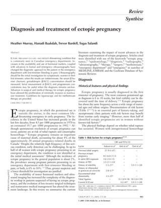
- 4. exception is in patients with ovulation induction and as- sisted conception, who are at risk of heterotopic pregnancy (dizygotic twins, 1 intrauterine and 1 extrauterine). Al- though this phenomenon is exceedingly rare in the general population (estimated frequency 1 per 3889 to 30 000 pregnancies),38 in the setting of assisted reproduction it may occur in 1 in 100 pregnancies.39 The spectrum of sonographic findings in ectopic preg- nancy is broad. Identification of an extrauterine gestational sac containing a yolk sac (with or without an embryo) con- firms the diagnosis. Suggestive findings include an empty uterus, cystic or solid adnexal or tubal masses (including the tubal-ring sign, representing a tubal gestational sac), hematosalpinx and echogenic or sonolucent cul-de-sac fluid (Fig. 3). Many prospective studies have shown that “formal” transvaginal ultrasound imaging (i.e., that performed by ul- trasound technicians and interpreted by radiologists) in the emergency department has high accuracy in confirming in- trauterine and ectopic pregnancy. Most protocols can es- tablish a diagnosis with the initial scan in more than 75% of emergency department patients.23,35,40 A diagnosis can of- ten be established even in the subgroup of patients with β- hCG levels below the discriminatory threshold. In some studies, transvaginal scanning has identified up to one-third of the patients with below-threshold β-hCG levels who had ectopic pregnancy.35,41 Given the likelihood of a definitive diagnosis, even with below-threshold β-hCG levels, ultra- sonography is the best initial investigation in problematic early pregnancy. Because expertise in transvaginal ultrasonography is not available in all hospitals and may not be quickly available in some larger centres, there have been several studies of ul- trasonography performed by emergency physicians in the assessment of patients with first-trimester bleeding or pain. Ultrasonography in the emergency department (ED-based ultrasonography) has evolved over the last decade and is now part of the diagnostic work-up for many clinical prob- lems in major Canadian centres, as well as in a large num- ber of smaller community emergency departments (Dr. Ray Wiss, Emergency Department Echo course director: personal communication, 2005). The evaluation involves 2 “Yes/No” questions: Can an intrauterine pregnancy be identified? Is there free pelvic or intra-abdominal fluid? This approach is in contrast to the goal of a “formal” pelvic ultrasound study, which is to describe the anatomic appear- ance and visible abnormalities of the uterus, adnexa and cul-de-sac. Absence of an intrauterine pregnancy translates to a risk of ectopic pregnancy of about 36%,42 and free fluid in the cul-de-sac represents an even higher risk. Several studies have documented the ability of emergency physi- cians to quickly and accurately identify both intrauterine pregnancy and intra-abdominal free fluid by means of ED- based ultrasonography after brief standardized train- ing.12,42,43 The addition of ED-based ultrasonography to structured protocols for assessing symptomatic patients in the 1st trimester of pregnancy has led to a dramatically de- creased stay in the emergency department44 as well as a de- crease in the incidence of complications associated with missed ectopic pregnancy and tubal rupture.12 Transvaginal ultrasonography should therefore be the initial investigation for pregnant patients presenting to the emergency department with first-trimester bleeding or pain. Not only is it highly accurate in identifying ectopic pregnancy, but also it offers patients what they are most ex- pecting from their visit: information about the health and viability of their pregnancy. No combination of history- taking, physical examination and laboratory tests can make the same claim. The use of ED-based ultrasonography of- fers rapid bedside detection of a viable intrauterine preg- nancy or a high risk of ectopic pregnancy. Emergency physicians without access to bedside ED-based ultrasonog- raphy should arrange formal ultrasonography for all pa- tients with early-pregnancy complaints. This investigation can be performed during the initial visit or, if the patient is stable and has minimal symptoms, the next day in an outpa- tient visit. However, in the case of outpatient investigation, mechanisms for timely follow-up, re-examination and fur- ther investigation must be in place. Fig. 4 outlines the recommended approach to imaging and follow-up. Treatment Expectant management Ectopic pregnancy can resolve spontaneously through regression or tubal abortion. However, about 90% of women with ectopic pregnancy and serum β-hCG levels greater than 2000 IU/L require operative intervention ow- ing to increasing symptoms or tubal rupture.3,4 Tubal rup- Murray et al 908 JAMC • 11 OCT. 2005; 173 (8) Fig. 3: Transvaginal ultrasound image, showing empty uterus and complex adnexal mass (ectopic pregnancy [EP]) separate from ovary.
- 5. ture can also occur when serum β-hCG levels are low or declining, or both.45 Expectant management should be of- fered only when transvaginal ultrasonography fails to show the location of the gestational sac and the serum levels of β- hCG and progesterone are low and declining. Because of the possibility of tubal rupture, these patients must be care- fully monitored until the serum β-hCG concentration falls below 15 IU/L; at this point almost all ectopic pregnancies resolve spontaneously, without rupture. Surgical management Surgical management of ectopic pregnancy should be reserved for patients who refuse or have contraindications to medical treatment, those in whom medical treatment has failed and those who are hemodynamically unstable. Three randomized studies have demonstrated that, compared with laparotomy, laparoscopic treatment of ec- topic pregnancy is associated with lower cost, shorter hos- Ectopic pregnancy CMAJ • OCT. 11, 2005; 173 (8) 909 Fig. 4: Recommended approach to investigating first-trimester pain or bleeding in the hemodynamically stable patient in the emergency department (ED). ββ-hCG = ββ human chorionic gonadotropin, US = ul- trasonography, IUP = intrauterine pregnancy, EP = ectopic pregnancy. • Discharge home • Follow-up with health care provider • Outpatient formal US High risk for EP • Expert consultation • Formal US Formal US Definite IUP (viable or nonviable) Indeterminate result Definite EP Positive β-hCG results ED-based US available? IUP No IUP No IUP; pelvic free fluid Serum β-hCG level above discriminatory threshold? High risk of EP • Expert consultation • 48-h follow-up with repeat β-hCG test • Consider measuring progesterone level • Consider expert consultation (particularly if adnexal findings are abnormal) No Expert consultation Yes NoYes
- 6. pital stay, less operative time, less blood loss, less analgesic requirement and faster recovery.46–48 Patients randomly as- signed to laparoscopy also had fewer adhesions than pa- tients treated with laparotomy (19% v. 64%).49 Tube-sparing salpingostomy (in which the gestational sac is removed, without the tube, through a 1-cm-long in- cision on the tubal wall) is preferred to salpingectomy (re- moval of the tube), as the former is less invasive but has comparable rates of subsequent fertility and ectopic preg- nancy.50–52 However, 8% of patients have persistent ectopic pregnancy after salpingostomy.50 Follow-up determina- tions are required until β-hCG is undetectable. Regardless of the type of surgery, contralateral tubal abnormalities predispose the patient to recurrent ectopic pregnancy. In a retrospective study of 276 women with ectopic pregnancy, the cumulative rates of spontaneous intrauterine preg- nancy over 7 year were 89% after conservative surgery and 66% after radical surgery.50 There was no significant dif- ference in the risk of repeat ectopic pregnancy (17% after conservative surgery and 16% after radical surgery). In summary, salpingostomy is preferred, particularly for women who wish to have another pregnancy. Salpingec- tomy may be necessary for women with uncontrolled bleeding, recurrent ectopic pregnancy in the same tube, a severely damaged tube or a tubal gestational sac greater than 5 cm in diameter.53 Medical treatment Methotrexate (MTX), a folic acid antagonist, inhibits DNA synthesis in actively dividing cells, including tro- phoblasts. Administered to properly selected patients, it has a success rate of up to 94%.53 The success in ectopic preg- nancy depends mainly on β-hCG concentration: a meta- analysis of data for 1327 women with ectopic pregnancy treated with MTX showed that resolution was inversely as- sociated with β-hCG level, and that increasing levels were significantly correlated with treatment failure. Fetal cardiac activity was also associated with MTX treatment failure. However, tubal diameter, a measure of fetal size, is unre- lated to outcome.54 The criteria for MTX treatment of ectopic pregnancy are as follows: Murray et al 910 JAMC • 11 OCT. 2005; 173 (8) Box 1: Protocol for methotrexate treatment of ectopic pregnancy Pretreatment investigations • Complete blood count • Blood group typing and antibody testing • Liver and renal function tests • Measurement of serum level of β human chorionic gonadotropin (β-hCG) • Transvaginal ultrasonography Treatment day 0 • Inject methotrexate (50 mg/m2 ) intramuscularly • Inject RhoGAM (300 µg) intramuscularly if needed • Discontinue folinic acid supplements • Advise patient to refrain from strenuous exercise and sexual intercourse Day 7 • Measure serum β-hCG concentration • Perform transvaginal ultrasonography • Inject second dose of methotrexate if decline in β-hCG level is < 25% Weekly • Measure serum β-hCG concentration until level is < 15 IU/L • Perform transvaginal ultrasonography Any time • Perform laparoscopy if patient has severe abdominal pain or acute abdomen or if ultrasonography reveals more than 100 mL of blood in the abdomen Side effects of methotrexate therapy are usually mild and self-limiting. Stomatitis and conjunctivitis are the most common. Pleuritis, dermatitis, alopecia, gastritis, enteritis, elevated liver enzyme concentrations and bone marrow suppression are rare. About 30% of patients will have side effects with a single dose and 40% with multiple doses.54 Table 4: Randomized studies comparing methotrexate (MTX) with laparoscopic salpingostomy (LS) for the treatment of ectopic pregnancy [abridged*] Treatment success rate, % Study (no. of patients) MTX LS Difference in rate Other outcomes Comments Hajenius et al, 199756 (100) 82 72 NS No difference in rates of tubal preservation All patients underwent laparoscopy for diagnosis or treatment. Four doses of MTX Fernandez et al, 199857 (100) 88 96 NS Higher rate of future pregnancy in MTX group (96% v. 62%, p < 0.05) but not of recurrent ectopic pregnancy One of a few centres using a scoring system and local MTX injection for ectopic pregnancy Saraj et al, 199858 (75) 95 91 NS No difference in rates of tubal patency or future pregnancy The study was underpowered Sowter et al, 200159 (62) 65 93 95% CI 10–47 Less time till β-hCG clearance in LS group: 15 (5–49) v. 28 (14–71) d This study represents the general clinical management of ectopic pregnancy Note: NS = not significant, CI = confidence interval. *An unabridged version of this table is available at www.cmaj.ca/cgi/content/full/173/8/905/DC1.
- 7. • Hemodynamic stability. • Ability and willingness of the patient to comply with post-treatment monitoring. • Pretreatment serum β-hCG concentration less than 5000 IU/L. • Absence of ultrasound evidence of fetal cardiac activity. Our protocol for using MTX in the management of ec- topic pregnancy is shown in Box 1. The overall success rate is greater with multiple-dose MTX therapy than with single-dose therapy (93% v. 88%); however, single-dose therapy is less expensive, has a lower rate of side effects (29% v. 48%), requires less in- tensive monitoring, does not require rescue with folinic acid and is effective for most women.54 The 2 regimens have not been directly compared in randomized trials. Patients in whom laparoscopy may be challenging (in- cluding those with many previous laparotomies and scar- ring) may have a better outcome with MTX treatment. In the presence of relative contraindications, such as high serum β-hCG levels (≥ 5000 IU/L) and the presence of fetal cardiac activity, multiple-dose treatment should be considered. Patients treated with MTX should be followed closely. The serum β-hCG concentration should be measured weekly. An increased level is uncommon 3 to 4 d after MTX administration. Patients may experience abdominal pain from tubal abortion or tubal distention due to hema- toma formation. Severe abdominal pain, however, can be a sign of actual or impending tubal rupture. If the serum β- hCG concentration has not declined by at least 25% 1 week after MTX administration, a second dose should be given. In general, a second dose is needed in 15% to 20% of patients.54,55 Only 1% of patients need more than 2 doses.54 The time for the serum β-hCG concentration to decline to less than 15 IU/L is 33.6 days on average but may be up to 109 days.55 Surgical versus medical treatment Several randomized studies found that MTX treatment in selected patients with ectopic pregnancy was as effective as laparoscopic treatment (Table 4).56–59 The 2 treatments were also equally effective in tubal preservation; however, the β-hCG concentration declined more quickly after la- paroscopic surgery.56 After MTX treatment the health- related quality of life may diminish, possibly owing to both long-term persistence of the ectopic pregnancy and the long treatment course. There were more physical symp- toms after 2 days and 2 weeks in those given MTX, although symptoms were increased in both treatment groups. However, because of the noninvasive nature of MTX treatment, most patients are willing to cope with this short-term burden.60 MTX treatment is less expensive than laparoscopic surgery,59,60 although in one study this was true only if the initial serum β-hCG level was less than 1500 IU/L.60 Conclusions Ectopic pregnancy is a common and serious problem, with a significant morbidity rate and the potential for ma- ternal death. Many patients have no documented risk fac- tors and no physical indications of ectopic pregnancy. Ultrasonography (either formal or ED-based) is the initial investigation that should be done in an ED patient with 1st-trimester bleeding or pain; indeterminate results may be clarified by measurement (single or serial) of the serum β-hCG and progesterone concentrations. Expert consulta- tion with radiologists and gynecologists is recommended whenever ectopic pregnancy is suspected. Management is dictated by the clinical presentation, serum β-hCG levels and transvaginal ultrasound findings. MTX, as a single intramuscular injection, can be given to women who are hemodynamically stable and compliant and have an initial serum β-hCG concentration of less than 5000 IU/L and no ultrasound evidence of fetal cardiac ac- tivity. Patients who do not meet these criteria should be treated surgically, in most cases by laparoscopy. Surgical treatment is particularly appropriate for women who are hemodynamically unstable or unlikely to be compliant with post-treatment monitoring and those who do not have im- mediate access to medical care. The choice of treatment should be guided by the patient’s preference, after a de- tailed discussion about monitoring, outcome, risks, and benefits of the 2 approaches. References 1. Goldner TE, Lawson HW, Xia Z, Atrash HK. Surveillance for ectopic pregnancy — United States, 1970–1989. MMWR CDC Surveill Summ 1993;42:73-85. 2. Ectopic pregnancy — United States, 1990–1992. MMWR Morb Mortal Wkly Rep 1995;44:46-8. 3. Shalev E, Peleg D, Tsabari A, Romano S, Bustan M. Spontaneous resolution of ectopic tubal pregnancy: natural history. Fertil Steril 1995;63:15-9. 4. Elson J, Tailor A, Banerjee S, Salim R, Hillaby K, Jurkovic D. Expectant management of tubal ectopic pregnancy: prediction of successful outcome us- ing decision tree analysis. Ultrasound Obstet Gynecol 2004;23:552-6. 5. Turner LA, Cyr M, Kinch RA, Liston R, Kramer MS, Fair M, et al.; Mater- nal Mortality and Morbidity Study Group of the Canadian Perinatal Surveil- lance System. Under-reporting of maternal mortality in Canada: a question of definition. Chronic Dis Can 2002;23:22-30. 6. Carson SA, Buster JE. Ectopic pregnancy. N Engl J Med 1993;329:1174-81. Comments in N Engl J Med 1994;330:712-3. 7. Barnhart K, Mennuti MT, Benjamin I, Jacobson S, Goodman D, Coutifaris C. Prompt diagnosis of ectopic pregnancy in an emergency department set- ting. Obstet Gynecol 1994;84:1010-5. 8. Buckley RG, King KJ, Disney JD, Gorman JD, Klausen JH. History and physical examination to estimate the risk of ectopic pregnancy: validation of a clinical prediction model. Ann Emerg Med 1999;34:589-94. Comment in Ann Emerg Med 1999;34:664-7. Ectopic pregnancy CMAJ • OCT. 11, 2005; 173 (8) 911 This article has been peer reviewed. Competing interests: None declared. Contributors: Heather Murray and Trevor Bardell reviewed the relevant literature and wrote the first draft of the diagnosis section of the manuscript. Hanadi Baak- dah and Togas Tulandi reviewed the relevant literature and wrote the first draft of the treatment section of the manuscript. All of the authors provided revisions and approved the final content of the manuscript. From the Departments of Emergency Medicine (Murray) and Surgery (Bardell), Queen’s University, Kingston, Ont., and the Department of Obstetrics and Gyne- cology, McGill University, Montréal, Que. (Baakdah, Tulandi)
- 8. 9. Dart RG, Kaplan B, Varaklis K. Predictive value of history and physical ex- amination in patients with suspected ectopic pregnancy. Ann Emerg Med 1999;33:283-90. 10. Durham B, Lane B, Burbridge L, Balasubramaniam S. Pelvic ultrasound per- formed by emergency physicians for the detection of ectopic pregnancy in complicated first-trimester pregnancies. Ann Emerg Med 1997;29:338-47. 11. Spandorfer SD, Barnhart KT. Role of previous ectopic pregnancy in altering the presentation of suspected ectopic pregnancy. J Reprod Med 2003;48:133-6. 12. Mateer JR, Valley VT, Aiman EJ, Phelan MB, Thoma ME, Kefer MP. Out- come analysis of a protocol including bedside endovaginal sonography in pa- tients at risk for ectopic pregnancy. Ann Emerg Med 1996;27:283-9. 13. Sauer MV, Rodi IA. Utility of an algorithm to diagnose ectopic pregnancy. Int J Gynaecol Obstet 1990;31:29-34. 14. Stovall TG, Kellerman AL, Ling FW, Buster JE. Emergency department di- agnosis of ectopic pregnancy. Ann Emerg Med 1990;19:1098-103. 15. Xiao GH, Chen DJ, Sun XF, She RQ, Mai YM. Abdominal pregnancy: full- term viable baby. Eur J Obstet Gynecol Reprod Biol 2005;118:117-8. 16. Ramachandran K, Kirk P. Massive hemorrhage in a previously undiagnosed abdominal pregnancy presenting for elective cesarean delivery. Can J Anaesth 2004;51:57-61. 17. Ankum WM, Mol BW, van der Veen F, Bossuyt PM. Risk factors for ectopic pregnancy: a meta-analysis. Fertil Steril 1996;65:1093-9. Comment in Fertil Steril 1997;67:791-2. 18. Mol BW, Ankum WM, Bossuyt PM, van der Veen F. Contraception and the risk of ectopic pregnancy: a meta-analysis. Contraception 1995;52:337-41. 19. Cacciatore B, Stenman UH, Ylostalo P. Early screening for ectopic preg- nancy in high-risk symptom-free women. Lancet 1994;343:517-8. 20. Mol BW, Hajenius PJ, Engelsbel S, Ankum WM, van der Veen F, Hemrika DJ, et al. Can noninvasive diagnostic tools predict tubal rupture or active bleeding in patients with tubal pregnancy? Fertil Steril 1999;71:167-73. 21. Mol BW, Hajenius PJ, Engelsbel S, Ankum WM, van der Veen F, Hemrika DJ, et al. Should patients who are suspected of having an ectopic pregnancy undergo physical examination? Fertil Steril 1999;71:155-7. 22. Brennan DF. Ectopic pregnancy — Part I: Clinical and laboratory diagnosis. Acad Emerg Med 1995;2:1081-9. 23. Kaplan BC, Dart RG, Moskos M, Kuligowska E, Chun B, Adel Hamid M, et al. Ectopic pregnancy: prospective study with improved diagnostic accuracy. Ann Emerg Med 1996;28:10-7. Comment in Ann Emerg Med 1997;29:295-6. 24. Kohn MA, Kerr K, Malkevich D, O’Neil N, Kerr MJ, Kaplan BC. Beta-hu- man chorionic gonadotropin levels and the likelihood of ectopic pregnancy in emergency department patients with abdominal pain or vaginal bleeding. Acad Emerg Med 2003;10:119-26. 25. Saxon D, Falcone T, Mascha EJ, Marino T, Yao M, Tulandi T. A study of ruptured tubal ectopic pregnancy. Obstet Gynecol 1997;90:46-9. Comment in Obstet Gynecol 1997;90:866-7. 26. Kadar N, Caldwell BV, Romero R. A method of screening for ectopic preg- nancy and its indications. Obstet Gynecol 1981;58:162-6. 27. Dart RG, Mitterando J, Dart LM. Rate of change of serial beta-human chori- onic gonadotropin values as a predictor of ectopic pregnancy in patients with in- determinate transvaginal ultrasound findings. Ann Emerg Med 1999;34:703-10. 28. Barnhart KT, Sammel MD, Rinaudo PF, Zhou L, Hummel AC, Guo W. Symptomatic patients with an early viable intrauterine pregnancy: HCG curves redefined. Obstet Gynecol 2004;104:50-5. 29. Mol BW, Lijmer JG, Ankum WM, van der Veen F, Bossuyt PM. The accu- racy of single serum progesterone measurement in the diagnosis of ectopic pregnancy: a meta-analysis. Hum Reprod 1998;13:3220-7. 30. Buckley RG, King KJ, Disney JD, Riffenburgh RH, Gorman JD, Klausen JH. Serum progesterone testing to predict ectopic pregnancy in symptomatic first-trimester patients. Ann Emerg Med 2000;36:95-100. 31. Dart R, Ramanujam P, Dart L. Progesterone as a predictor of ectopic preg- nancy when the ultrasound is indeterminate. Am J Emerg Med 2002;20:575-9. 32. Albayram F, Hamper UM. First-trimester obstetric emergencies: spectrum of sonographic findings. J Clin Ultrasound 2002;30:161-77. 33. Kadar N, DeVore G, Romero R. Discriminatory hCG zone: its use in the sonographic evaluation for ectopic pregnancy. Obstet Gynecol 1981;58:156-61. Comment in Obstet Gynecol 2003;102:672. 34. Mehta TS, Levine D, Beckwith B. Treatment of ectopic pregnancy: Is a hu- man chorionic gonadotropin level of 2,000 mIU/mL a reasonable threshold? Radiology 1997;205:569-73. 35. Barnhart KT, Simhan H, Kamelle SA. Diagnostic accuracy of ultrasound above and below the beta-hCG discriminatory zone. Obstet Gynecol 1999;94:583-7. Comment in Obstet Gynecol 2000;95:475-6. 36. Braffman BH, Coleman BG, Ramchandani P, Arger PH, Nodine CF, Dins- more BJ, et al. Emergency department screening for ectopic pregnancy: a prospective US study. Radiology 1994;190:797-802. 37. Kadar N, Bohrer M, Kemmann E, Shelden R. The discriminatory human chorionic gonadotropin zone for endovaginal sonography: a prospective, ran- domized study. Fertil Steril 1994;61:1016-20. Comment in Fertil Steril 1995;63:683-4. 38. Habana A, Dokras A, Giraldo JL, Jones EE. Cornual heterotopic pregnancy: contemporary management options. Am J Obstet Gynecol 2000;182:1264-70. Comment in Am J Obstet Gynecol 2001;185:522. 39. Tal J, Haddad S, Gordon N, Timor-Tritsch I. Heterotopic pregnancy after ovulation induction and assisted reproductive technologies: a literature review from 1971 to 1993. Fertil Steril 1996;66:1-12. 40. Mertz HL, Yalcinkaya TM. Early diagnosis of ectopic pregnancy. Does use of a strict algorithm decrease the incidence of tubal rupture? J Reprod Med 2001;46:29-33. 41. Dart RG, Kaplan B, Cox C. Transvaginal ultrasound in patients with low beta- human chorionic gonadotropin values: How often is the study diagnostic? Ann Emerg Med 1997;30:135-40. Comment in Ann Emerg Med 1997;30:206-9. 42. Mateer JR, Aiman EJ, Brown MH, Olson DW. Ultrasonographic examina- tion by emergency physicians of patients at risk for ectopic pregnancy. Acad Emerg Med 1995;2:867-73. 43. Mateer J, Plummer D, Heller M, Olson D, Jehle D, Overton D, et al. Model curriculum for physician training in emergency ultrasonography. Ann Emerg Med 1994;23:95-102. 44. Shih CH. Effect of emergency physician-performed pelvic sonography on length of stay in the emergency department [discussion 352]. Ann Emerg Med 1997;29:348-51. 45. Tulandi T, Hemmings R, Khalifa F. Rupture of ectopic pregnancy in women with low and declining serum beta-human chorionic gonadotropin concentra- tions. Fertil Steril 1991;56:786-7. 46. Lundorff P, Hahlin M, Kallfelt B, Thorburn J, Lindblom B. Adhesion forma- tion after laparoscopic surgery in tubal pregnancy: a randomized trial versus laparotomy. Fertil Steril 1991;55:911-5. 47. Murphy AA, Nager CW, Wujek JJ, Kettel LM, Torp VA, Chin HG. Opera- tive laparoscopy versus laparotomy for the management of ectopic pregnancy: a prospective trial. Fertil Steril 1992;57:1180-5. 48. Vermesh M, Silva PD, Rosen GF, Stein AL, Fossum GT, Sauer MV. Man- agement of unruptured ectopic gestation by linear salpingostomy: a prospec- tive, randomized clinical trial of laparoscopy versus laparotomy. Obstet Gynecol 1989;73(3 pt 1):400-4. Comment in Obstet Gynecol 1989;74:282-3. 49. Lundorff P, Thorburn J, Hahlin M, Kallfelt B, Lindblom B. Laparoscopic surgery in ectopic pregnancy. A randomized trial versus laparotomy. Acta Ob- stet Gynecol Scand 1991;70:343-8. 50. Bangsgaard N, Lund CO, Ottesen B, Nilas L. Improved fertility following conservative surgical treatment of ectopic pregnancy. BJOG 2003;110:765-70. Comment in BJOG 2004;111:635-6. 51. Dubuisson JB, Morice P, Chapron C, De Gayffier A, Mouelhi T. Salpingec- tomy — the laparoscopic surgical choice for ectopic pregnancy. Hum Reprod 1996;11:1199-203. 52. Fernandez H, Marchal L, Vincent Y. Fertility after radical surgery for tubal pregnancy. Fertil Steril 1998;70:680-6. 53. Yao M, Tulandi T. Current status of surgical and nonsurgical management of ectopic pregnancy. Fertil Steril 1997;67:421-33. Comment in Fertil Steril 1997;68:945-7. 54. Barnhart KT, Gosman G, Ashby R, Sammel M. The medical management of ectopic pregnancy: a meta-analysis comparing “single dose” and “multidose” regimens. Obstet Gynecol 2003;101:778-84. 55. Lipscomb GH, Bran D, McCord ML, Portera JC, Ling FW. Analysis of three hundred fifteen ectopic pregnancies treated with single-dose methotrex- ate. Am J Obstet Gynecol 1998;178:1354-8. 56. Hajenius PJ, Engelsbel S, Mol BW, van der Veen F, Ankum WM, Bossuyt PM, et al. Randomised trial of systemic methotrexate versus laparoscopic salpingostomy in tubal pregnancy. Lancet 1997;350:774-9. Comments in Lancet 1997;350:1554-5. 57. Fernandez H, Yves Vincent SC, Pauthier S, Audibert F, Frydman R. Ran- domized trial of conservative laparoscopic treatment and methotrexate ad- ministration in ectopic pregnancy and subsequent fertility. Hum Reprod 1998;13:3239-43. 58. Saraj AJ, Wilcox JG, Najmabadi S, Stein SM, Johnson MB, Paulson RJ. Reso- lution of hormonal markers of ectopic gestation: a randomized trial compar- ing single-dose intramuscular methotrexate with salpingostomy. Obstet Gy- necol 1998;92:989-94. 59. Sowter MC, Farquhar CM, Petrie KJ, Gudex G. A randomised trial compar- ing single dose systemic methotrexate and laparoscopic surgery for the treat- ment of unruptured tubal pregnancy. BJOG 2001;108:192-203. 60. Nieuwkerk PT, Hajenius PJ, Ankum WM, van der Veen F, Wijker W, Bossuyt PM. Systemic methotrexate therapy versus laparoscopic salpingos- tomy in patients with tubal pregnancy. Part I. Impact on patients’ health- related quality of life. Fertil Steril 1998;70:511-7. Murray et al 912 JAMC • 11 OCT. 2005; 173 (8) Correspondence to: Dr. Heather Murray, Department of Emergency Medicine, Queen’s University, Kingston General Hospital, 76 Stuart St., Kingston ON K7L 2V7; fax 613 548-1374; hm9@post.queensu.ca