
Recomendados
Más contenido relacionado
La actualidad más candente
La actualidad más candente (20)
Similar a Oxphos
Similar a Oxphos (20)
Más de obanbrahma
Último
Último (20)
Oxphos
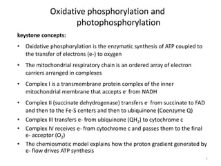
- 1. 1 Oxidative phosphorylation and photophosphorylation keystone concepts: • Oxidative phosphorylation is the enzymatic synthesis of ATP coupled to the transfer of electrons (e-) to oxygen • The mitochondrial respiratory chain is an ordered array of electron carriers arranged in complexes • Complex I is a transmembrane protein complex of the inner mitochondrial membrane that accepts e- from NADH • Complex II (succinate dehydrogenase) transfers e- from succinate to FAD and then to the Fe-S centers and then to ubiquinone (Coenzyme Q) • Complex III transfers e- from ubiquinone (QH2) to cytochrome c • Complex IV receives e- from cytochrome c and passes them to the final e- acceptor (O2) • The chemiosmotic model explains how the proton gradient generated by e- flow drives ATP synthesis
- 2. Impermeable to ions and most other compounds In inner membrane mitochondrion the mitochondrion contained the enzymes responsible for electron transport and oxidative phosphorylation
- 3. 3 anatomy of a mitochondrion Where is ATP synthesized? Where are the citric acid cycle and β-oxidation pathways? Which membranes are permeable?
- 4. Complexes of the respiratory chain
- 5. 5 Where do the electrons come from? Where do they go? • All the dehydrogenase reactions in the citric acid cycle, b- oxidation and amino acid oxidation and glycolysis – Hydride ion transfers + hydrogen atom transfers • Electron carriers in addition to NAD and flavoproteins: 1. Ubiquinone (aka. Coenzyme Q) - a fat soluble mobile protein 2. Cytochromes – iron containing e- transfer proteins (in heme) 3. Iron sulfur proteins – (not in heme) but where iron is directly associated with inorganic sulfur or the sulfur on cysteine residues
- 6. NAD+, flavins and Q carry electrons and H+ Cytochromes and non-heme iron proteins carry only electrons NAD+, FAD undergoes only a 2 e- reaction; cytochromes undergo only 1e- reactions FMN, Q undergoes 1e- and 2 e- reaction Electron carriers
- 7. Ubiquinone • Coenzyme Q (CoQ, or Q) is lipid-soluble. It dissolves in the hydrocarbon core of a membrane. • the only electron carrier not bound to a protein. • it can accept/donate 1 or 2 e-. Q can mediate e- transfer between 2 e- and 1 e- carriers
- 8. 8 ubiquinone (Q, or coenzyme Q)
- 9. Cytochromes are electron carriers containing hemes . Hemes in 3 classes of cytochrome (a, b, c) differ in substituents on the porphyrin ring. Some cytochromes(b,c1,a,a3) are part of large integral membrane protein complexes (such as complex III). Cytochrome c is a small, water-soluble protein. Cytochromes
- 10. 10 cytochromes
- 11. The heme iron can undergo 1 e- transition between ferric and ferrous states: Fe3+ + e- Fe2+ Copper ions besides two heme A groups (a and a3) act as electron carriers in CuB, accepting electrons from heme a Cu2++e- Cu+ Heme is a prosthetic group of cytochromes. Heme contains an iron atom in a porphyrin ring system.
- 12. Cytochromes NAD+ FMN FeS ubiquinoneFAD FeS Cyt b FeS Cyt c1 Cyt c Cyt a Cyt a3 1/2 O2 ubiquinone proteins that accept electrons from QH2 or FeS Ultimately transfers the electrons to oxygen
- 13. Iron-sulfur centers (Fe-S) are prosthetic groups containing 1-4 iron atoms Iron-sulfur centers transfer only one electron, even if they contain two or more iron atoms. E.g., a 4-Fe center might cycle between redox states: 3Fe+++ + Fe++ + 1 e- 2Fe+++ + 2Fe++ Iron-sulfur Centers
- 15. 15 OVERVIEW
- 16. Electron Transport chain • The electron transport chain in the inner mitochondrial membrane can be isolated in four proteins complexes(I, II, III, IV). • A lipid soluble coenzyme (Q) and a water soluble protein (cyt c) shuttle between protein complexes • Electrons transfer through the chain - from complexes I to complex IV
- 17. NAD+ FMN FeS ubiquinoneFAD FeS Cyt b FeS Cyt c1 Cyt c Cyt a Cyt a3 1/2 O2 ubiquinone I II III IV Mitochondrial Complexes NADH Dehydrogenase Succinate dehydrogenase CoQ-cyt c Reductase Cytochrome Oxidase
- 18. 18 How are electron transport complexes studied?
- 19. Support for this order of events 1. Energetically favorable. electrons pass from lower to higher standard reduction potentials 2. Spectra: the absorption spectrum for the reduced carrier differs from that of its oxidized form. carriers closer to oxygen are more oxidized. 3. Specific inhibitors. Those before the blocked step should be reduced and those after be oxidized. 1. Assay of individual complexes: NADH can reduce complex I but not the other complexes.
- 20. H+ Transport Complex I, III, IV drive H+ transport from matrix to the cytosol when e- flow through, which creates proton gradient Creates an electrochemical potential across the inner membrane
- 21. 1.Electrons are transported along the inner mitochondrial membrane, through a series of electron carriers 2.Protons (indicated by + charge) are translocated across the membrane, from the matrix to the intermembrane space 3.Oxygen is the terminal electron acceptor, combining with electrons and H+ ions to produce water 4. As NADH delivers more H+ and electrons into the ETS, the proton gradient increases, with H+ building up outside the inner mitochondrial membrane
- 22. Complex I: NADH dehydrogenase • NADH binds complex I & passes 2 electrons to a flavin momonucleotide (FMN) prosthetic group. • The FMN is reduced to FMNH2. Each electron is transferred with a proton. • The electrons are then passed to iron-sulphur proteins (FeS) in complex I (this is non-heme iron). The electron is accepted by Fe3+ which is reduced to Fe2+
- 24. Complex I • Two electrons from the reduced FeS proteins are then passed to CoQ along with 2 protons. • The CoQ is thus reduced to CoQH2 (ubiquinol) while the FeS proteins are oxidized back to Fe3+ state.
- 25. CoQ is small and lipid soluble so it is mobile in the mitochondrial membrane. It diffuses easily and shuttles the electrons to complex III
- 26. 26 complex I: NADH to ubiquinone
- 27. Complex II: Succinate dehydrogenase • Complex II actually contains the enzyme succinate dehydrogenase which catalyses the reduction of succinate to fumarate (reaction of the citric acid cycle). • FAD oxidizes succinate to fumarate (FAD becoming reduced to FADH2 as it picks up 2 electrons and 2 protons). • FADH2 is oxidized back to FAD by passing the electrons on to FeS proteins in complex II. The electrons are then passed to CoQ and are passed on to complex III
- 28. 28 complex II: FADH2 to ubiquinone
- 29. Complex III: cytochrome reductase • Complex III contains cytochrome b, cytochrome c1 and FeS proteins. • Like FeS proteins, cytochromes contain bound Fe atoms in heme. • The iron atoms alternate between +3 and +2 oxidation states as they pass on the electrons. • CoQH2 passes 2 electrons to cyt b causing the Fe3+ to be reduced to Fe2+. • The electrons are passed to the FeS protein and then to cyt c1.
- 31. 31 Complex III: QH2 to cytochrome c -not a direct proton path across membrane
- 32. Cytochrome C • Cyt c is another small mobile protein. • It accepts electrons from complex III (Fe3+ is reduced to Fe2+) and shuttles them to the last electron transport protein in the chain (complex IV).
- 34. Complex IV: cytochrome oxidase • Complex IV contains cytochrome a and cytochrome a3 (both use Fe and Cu atoms to handle the electrons). • Four cytochrome c molecules pass on 4 electrons to complex IV. • These are eventually transferred with 4 H+ to O2 to form 2 water molecules.
- 35. 35 complex IV: cytochrome oxidase
- 36. Cytochrome C oxidase combines electrons with oxygen and 4H+ to form 2 water Oxygen = final electron acceptor
- 37. Paths of H+ and e- transfer in cytochrome c oxidase Blue- chemical reaction of O2 reduction to water coupled to Red- translocation of four protons e- flow from red cyt c in inner mem space CuA center heme a3-CuB center reduce O2 H+ from matrix: -shuttled to heme a3-CuB site and consumed in production of H2O Or -transloacted across mem Intermembrane space Matrix
- 38. 38 summary of e- flow from complex I-IV • Transfer of e- from NADH, energy is conserved in the proton gradient (called the proton motive force) • Energy is used to pump protons across the membrane, which can then be used for work (ATP synthesis)
- 39. The proton pumps are Complexes I, III and IV. Protons return thru ATP synthase
- 40. Chemiosmotic model • Electron transport linked to ATP synthesis • Protons “trapped” in intermembrane space form electrochemical gradient • Protons flow down gradient through ATP synthase complex – phosphorylates ADP and Pi to form ATP
- 41. -- The protons have a thermodynamic tendency to return to the matrix = Proton-motive force The proton move back into the matrix through the FoF1ATP synthase driving ATP synthesis.
- 42. ATP Synthase • aka F1F2- ATPase • Couples the flow of E- across the inner mitochondrial membrane to synthesis of ATP (reverse reaction also possible) • 18 subunits (mammals) “molecular machine”
- 43. 43 mitochondrial ATP synthase complex: FoF1 ATP synthase complex • Couples the flow of e- across the inner mitochondrial membrane to synthesize of ATP (reverse reaction also possible) • 18 subunits (mammals)
- 44. H+ catalytic head rod rotor H+ H+ H+ H+ H+ H+H+ H+ FoF1 ATP synthase ATP ADP P+ • Enzyme channel in mitochondrial membrane – permeable to H+ – H+ flow down concentration gradient • flow like water over water wheel • flowing H+ cause change in shape of ATP synthase enzyme • powers binding of Pi to ADP ADP + Pi ATP
- 45. FoF1 ATP synthase -- ATP synthesized on matrix side. -- electron transport complexes and FoF1 ATP synthase arranged on the inner membrane of the mitochondrion facing in and lining the membranes.
- 46. The return of protons “downhill” through Fo rotates Fo relative to F1, driving ATP synthesis. -Note: Subunit rotates through F1. -Catalytic sites are located in the α/β interfaces
- 47. 47 Where do the substrates come from? Where do the products go?
- 48. Mitochondrial ATP transport • Charge difference between ATP4- and ADP3- provides driving force for translocation – ATP moves from more negative matrix to more positive intermembrane space – ADP moves in opposite direction – Reduces charge gradient across inner membrane by 1
- 49. Respiratory Control -- Most mitochondria are said to be tightly coupled. That is there is no electron flow without phosphorylation and no phosphorylation without electron flow. -- Substrate ADP, Pi and O2 are all necessary for oxidative phosphorylation.
- 50. For example, in the absence of ADP or O2 electron flow stops, reduced substrate is not consumed and no ATP is made = acceptor control. Under certain conditions, coupling can be lost.
- 51. -- Brown adipose (fat) cells contain natural uncouplers to warm animals - cold adaptation and hibernation.
- 52. 52 thermogenesis Thermogenin or uncoupling proteins provide a path for protons to return to the matrix without passing through ATP synthase
- 53. NADH shuttles • NADH produced in cytosol during glycolysis • Mitochondrial membranes impermeable to NADH – Reducing equivalents shuttled into mitochondria to ETC • Two shuttles operate: – Glycerol phosphate shuttle – Malate-aspartate shuttle
- 54. Glycerol phosphate shuttle • Operates to minor extent in variety of tissues, but very important in Skeletal muscle and brain • Transfers reducing equivalents held by cytosolic NADH to FAD in ETC
- 55. Malate-aspartate shuttle • Dominant shuttle in liver, kidney and heart • Transfers reducing equivalents held by cytosolic NADH to NAD in ETC