SlideShare una empresa de Scribd logo
1 de 66
What is cell signaling?
• All cells receive and respond to signals from their surroundings
• Communication between cells.
Hormone
Growth factor
Cytokine
Steroid
Polypeptide
Signal from where?
The ligand is the signaling molecule. ligand may be
Signaling molecules, which
are released by signal-
producing cells, reach and
transfer biological signals to
their target cells to initiate
specific cellular responses
CONDITION FOR SIGNALING
Receptor activated by ligand, the receptor
causes a change in the target cell in which
it is expressed
 Extracellular molecules
Protein, peptides, Hormone, cytokine
adrenaline, thyroxine .prostaglandin
 Intracellular molecules
CCa2+, carbohydrate, CAMP/CGMP
Intercellular signaling
Communication between cells
Intracellular signaling
Signaling chains within the cell
Intercellular signalling Controls
Metabolic fluxes
Cell division
Growth
Development
Processes sensory information
Differentiation
Cells communicate via
Messenger
substances
Gap junctions
Surface proteins
Electrical
signals
Hormones
As a Signal molecules
Binding with
Receptor
Activated Enzyme
Second
messenger
Transcription
Effect on
cellular
function
The signalling molecules produce an effect on same cell that produce it
Autocrine signalling
Autocrine signalling the cell that is
producing the messenger expresses
receptors on its surface that can respond
to that messenger Consequently, cells
releasing the message will stimulate (or
inhibit) themselves.
The cytokine interleukin-1 in monocytes.
When interleukin-1 is produced in
response to external stimuli, it can bind
to cell-surface receptors on the same cell
that produced it.
Activated T lymphocytes T lymphocyte
respond to antigenic stimulation by
synthesizing a growth factor that derives their
own proliferation, and there by increasing the
number of responsive T-Lymphocytes
Cancer development- Autocrine
signaling plays critical roles in cancer
activation and also in providing self-
sustaining growth signals tumors
EXAMPLES OF
AUTOCRINE SIGNALING
•Paracrine signaling is a form of cell-to-cell
communication in which a cell produces a signal to
induce changes in nearby cells(close proximity)
• Signaling molecules known as paracrine factors diffuse
over a relatively short distance (local action)
• Cells that produce paracrine factors secrete them into
the Immediate extracellular environment.
• Follow paracrine signaling pathway-Fibroblast family,
Hedgehog family, Wnt family, and TGF-B superfamily.
Growth Factor(FGF)
Receptors available on the cell membrane to receive the
signals, also known as Being competent
PARACRINE SIGNALING
• The signaling molecule act on target cells distantly
located from the Site of synthesis.
• It is a long range signaling and signal molecules is
transported by Blood streams.
The pancreas is an endocrine gland and produces the
hormone insulin, which regulates the uptake of glucose
in cells all over the body.
Hormones that function in an endocrine manner
include testosterone, progesterone and gonadotropins.
ENDOCRINE SIGNALING
EXAMPLES OF ENDOCRINE SIGNALING
JUXTACRINE SIGNALING
• This kind of signaling require physical contact between the cells. Also known as contact-
dependent signaling.
A membrane ligand (protein, oligosaccharide, lipid)
and a membrane protein of two adjacent cells
interact.
A communicating junction links the intracellular
compartments adjacent cells, allowing transit of
relatively small molecules.
An extracellular matrix glycoprotein and a
membrane protein interact.
3 types of juxtacrine signaling molecules.
TYPES OF LIGANDS
1. Membrane bound signal molecule – signal molecule remain surface of the cell and mediate
contact dependent signaling.
2. Secretory signal molecules- secreted from signaling cell and binds to receptor.
Secretory signal molecules are extracellular and
divided in 3 categories depending on distance
1
2
3
Endocrine
Paracrine
Autocrine
Depending On The Nature Of Extracellular Signal
Molecules It Is Divided In 2 More Categories
LIPOPHILIC MOLECULES
HYDROPHILIC MOLECULES
EXAMPLE OF SIGNAL MOLECULES THAT INTRACT WITH CELL SURFACE RECEPTOR
Epinephrine
Glucagon
Insulin
Gastrin
Secretin
Adrenocorticotropic hormone
EXAMPLE OF SIGNAL MOLECULES THAT INTRACT WITH CYTOSOLIC OR NUCLEAR
RECEPTOR
Steroid hormones-
Progesterone, testosterone,
cortisol, aldosterone.
Non steroid hormones-
thyroid hormone and retinoic
acid.
• Receptors are protein molecules inside the target cell or on its surface that receive a chemical
signal.
• Chemical signals are released by signaling cells in the form of small, or soluble molecules called
ligands.
RECEPTOR AND LIGAND
Orphan receptor
Orphan receptor is a protein that has a similar structure to other identified
receptors but whose ligand has not yet been identified.
Intracellular receptors
Intracellular receptors are receptor proteins found on the
inside of the cell, typically in the cytoplasm or nucleus.
The ligands of intracellular receptors are small,
hydrophobic{water hating) molecules, since they must be able to
cross the plasma membrane in order to reach their receptors.
For example, receptors for hydrophobic steroid hormones, such as
the sex hormones -an oestrogen and testosterone, thyroid
hormone, retinoid and vitamin D are intracellular.
Also contain orphan receptors.
Intracellular receptors
Intracellular receptor belongs to Nuclear
receptor superfamily
Nuclear receptor superfamily is a family of highly
conserved transcription factors that regulates transcription
in response to small lipophilic signal molecules.
Members of the Nuclear receptor superfamily can
both positively and Negatively regulate transcription.
Nuclear receptor superfamily
N-terminal activation
domain, called the A/B
region,
A central DNA binding
domain(DBD) and
A C-terminal ligand
binding domain (LBD)
Example Steroid hormone receptor
Location-cytosol
Homodimer receptor
In cytosol= receptor +HSP - INACTIVE
In cytosol= receptor+ligand receptor-ligand complex
• It can be homodimer-example ----steroid receptor
• It can be heterodimer- example--- retinoic acid receptor, thyroid
hormone receptor, vitamin D receptor and orphan receptor
Nuclear receptor
Thyroid hormone receptor
Heterodimer receptor
Location of receptor= nucleus
Cell-surface receptors
Cell-surface receptors, also known as
transmembrane receptors, are
proteins that are found attached to
the cell membrane.
These receptors bind to external
ligand molecules because ligands
do not travel across the cell
membrane.
This type of receptor spans the
plasma membrane and performs
signal transduction, in which an
extracellular signal is Converted into
an intercellular signal.
Cell-surface receptors are also called
cell-specific proteins or markers
because they are specific to
individual cell types.
Cytosol
• Each cell-surface receptor has three main
components:
1. An external ligand-binding domain
2. A hydrophobic membrane-spanning region, and
3. An intracellular domain inside the
cell.
Cell-surface receptors
1. Dimerization
2. Conformational changes
Mechanism of working
Categories of cell-surface receptors
Ion channel-linked receptors,
G-protein-linked receptors, and
Enzyme-linked receptors.
Ion channel linked receptors
Also called transmitter gated ion
channel or inotropic receptors.
lon channel-linked receptors bind a
ligand and open a channel through
the membrane that allows specific
ions to pass through.
To form a channel, this type of
cell-surface receptor has an
extensive membrane-spanning
region.
When a ligand binds to the
extracellular region of the channel,
there is a conformational change in
the proteins structure
Example- ligand gated channel for NTMs-Acetycholine
• Acetylcholine receptor is a receptor
linked to a cation channel.
• As two molecules of acetylcholine bind
to the binding sites on a subunits, the
conformation of the receptor is altered
and the gate is opened, allowing for the
entry of many ions and small molecules.
G-protein-coupled receptors
It is a G-protein linked receptor
G-protein-coupled receptors bind a ligand and activate a membrane protein called a G-
protein.
The activated G-protein then interacts with an enzyme in the membrane
Before the ligand binds, the inactive G-protein can bind to a site on a specific receptor. Once the G-protein
binds to the receptor, the G-protein changes shape, becomes active, and splits into two different subunits.
One or both of these subunits may be able to activate other proteins as a result.
G- Protein Coupled Receptor
Enzyme-linked receptors
Enzyme-linked receptors are cell-surface receptors with
intracellular domains that are associated with an enzyme.
In some cases, the intracellular domain of the receptor itself is
an enzyme.
Other enzyme-linked receptors have a small intracellular
domain that interacts directly with an enzyme.
When a ligand binds to the extracellular domain, a signal is
transferred through the membrane, activating the enzyme.
Activation of the enzyme sets off a chain of events within the cell
that eventually leads to a response
G-PROTEIN LINKED RECEPTOR
What is GPCR?
What is G
Protein and its
mode of action?
What is GTP? What is GTPase?
What is
GEF(Guanine
exchange factor)?
What is
stimulatory G-
protein?
What is
secondary
messenger?
What is GPCR?
G-Protein coupled receptor(GPCR) or G-
Protein linked receptor.
Have seven transmembrane domain, so
it is also called serpentine Extracellular
receptor.
Largest family of cell surface receptor.
Multiple single polypeptide.
GPCR present is all eukaryotes
GPCR are the largest group of cell
surface receptor that transmit signal
inside the cell with the action of
guanine nucleotide binding protein
called G-protein.
What is G Protein?
• G proteins, also known as guanine nucleotide-binding proteins, are a family of
proteins that act as molecular switches inside cells, and are involved in
transmitting signals from outside a cell to its interior.
• G proteins belong to the larger group of enzymes called GTPases.
What is GTPase?
GTPases are large family a of hydrolase enzymes that can bind and hydrolyze
guanosine Triphosphate (GTP)
What is GTP?
It also has the role of a source of energy or an activator of substrates in metabolic reactions, like
that of ATP. GTP is essential to signal transduction, with G-proteins. It is converted to guanosine
diphosphate (GDP) through the action of GTPases.
Mode of Action
The inactive form of
GTPase (GDP) are
activated by a class of
protein called guanosine
nucleotide exchange factor
(GEFs)
GPCR has receptors for
1. NTMS
2. Neuropeptides
3. Peptide hormone
4. Immune cells
5. Disease factors
6. Adrenaline (epinephrine)
7. Smell
8. Light
9. Taste
What is stimulatory G- Protein
When G- protein involved in activation of enzyme like effector, Adenylate cyclase, is
called stimulatory G-protein
What is Primary messenger?
Ligand are termed as primary/first messengers. First messengers are
extracellular factors, often hormones, Growth Factor, neurotransmitters,
Peptides etc.
What is secondary messenger?
Second messengers are intracellular signaling molecules released by the cell in
response to exposure to extracellular signaling molecules – the first messengers.
They triggers intracellular signal transduction cascades.
Removal or degradation of second messenger terminate the
cellular response
Four classes of second messengers
Cyclic nucleotides
Membrane lipid derivatives
Ca2+
Nitric oxide/carbon monoxide
Examples of second
messenger molecules include
cyclic AMP, cyclic GMP,
inositol trisphosphate (IP3),
diacylglycerol, and calcium
Which of the following is NOT a second messenger?
1. Cyclic GMP
2. Diacylglycerol
3. Inositol triphosphate
4. Phosphatidyl inositol
G-protein coupled receptors (GPCR) consist of three protein subunits a. B and y. In unstimulated
state, a subunit is GDP bound and GPCR is inactive. When GPCR gets activated, it acts like
guanine nucleotide exchange (GEF) factor and induces a-subunit to release its bound GDP
allowing GTP to bind in its place. In order to regulate G protein activity by regulating GDP/GTP
concentration, a subunit acts as
GTPase
GDP kinase
CGMP-specific phosphodiesterase
CAMP-specific phosphodiesterase
What is cAMP?
 CAMP is a derivative of adenosine triphosphate (ATP) and
used for intracellular signal transduction in many different
organisms, conveying the CAMP-dependent pathway
 Cyclic adenosine monophosphate (CAMP, cyclic AMP, or
3,5'-cyclic adenosine monophosphate) is a second
messenger important in many biological processes.
 Cyclic AMP is synthesized from ATP by adenylate cyclase
located on the inner side of the plasma membrane
 CAMP decomposition into AMP is catalyzed by the enzyme
phosphodiesterase
Adenylate
Cyclase
PPi
ATP
cAMP
Cyclic AMP Synthesis
Molecules that activate cAMP pathway include:
Cholera toxin
Caffeine
Pertussis toxin
Molecules that inhibit the cAMP pathway include:
 cAMP phosphodiesterase converts cAMP into AMP by breaking the phosphodiester bond, in
turn reducing the cAMP levels
 Gi protein, which is a G protein that inhibits adenylyl cyclase, reducing CAMP levels.
 Inactive G-protein
 Ligand bind to GPCR
 Conformational change in GPCR
 Active G-protein
 GDP replaced by GTP
 Active G-protein activate adenylate cyclase
 Adenylate cyclase convert ATP to CAMP
 CAMP Dependent signal cascade
 Protein kinase A activation
 Action of catalytic unit for gene
The intracellular levels of cAMP are regulated by
the balance between the activities of two enzymes
adenylyl cyclase (AC) and cyclic nucleotide
phosphodiesterase (PDE)
 CAMP-dependent pathway is necessary for many living organisms and life processes
 Many different cell responses are mediated by CAMP; these include increase in heart rate,
cortisol secretion, and breakdown of glycogen and fat.
 CAMP is essential for the maintenance of memory in the brain, relaxation in the heart, and
water absorbed in the kidney.
 This pathway can activate enzymes and regulate gene expression.
 If CAMP-dependent pathway is not controlled, it can ultimately lead to hyper-proliferation,
which may contribute to the development and/or progression of cancer.
The PKA enzyme is also known as CAMP-dependent enzyme because it gets
activated only if cAMP is present. Once PKA is activated, it phosphorylates a
number of other proteins including:
 Enzymes that convert glycogen into glucose
Enzymes that promote muscle contraction in the heart leading to an Increase in
heart rate.
 Transcription factors, which regulate gene expression.
CHOLERA
TOXIN
Cholera toxin (also known as choleragen and
sometimes abbreviated to CTX, Ctx or CT)
It is a enterotoxin secreted by bacterium
Vibrio cholerae.
Genes for cholera toxin are present on the
integrated phage genome.
The gene encoding the cholera toxin is
introduced into V. cholerae by horizontal
gene transfer.
CHOLERA
TOXIN
Cholera toxin is a oligomeric/multimeric
complex made up of six protein subunits-A-
Subunits (A1 and A2 Part) and Five B Subunit.
A-Subunit has catalytic property (enzymatic).
Result of cholera- massive, watery diarrhea.
Single copy of the A subunit(enzymatic), and
five copies of the B subunit (receptor binding),
denoted as AB,.
MODE OF ACTION OF CHOLERA TOXIN
• Ligand-cholera toxin
• Receptor-Gangliosides
• Target cell-intestinal epithelial cell on which Gangliosides Receptor is present(
Gangliosides are present and concentrated on cell surfaces)
WHAT IS GANGLIOSIDES?
A ganglioside is a molecule composed of a
glycosphingolipid with one or more sialic
acids linked on the sugar chain.
Common type
of gangliosides.
GM1 acts as the site of binding for both Cholera toxin
and E. Coli heat labile enterotoxin (Traveller’s Diarrhea)
Galactosidases are enzymes that breakdown GM1
MODE OF ACTION OF CHOLERA TOXIN
PHOSPHATASE
Enzyme that remove a phosphate group from its substrate by Hydrolyzing
phosphoric acid monoester into phosphate ion and molecule with free hydroxyl
group.
• It is a subcategory of hydrolases.
• Dephosphorylation is done by phosphatases.
KINASE (Phosphotransferase)
A kinase is an enzyme that catalyzes the transfer of phosphate groups from high-energy,
phosphate-donating molecules, such as ATP.
PHOSPHODIESTERASE
 Enzyme that break phosphodiester bond.
PHOSPHORYLASE
 Enzyme that catalyze the addition of inorganic phosphate to acceptor.
ADP- ribosylation is the addition of one or more ADP-ribose moieties to a
protein. It is a reversible post-translational modification that is involved in many
cellular processes, including cell signaling, DNA repair, gene regulation and
apoptosis
Mode of Action
of Cholera Toxin
Gangliosidosis
• Gangliosides are continuously synthesized and degraded in cells. They are degraded to
ceramides by sequential removal of sugar units in the oligosaccharide group, catalyzed by a
set of highly specific lysosomal enzymes.
• Mutations in genes coding for these enzymes leads to the accumulation of partially broken
down gangliosides in lysosomes, which results in a group of diseases called gangliosidosis.
• For example, the fatal Tay-Sachs disease arises as a genetic defect which leads to no
functional hexosaminidase A produced, causing GM2 to accumulate in lysosomes.
Ultimately the ganglion cells in the nervous system swell enormously, disturbing the normal
functions of neurons.
Gangliosides are also
involved in several
diseases:
Influenza, in which haemagglutinin
of influenza virus exploits certain
gangliosides to enter and infect the
cells expressing them.
 When cholera toxin is released from the bacteria in the infected intestine, it binds to the
intestinal cells known as enterocytes (epithelial cell )through the interaction of the
pentameric B subunit of the toxin with the GM1 ganglioside receptor on the intestinal cell.
 Occurrence of endocytosis of the toxin.
 Next, the A/B cholera toxin must undergo cleavage of the A1 domain from the A2 domain in
order for A1 to become an active enzyme.
 Once inside the enterocyte, the enzymatic A1 fragment of the toxin A subunit enters the
cytosol, where it activates the G protein G alpha (Gα,) through an ADP- ribosylation reaction
that acts to lock the G protein in its GTP-bound form,
STEPS:
 The ADP-ribosylation causes the Gα, subunit to lose its catalytic activity of GTP hydrolysis
into GDP + P Thus maintaining Gα, in its activated state. Thereby leading continually
stimulating adenylate cyclase to produce CAMP.
 The high cAMP levels activate the cystic fibrosis transmembrane conductance regulator
(CFTR), causing A dramatic efflux of ions and water from infected enterocytes, leading to
watery diarrhea.
STEPS:
When the cholera toxin (protein of Mr 90,000 Da) gains
access to the human intestinal tract it binds tightly to
specific receptors in the plasma membrane of the epithelial
cells lining the small intestine, causing membrane bound
adenylyl cyclase to undergo prolonged activation resulting
in extensive loss of H2O and Na+ Pretreatment of the
epithelial cells with various phospholipases and proteases
failed to inhibit the binding of cholera toxin to its receptor
and the fluid loss but treatment with exoglycosidase, prior
to binding significantly reduces these effects Which of the
following molecule could be the receptor For this toxin?
CSIR NET-2017-JUNE
Phosphatidyl
choline
Sodium-potassium
ATPase
Ganglioside
Chloride
bicarbonate
exchanger
IP3 DAG PATHWAY
DAG
PIP2
IP3
G Protein-Coupled
receptor
Ligand
Cytosol
GTP
PLC-β
PIP2 DAG DAG
Ca++
IP3
PKC
Active
ER
Open IP3
Gated ion
Channel
Ligand
Activated G-Protein
Exterior
Significance
 IP3 has main functions are to mobilize Ca²+ from storage organelles and to Regulate cell proliferation and
other cellular reactions that require free calcium.
 In smooth muscle cells, for example, an increase in concentration of cytoplasmic Ca²+ results in the contraction
of the muscle cell.
 In the nervous system, IP3 serves as a second messenger, with the cerebellum containing the highest
concentration of IP3 receptors. There is evidence that IP3 receptors play an important role in the induction of
plasticity in cerebellar Purkinje cells.
 The slow block to polyspermy in the sea urchin is mediated by the PIP2 secondary messenger system.
Activation of the binding receptors activates PLC, which cleaves PIP2 in the egg plasma membrane, releasing
IP3 into the egg cell cytoplasm. IP3 diffuses to the ER, where it opens Ca²+ channels.
? What is Protein kinase C
• Protein kinase C, commonly abbreviated to PKC
is a family of protein kinase enzymes that are
involved in controlling the function of other
proteins through the phosphorylation of hydroxyl
groups of serine and threonine amino acid
residues on these proteins, or a member of this
family
What is IP3 and DAG
?
 Inositol triphosphate (IP3) and diacylglycerol (DAG)
are important secondary messenger molecule used in
signal transduction and lipid signaling in biological
cells.
 They are the product of membrane phospholipid PIP2
Breakdown.
STEPS OF IP3-DAG PATHWAY
 Ligand binds to GPCR
 GDP Replaces GTP
 The a subunit of the G protein interacts with the membrane bound enzyme called
phospholipase C; this activates the enzyme.
 Once activated phospholipase C (PKC) catalyzes (breaks down) phospholipids (P1P2) within
the cell membrane into two specific molecules: DAG and iP3. .
 Although inositol trisphosphate diffuses into the cytosol, diacylglycerol remains within the
plasma Membrane, due to its hydrophobic properties.
 IP3 stimulates the release of calcium ions from the smooth endoplasmic
reticulum, whereas DAG is a physiological activator of protein kinase C (PKC).
 The production of DAG in the membrane facilitates translocation of PKC from
the cytosol to the plasma membrane
 PKC enzymes in turn are activated by signals such as increases in the
concentration of diacylglycerol (DAG) Or calcium ions (Ca++).
STEPS OF IP3-DAG PATHWAY
 Calmodulin (CaM) (an abbreviation for calcium modulated
protein)
 Calmodulin is a small, highly conserved calcium binding
cytosolic acidic protein found in all eukaryotic cells.
 It is An Intracellular target/receptor of The secondary
messenger Ca2+, and the binding Ca²+ is required for the
activation of calmodulin.
 Once bound to Ca²+, calmodulin acts as part of a calcium
signal transduction pathway by modifying its interactions
with various target proteins such as kinases or phosphatases
CALCIUM CALMODULIN COMPLEX
Inactivate
calmodulin
activate
calmodulin
Target Protein
1.
2.
3.
Ca++
CALCIUM CALMODULIN COMPLEX
 Calmodulin has total of four Ca²+ binding sites.
 Calmodulin + four Ca²+ = conformational
change in calmodulin.
 Calmodulin’s structure is very similar to the
structure of troponin C (which is another
calcium binding protein). Calmodulin is smaller
than that of Troponin C.

Más contenido relacionado

La actualidad más candente

Cell Signaling | Steps Involved | Types | Receptors | Signal Transduction | ...
Cell Signaling | Steps Involved | Types |  Receptors | Signal Transduction | ...Cell Signaling | Steps Involved | Types |  Receptors | Signal Transduction | ...
Cell Signaling | Steps Involved | Types | Receptors | Signal Transduction | ...Chetan Prakash
 
Receptor tyrosine kinases.ppt
Receptor tyrosine kinases.pptReceptor tyrosine kinases.ppt
Receptor tyrosine kinases.pptDr. Khuram Aziz
 
Chemical Signalling.pptx
Chemical Signalling.pptxChemical Signalling.pptx
Chemical Signalling.pptxFarazaJaved
 
Signal transduction mechanism
Signal transduction mechanismSignal transduction mechanism
Signal transduction mechanismKAUSHAL SAHU
 
Cell adhesion molecules
Cell adhesion moleculesCell adhesion molecules
Cell adhesion moleculesNahla Imbarak
 
Signal transduction
Signal transduction Signal transduction
Signal transduction KAUSHAL SAHU
 
Cell signaling
Cell signalingCell signaling
Cell signalingPavana K A
 
Cell to cell communication
Cell to cell communicationCell to cell communication
Cell to cell communicationLawrence James
 
Signal Transduction Revised
Signal Transduction RevisedSignal Transduction Revised
Signal Transduction RevisedMD Specialclass
 
Signal transduction process
Signal transduction processSignal transduction process
Signal transduction processKAUSHAL SAHU
 
Mechanism of action of hormones
Mechanism of action of hormonesMechanism of action of hormones
Mechanism of action of hormonesDr.Nusrat Tariq
 
Receptor tyrosine kinase
Receptor tyrosine kinaseReceptor tyrosine kinase
Receptor tyrosine kinaseSobia
 
Cell signaling, cell biology
Cell signaling, cell biologyCell signaling, cell biology
Cell signaling, cell biologykcyaadav
 

La actualidad más candente (20)

Cell Signaling | Steps Involved | Types | Receptors | Signal Transduction | ...
Cell Signaling | Steps Involved | Types |  Receptors | Signal Transduction | ...Cell Signaling | Steps Involved | Types |  Receptors | Signal Transduction | ...
Cell Signaling | Steps Involved | Types | Receptors | Signal Transduction | ...
 
Receptor tyrosine kinases.ppt
Receptor tyrosine kinases.pptReceptor tyrosine kinases.ppt
Receptor tyrosine kinases.ppt
 
Cyclic amp
Cyclic ampCyclic amp
Cyclic amp
 
Cell signaling
Cell signalingCell signaling
Cell signaling
 
Chemical Signalling.pptx
Chemical Signalling.pptxChemical Signalling.pptx
Chemical Signalling.pptx
 
Signal transduction mechanism
Signal transduction mechanismSignal transduction mechanism
Signal transduction mechanism
 
Cell signaling
Cell signalingCell signaling
Cell signaling
 
Cell adhesion molecules
Cell adhesion moleculesCell adhesion molecules
Cell adhesion molecules
 
Signal transduction
Signal transduction Signal transduction
Signal transduction
 
Cell signalling -
Cell signalling  -Cell signalling  -
Cell signalling -
 
Cell signaling
Cell signalingCell signaling
Cell signaling
 
Cell to cell communication
Cell to cell communicationCell to cell communication
Cell to cell communication
 
Signal Transduction Revised
Signal Transduction RevisedSignal Transduction Revised
Signal Transduction Revised
 
Signal transduction process
Signal transduction processSignal transduction process
Signal transduction process
 
Mechanism of action of hormones
Mechanism of action of hormonesMechanism of action of hormones
Mechanism of action of hormones
 
Cell adhesion molecules
Cell adhesion moleculesCell adhesion molecules
Cell adhesion molecules
 
Hormones
HormonesHormones
Hormones
 
Receptor tyrosine kinase
Receptor tyrosine kinaseReceptor tyrosine kinase
Receptor tyrosine kinase
 
Cell communication
Cell communicationCell communication
Cell communication
 
Cell signaling, cell biology
Cell signaling, cell biologyCell signaling, cell biology
Cell signaling, cell biology
 

Similar a Cell signaling by Vidan Biology

Cell signaling Part-1
Cell signaling Part-1Cell signaling Part-1
Cell signaling Part-1227777222an
 
Signaling by hormones using surface receptors and second.pptx
Signaling by hormones using surface receptors and second.pptxSignaling by hormones using surface receptors and second.pptx
Signaling by hormones using surface receptors and second.pptxMkindi Mkindi
 
Cell signaling(signaling through g protien coupled receptors,signal transduct...
Cell signaling(signaling through g protien coupled receptors,signal transduct...Cell signaling(signaling through g protien coupled receptors,signal transduct...
Cell signaling(signaling through g protien coupled receptors,signal transduct...Senthura Pandi
 
_Cell Signalling .pptx
_Cell Signalling .pptx_Cell Signalling .pptx
_Cell Signalling .pptxAlisha Shaikh
 
Cellular communication (signal transduction)
Cellular communication (signal transduction)Cellular communication (signal transduction)
Cellular communication (signal transduction)Hara O.
 
Mode of cell signaling Mechanism
Mode of cell signaling MechanismMode of cell signaling Mechanism
Mode of cell signaling MechanismLokeshP38
 
Membranes pt. 2
Membranes pt. 2Membranes pt. 2
Membranes pt. 2Jolie Yu
 
Molecular interaction, Regulation and Signalling receptors and vesicles
Molecular interaction, Regulation and Signalling receptors and vesiclesMolecular interaction, Regulation and Signalling receptors and vesicles
Molecular interaction, Regulation and Signalling receptors and vesiclesAnantha Kumar
 
Signal transduction
Signal transductionSignal transduction
Signal transductionAyman Hany
 
Cell communications in Cell Biology Becker's Textbook
Cell communications in Cell Biology Becker's TextbookCell communications in Cell Biology Becker's Textbook
Cell communications in Cell Biology Becker's Textbookp6315
 
Trans membrane signaling
Trans membrane signalingTrans membrane signaling
Trans membrane signalingkhuram sajjad
 
Cell signal lecture
Cell signal lectureCell signal lecture
Cell signal lecturesafaa eid
 
SIGNAL TRANSDUCTION (1).pptx
SIGNAL TRANSDUCTION (1).pptxSIGNAL TRANSDUCTION (1).pptx
SIGNAL TRANSDUCTION (1).pptxPriyanka Saroj
 
CELL SIGNALING AND PATHWAYS INVOLVED IN CELL SIGNALING AND SOLID TUMORS.
CELL SIGNALING AND PATHWAYS INVOLVED IN CELL SIGNALING AND SOLID TUMORS.CELL SIGNALING AND PATHWAYS INVOLVED IN CELL SIGNALING AND SOLID TUMORS.
CELL SIGNALING AND PATHWAYS INVOLVED IN CELL SIGNALING AND SOLID TUMORS.Sunny Rathee
 
overview signaltransductionpathways
 overview signaltransductionpathways overview signaltransductionpathways
overview signaltransductionpathwaysANU RAJ
 

Similar a Cell signaling by Vidan Biology (20)

Cell signaling Part-1
Cell signaling Part-1Cell signaling Part-1
Cell signaling Part-1
 
Signaling by hormones using surface receptors and second.pptx
Signaling by hormones using surface receptors and second.pptxSignaling by hormones using surface receptors and second.pptx
Signaling by hormones using surface receptors and second.pptx
 
Cell signaling
Cell signalingCell signaling
Cell signaling
 
Cell signaling(signaling through g protien coupled receptors,signal transduct...
Cell signaling(signaling through g protien coupled receptors,signal transduct...Cell signaling(signaling through g protien coupled receptors,signal transduct...
Cell signaling(signaling through g protien coupled receptors,signal transduct...
 
_Cell Signalling .pptx
_Cell Signalling .pptx_Cell Signalling .pptx
_Cell Signalling .pptx
 
Cellular communication (signal transduction)
Cellular communication (signal transduction)Cellular communication (signal transduction)
Cellular communication (signal transduction)
 
Mode of cell signaling Mechanism
Mode of cell signaling MechanismMode of cell signaling Mechanism
Mode of cell signaling Mechanism
 
Cell signalling
Cell signallingCell signalling
Cell signalling
 
Membranes pt. 2
Membranes pt. 2Membranes pt. 2
Membranes pt. 2
 
Molecular interaction, Regulation and Signalling receptors and vesicles
Molecular interaction, Regulation and Signalling receptors and vesiclesMolecular interaction, Regulation and Signalling receptors and vesicles
Molecular interaction, Regulation and Signalling receptors and vesicles
 
Cell Signaling
Cell SignalingCell Signaling
Cell Signaling
 
Introduction to cell signalling
Introduction to cell signallingIntroduction to cell signalling
Introduction to cell signalling
 
ubaid afzal
ubaid afzalubaid afzal
ubaid afzal
 
Signal transduction
Signal transductionSignal transduction
Signal transduction
 
Cell communications in Cell Biology Becker's Textbook
Cell communications in Cell Biology Becker's TextbookCell communications in Cell Biology Becker's Textbook
Cell communications in Cell Biology Becker's Textbook
 
Trans membrane signaling
Trans membrane signalingTrans membrane signaling
Trans membrane signaling
 
Cell signal lecture
Cell signal lectureCell signal lecture
Cell signal lecture
 
SIGNAL TRANSDUCTION (1).pptx
SIGNAL TRANSDUCTION (1).pptxSIGNAL TRANSDUCTION (1).pptx
SIGNAL TRANSDUCTION (1).pptx
 
CELL SIGNALING AND PATHWAYS INVOLVED IN CELL SIGNALING AND SOLID TUMORS.
CELL SIGNALING AND PATHWAYS INVOLVED IN CELL SIGNALING AND SOLID TUMORS.CELL SIGNALING AND PATHWAYS INVOLVED IN CELL SIGNALING AND SOLID TUMORS.
CELL SIGNALING AND PATHWAYS INVOLVED IN CELL SIGNALING AND SOLID TUMORS.
 
overview signaltransductionpathways
 overview signaltransductionpathways overview signaltransductionpathways
overview signaltransductionpathways
 

Más de vidan biology

ABC Model of Flower development
ABC Model of Flower developmentABC Model of Flower development
ABC Model of Flower developmentvidan biology
 
Cytokinin Plant Hormones
Cytokinin Plant HormonesCytokinin Plant Hormones
Cytokinin Plant Hormonesvidan biology
 
POLYMORPHISM IN COELENTERATA
POLYMORPHISM IN COELENTERATAPOLYMORPHISM IN COELENTERATA
POLYMORPHISM IN COELENTERATAvidan biology
 
Larval Stage in Different Animal
Larval Stage  in Different AnimalLarval Stage  in Different Animal
Larval Stage in Different Animalvidan biology
 
Biodiversity Conservation
Biodiversity ConservationBiodiversity Conservation
Biodiversity Conservationvidan biology
 
Lichens and its types
Lichens and its typesLichens and its types
Lichens and its typesvidan biology
 
Terminology of plant Taxonomy.pdf
Terminology of plant Taxonomy.pdfTerminology of plant Taxonomy.pdf
Terminology of plant Taxonomy.pdfvidan biology
 
Vernalization (Revernalization, Devernalization) gene
Vernalization (Revernalization, Devernalization) geneVernalization (Revernalization, Devernalization) gene
Vernalization (Revernalization, Devernalization) genevidan biology
 
Invertebrates (Plant Kingdom) complete
Invertebrates (Plant Kingdom) completeInvertebrates (Plant Kingdom) complete
Invertebrates (Plant Kingdom) completevidan biology
 
Cytokinin plant hormones complete
Cytokinin plant hormones completeCytokinin plant hormones complete
Cytokinin plant hormones completevidan biology
 
Gibberellic Acid or Gibberellin Hormones
Gibberellic Acid or Gibberellin HormonesGibberellic Acid or Gibberellin Hormones
Gibberellic Acid or Gibberellin Hormonesvidan biology
 
Animal Kingdom phylum Detail
Animal Kingdom phylum DetailAnimal Kingdom phylum Detail
Animal Kingdom phylum Detailvidan biology
 
Innate Behavior (Animal Behavior) Taxis, Fixed Action Pattern, reflex.
Innate Behavior (Animal Behavior) Taxis, Fixed Action Pattern, reflex.Innate Behavior (Animal Behavior) Taxis, Fixed Action Pattern, reflex.
Innate Behavior (Animal Behavior) Taxis, Fixed Action Pattern, reflex.vidan biology
 
Fish migration (Animal Behavior) Catadromous, Anadromous
Fish migration (Animal Behavior) Catadromous, AnadromousFish migration (Animal Behavior) Catadromous, Anadromous
Fish migration (Animal Behavior) Catadromous, Anadromousvidan biology
 
BIRD MIGRATION (ANIMAL BEHAVIOR)
BIRD MIGRATION (ANIMAL BEHAVIOR) BIRD MIGRATION (ANIMAL BEHAVIOR)
BIRD MIGRATION (ANIMAL BEHAVIOR) vidan biology
 
Photoperiodism Detail slide
Photoperiodism Detail slidePhotoperiodism Detail slide
Photoperiodism Detail slidevidan biology
 

Más de vidan biology (20)

ABC Model of Flower development
ABC Model of Flower developmentABC Model of Flower development
ABC Model of Flower development
 
Cytokinin Plant Hormones
Cytokinin Plant HormonesCytokinin Plant Hormones
Cytokinin Plant Hormones
 
RNA Interference
RNA InterferenceRNA Interference
RNA Interference
 
Apoptosis Pathway
 Apoptosis Pathway Apoptosis Pathway
Apoptosis Pathway
 
Abscisic Acid.pptx
Abscisic Acid.pptxAbscisic Acid.pptx
Abscisic Acid.pptx
 
POLYMORPHISM IN COELENTERATA
POLYMORPHISM IN COELENTERATAPOLYMORPHISM IN COELENTERATA
POLYMORPHISM IN COELENTERATA
 
Larval Stage in Different Animal
Larval Stage  in Different AnimalLarval Stage  in Different Animal
Larval Stage in Different Animal
 
Biodiversity Conservation
Biodiversity ConservationBiodiversity Conservation
Biodiversity Conservation
 
Lichens and its types
Lichens and its typesLichens and its types
Lichens and its types
 
Terminology of plant Taxonomy.pdf
Terminology of plant Taxonomy.pdfTerminology of plant Taxonomy.pdf
Terminology of plant Taxonomy.pdf
 
Root
RootRoot
Root
 
Vernalization (Revernalization, Devernalization) gene
Vernalization (Revernalization, Devernalization) geneVernalization (Revernalization, Devernalization) gene
Vernalization (Revernalization, Devernalization) gene
 
Invertebrates (Plant Kingdom) complete
Invertebrates (Plant Kingdom) completeInvertebrates (Plant Kingdom) complete
Invertebrates (Plant Kingdom) complete
 
Cytokinin plant hormones complete
Cytokinin plant hormones completeCytokinin plant hormones complete
Cytokinin plant hormones complete
 
Gibberellic Acid or Gibberellin Hormones
Gibberellic Acid or Gibberellin HormonesGibberellic Acid or Gibberellin Hormones
Gibberellic Acid or Gibberellin Hormones
 
Animal Kingdom phylum Detail
Animal Kingdom phylum DetailAnimal Kingdom phylum Detail
Animal Kingdom phylum Detail
 
Innate Behavior (Animal Behavior) Taxis, Fixed Action Pattern, reflex.
Innate Behavior (Animal Behavior) Taxis, Fixed Action Pattern, reflex.Innate Behavior (Animal Behavior) Taxis, Fixed Action Pattern, reflex.
Innate Behavior (Animal Behavior) Taxis, Fixed Action Pattern, reflex.
 
Fish migration (Animal Behavior) Catadromous, Anadromous
Fish migration (Animal Behavior) Catadromous, AnadromousFish migration (Animal Behavior) Catadromous, Anadromous
Fish migration (Animal Behavior) Catadromous, Anadromous
 
BIRD MIGRATION (ANIMAL BEHAVIOR)
BIRD MIGRATION (ANIMAL BEHAVIOR) BIRD MIGRATION (ANIMAL BEHAVIOR)
BIRD MIGRATION (ANIMAL BEHAVIOR)
 
Photoperiodism Detail slide
Photoperiodism Detail slidePhotoperiodism Detail slide
Photoperiodism Detail slide
 

Último

Z Score,T Score, Percential Rank and Box Plot Graph
Z Score,T Score, Percential Rank and Box Plot GraphZ Score,T Score, Percential Rank and Box Plot Graph
Z Score,T Score, Percential Rank and Box Plot GraphThiyagu K
 
Introduction to AI in Higher Education_draft.pptx
Introduction to AI in Higher Education_draft.pptxIntroduction to AI in Higher Education_draft.pptx
Introduction to AI in Higher Education_draft.pptxpboyjonauth
 
Measures of Central Tendency: Mean, Median and Mode
Measures of Central Tendency: Mean, Median and ModeMeasures of Central Tendency: Mean, Median and Mode
Measures of Central Tendency: Mean, Median and ModeThiyagu K
 
Presentation by Andreas Schleicher Tackling the School Absenteeism Crisis 30 ...
Presentation by Andreas Schleicher Tackling the School Absenteeism Crisis 30 ...Presentation by Andreas Schleicher Tackling the School Absenteeism Crisis 30 ...
Presentation by Andreas Schleicher Tackling the School Absenteeism Crisis 30 ...EduSkills OECD
 
microwave assisted reaction. General introduction
microwave assisted reaction. General introductionmicrowave assisted reaction. General introduction
microwave assisted reaction. General introductionMaksud Ahmed
 
Software Engineering Methodologies (overview)
Software Engineering Methodologies (overview)Software Engineering Methodologies (overview)
Software Engineering Methodologies (overview)eniolaolutunde
 
Separation of Lanthanides/ Lanthanides and Actinides
Separation of Lanthanides/ Lanthanides and ActinidesSeparation of Lanthanides/ Lanthanides and Actinides
Separation of Lanthanides/ Lanthanides and ActinidesFatimaKhan178732
 
Beyond the EU: DORA and NIS 2 Directive's Global Impact
Beyond the EU: DORA and NIS 2 Directive's Global ImpactBeyond the EU: DORA and NIS 2 Directive's Global Impact
Beyond the EU: DORA and NIS 2 Directive's Global ImpactPECB
 
Introduction to ArtificiaI Intelligence in Higher Education
Introduction to ArtificiaI Intelligence in Higher EducationIntroduction to ArtificiaI Intelligence in Higher Education
Introduction to ArtificiaI Intelligence in Higher Educationpboyjonauth
 
Grant Readiness 101 TechSoup and Remy Consulting
Grant Readiness 101 TechSoup and Remy ConsultingGrant Readiness 101 TechSoup and Remy Consulting
Grant Readiness 101 TechSoup and Remy ConsultingTechSoup
 
Advanced Views - Calendar View in Odoo 17
Advanced Views - Calendar View in Odoo 17Advanced Views - Calendar View in Odoo 17
Advanced Views - Calendar View in Odoo 17Celine George
 
Mastering the Unannounced Regulatory Inspection
Mastering the Unannounced Regulatory InspectionMastering the Unannounced Regulatory Inspection
Mastering the Unannounced Regulatory InspectionSafetyChain Software
 
Interactive Powerpoint_How to Master effective communication
Interactive Powerpoint_How to Master effective communicationInteractive Powerpoint_How to Master effective communication
Interactive Powerpoint_How to Master effective communicationnomboosow
 
The Most Excellent Way | 1 Corinthians 13
The Most Excellent Way | 1 Corinthians 13The Most Excellent Way | 1 Corinthians 13
The Most Excellent Way | 1 Corinthians 13Steve Thomason
 
Paris 2024 Olympic Geographies - an activity
Paris 2024 Olympic Geographies - an activityParis 2024 Olympic Geographies - an activity
Paris 2024 Olympic Geographies - an activityGeoBlogs
 
Nutritional Needs Presentation - HLTH 104
Nutritional Needs Presentation - HLTH 104Nutritional Needs Presentation - HLTH 104
Nutritional Needs Presentation - HLTH 104misteraugie
 
Web & Social Media Analytics Previous Year Question Paper.pdf
Web & Social Media Analytics Previous Year Question Paper.pdfWeb & Social Media Analytics Previous Year Question Paper.pdf
Web & Social Media Analytics Previous Year Question Paper.pdfJayanti Pande
 

Último (20)

Z Score,T Score, Percential Rank and Box Plot Graph
Z Score,T Score, Percential Rank and Box Plot GraphZ Score,T Score, Percential Rank and Box Plot Graph
Z Score,T Score, Percential Rank and Box Plot Graph
 
Staff of Color (SOC) Retention Efforts DDSD
Staff of Color (SOC) Retention Efforts DDSDStaff of Color (SOC) Retention Efforts DDSD
Staff of Color (SOC) Retention Efforts DDSD
 
Introduction to AI in Higher Education_draft.pptx
Introduction to AI in Higher Education_draft.pptxIntroduction to AI in Higher Education_draft.pptx
Introduction to AI in Higher Education_draft.pptx
 
Measures of Central Tendency: Mean, Median and Mode
Measures of Central Tendency: Mean, Median and ModeMeasures of Central Tendency: Mean, Median and Mode
Measures of Central Tendency: Mean, Median and Mode
 
Presentation by Andreas Schleicher Tackling the School Absenteeism Crisis 30 ...
Presentation by Andreas Schleicher Tackling the School Absenteeism Crisis 30 ...Presentation by Andreas Schleicher Tackling the School Absenteeism Crisis 30 ...
Presentation by Andreas Schleicher Tackling the School Absenteeism Crisis 30 ...
 
microwave assisted reaction. General introduction
microwave assisted reaction. General introductionmicrowave assisted reaction. General introduction
microwave assisted reaction. General introduction
 
Software Engineering Methodologies (overview)
Software Engineering Methodologies (overview)Software Engineering Methodologies (overview)
Software Engineering Methodologies (overview)
 
Separation of Lanthanides/ Lanthanides and Actinides
Separation of Lanthanides/ Lanthanides and ActinidesSeparation of Lanthanides/ Lanthanides and Actinides
Separation of Lanthanides/ Lanthanides and Actinides
 
Beyond the EU: DORA and NIS 2 Directive's Global Impact
Beyond the EU: DORA and NIS 2 Directive's Global ImpactBeyond the EU: DORA and NIS 2 Directive's Global Impact
Beyond the EU: DORA and NIS 2 Directive's Global Impact
 
INDIA QUIZ 2024 RLAC DELHI UNIVERSITY.pptx
INDIA QUIZ 2024 RLAC DELHI UNIVERSITY.pptxINDIA QUIZ 2024 RLAC DELHI UNIVERSITY.pptx
INDIA QUIZ 2024 RLAC DELHI UNIVERSITY.pptx
 
Introduction to ArtificiaI Intelligence in Higher Education
Introduction to ArtificiaI Intelligence in Higher EducationIntroduction to ArtificiaI Intelligence in Higher Education
Introduction to ArtificiaI Intelligence in Higher Education
 
Grant Readiness 101 TechSoup and Remy Consulting
Grant Readiness 101 TechSoup and Remy ConsultingGrant Readiness 101 TechSoup and Remy Consulting
Grant Readiness 101 TechSoup and Remy Consulting
 
Advanced Views - Calendar View in Odoo 17
Advanced Views - Calendar View in Odoo 17Advanced Views - Calendar View in Odoo 17
Advanced Views - Calendar View in Odoo 17
 
Mastering the Unannounced Regulatory Inspection
Mastering the Unannounced Regulatory InspectionMastering the Unannounced Regulatory Inspection
Mastering the Unannounced Regulatory Inspection
 
Interactive Powerpoint_How to Master effective communication
Interactive Powerpoint_How to Master effective communicationInteractive Powerpoint_How to Master effective communication
Interactive Powerpoint_How to Master effective communication
 
Mattingly "AI & Prompt Design: The Basics of Prompt Design"
Mattingly "AI & Prompt Design: The Basics of Prompt Design"Mattingly "AI & Prompt Design: The Basics of Prompt Design"
Mattingly "AI & Prompt Design: The Basics of Prompt Design"
 
The Most Excellent Way | 1 Corinthians 13
The Most Excellent Way | 1 Corinthians 13The Most Excellent Way | 1 Corinthians 13
The Most Excellent Way | 1 Corinthians 13
 
Paris 2024 Olympic Geographies - an activity
Paris 2024 Olympic Geographies - an activityParis 2024 Olympic Geographies - an activity
Paris 2024 Olympic Geographies - an activity
 
Nutritional Needs Presentation - HLTH 104
Nutritional Needs Presentation - HLTH 104Nutritional Needs Presentation - HLTH 104
Nutritional Needs Presentation - HLTH 104
 
Web & Social Media Analytics Previous Year Question Paper.pdf
Web & Social Media Analytics Previous Year Question Paper.pdfWeb & Social Media Analytics Previous Year Question Paper.pdf
Web & Social Media Analytics Previous Year Question Paper.pdf
 

Cell signaling by Vidan Biology

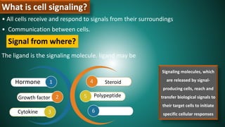
  • 1. What is cell signaling? • All cells receive and respond to signals from their surroundings • Communication between cells. Hormone Growth factor Cytokine Steroid Polypeptide Signal from where? The ligand is the signaling molecule. ligand may be Signaling molecules, which are released by signal- producing cells, reach and transfer biological signals to their target cells to initiate specific cellular responses
  • 2. CONDITION FOR SIGNALING Receptor activated by ligand, the receptor causes a change in the target cell in which it is expressed  Extracellular molecules Protein, peptides, Hormone, cytokine adrenaline, thyroxine .prostaglandin  Intracellular molecules CCa2+, carbohydrate, CAMP/CGMP Intercellular signaling Communication between cells Intracellular signaling Signaling chains within the cell
  • 3. Intercellular signalling Controls Metabolic fluxes Cell division Growth Development Processes sensory information Differentiation Cells communicate via Messenger substances Gap junctions Surface proteins Electrical signals
  • 4. Hormones As a Signal molecules Binding with Receptor Activated Enzyme Second messenger Transcription Effect on cellular function
  • 5. The signalling molecules produce an effect on same cell that produce it Autocrine signalling Autocrine signalling the cell that is producing the messenger expresses receptors on its surface that can respond to that messenger Consequently, cells releasing the message will stimulate (or inhibit) themselves.
  • 6. The cytokine interleukin-1 in monocytes. When interleukin-1 is produced in response to external stimuli, it can bind to cell-surface receptors on the same cell that produced it. Activated T lymphocytes T lymphocyte respond to antigenic stimulation by synthesizing a growth factor that derives their own proliferation, and there by increasing the number of responsive T-Lymphocytes Cancer development- Autocrine signaling plays critical roles in cancer activation and also in providing self- sustaining growth signals tumors EXAMPLES OF AUTOCRINE SIGNALING
  • 7. •Paracrine signaling is a form of cell-to-cell communication in which a cell produces a signal to induce changes in nearby cells(close proximity) • Signaling molecules known as paracrine factors diffuse over a relatively short distance (local action) • Cells that produce paracrine factors secrete them into the Immediate extracellular environment. • Follow paracrine signaling pathway-Fibroblast family, Hedgehog family, Wnt family, and TGF-B superfamily. Growth Factor(FGF) Receptors available on the cell membrane to receive the signals, also known as Being competent PARACRINE SIGNALING
  • 8. • The signaling molecule act on target cells distantly located from the Site of synthesis. • It is a long range signaling and signal molecules is transported by Blood streams. The pancreas is an endocrine gland and produces the hormone insulin, which regulates the uptake of glucose in cells all over the body. Hormones that function in an endocrine manner include testosterone, progesterone and gonadotropins. ENDOCRINE SIGNALING EXAMPLES OF ENDOCRINE SIGNALING
  • 9. JUXTACRINE SIGNALING • This kind of signaling require physical contact between the cells. Also known as contact- dependent signaling. A membrane ligand (protein, oligosaccharide, lipid) and a membrane protein of two adjacent cells interact. A communicating junction links the intracellular compartments adjacent cells, allowing transit of relatively small molecules. An extracellular matrix glycoprotein and a membrane protein interact. 3 types of juxtacrine signaling molecules.
  • 10. TYPES OF LIGANDS 1. Membrane bound signal molecule – signal molecule remain surface of the cell and mediate contact dependent signaling. 2. Secretory signal molecules- secreted from signaling cell and binds to receptor. Secretory signal molecules are extracellular and divided in 3 categories depending on distance 1 2 3 Endocrine Paracrine Autocrine Depending On The Nature Of Extracellular Signal Molecules It Is Divided In 2 More Categories LIPOPHILIC MOLECULES HYDROPHILIC MOLECULES
  • 11. EXAMPLE OF SIGNAL MOLECULES THAT INTRACT WITH CELL SURFACE RECEPTOR Epinephrine Glucagon Insulin Gastrin Secretin Adrenocorticotropic hormone
  • 12. EXAMPLE OF SIGNAL MOLECULES THAT INTRACT WITH CYTOSOLIC OR NUCLEAR RECEPTOR Steroid hormones- Progesterone, testosterone, cortisol, aldosterone. Non steroid hormones- thyroid hormone and retinoic acid.
  • 13. • Receptors are protein molecules inside the target cell or on its surface that receive a chemical signal. • Chemical signals are released by signaling cells in the form of small, or soluble molecules called ligands. RECEPTOR AND LIGAND Orphan receptor Orphan receptor is a protein that has a similar structure to other identified receptors but whose ligand has not yet been identified.
  • 14. Intracellular receptors Intracellular receptors are receptor proteins found on the inside of the cell, typically in the cytoplasm or nucleus. The ligands of intracellular receptors are small, hydrophobic{water hating) molecules, since they must be able to cross the plasma membrane in order to reach their receptors. For example, receptors for hydrophobic steroid hormones, such as the sex hormones -an oestrogen and testosterone, thyroid hormone, retinoid and vitamin D are intracellular. Also contain orphan receptors.
  • 15. Intracellular receptors Intracellular receptor belongs to Nuclear receptor superfamily Nuclear receptor superfamily is a family of highly conserved transcription factors that regulates transcription in response to small lipophilic signal molecules. Members of the Nuclear receptor superfamily can both positively and Negatively regulate transcription.
  • 16. Nuclear receptor superfamily N-terminal activation domain, called the A/B region, A central DNA binding domain(DBD) and A C-terminal ligand binding domain (LBD)
  • 17. Example Steroid hormone receptor Location-cytosol Homodimer receptor In cytosol= receptor +HSP - INACTIVE In cytosol= receptor+ligand receptor-ligand complex • It can be homodimer-example ----steroid receptor • It can be heterodimer- example--- retinoic acid receptor, thyroid hormone receptor, vitamin D receptor and orphan receptor Nuclear receptor Thyroid hormone receptor Heterodimer receptor Location of receptor= nucleus
  • 18. Cell-surface receptors Cell-surface receptors, also known as transmembrane receptors, are proteins that are found attached to the cell membrane. These receptors bind to external ligand molecules because ligands do not travel across the cell membrane. This type of receptor spans the plasma membrane and performs signal transduction, in which an extracellular signal is Converted into an intercellular signal. Cell-surface receptors are also called cell-specific proteins or markers because they are specific to individual cell types.
  • 19. Cytosol • Each cell-surface receptor has three main components: 1. An external ligand-binding domain 2. A hydrophobic membrane-spanning region, and 3. An intracellular domain inside the cell. Cell-surface receptors
  • 20. 1. Dimerization 2. Conformational changes Mechanism of working Categories of cell-surface receptors Ion channel-linked receptors, G-protein-linked receptors, and Enzyme-linked receptors.
  • 21. Ion channel linked receptors Also called transmitter gated ion channel or inotropic receptors. lon channel-linked receptors bind a ligand and open a channel through the membrane that allows specific ions to pass through. To form a channel, this type of cell-surface receptor has an extensive membrane-spanning region. When a ligand binds to the extracellular region of the channel, there is a conformational change in the proteins structure
  • 22. Example- ligand gated channel for NTMs-Acetycholine • Acetylcholine receptor is a receptor linked to a cation channel. • As two molecules of acetylcholine bind to the binding sites on a subunits, the conformation of the receptor is altered and the gate is opened, allowing for the entry of many ions and small molecules.
  • 23. G-protein-coupled receptors It is a G-protein linked receptor G-protein-coupled receptors bind a ligand and activate a membrane protein called a G- protein. The activated G-protein then interacts with an enzyme in the membrane Before the ligand binds, the inactive G-protein can bind to a site on a specific receptor. Once the G-protein binds to the receptor, the G-protein changes shape, becomes active, and splits into two different subunits. One or both of these subunits may be able to activate other proteins as a result.
  • 24. G- Protein Coupled Receptor
  • 25. Enzyme-linked receptors Enzyme-linked receptors are cell-surface receptors with intracellular domains that are associated with an enzyme. In some cases, the intracellular domain of the receptor itself is an enzyme. Other enzyme-linked receptors have a small intracellular domain that interacts directly with an enzyme. When a ligand binds to the extracellular domain, a signal is transferred through the membrane, activating the enzyme. Activation of the enzyme sets off a chain of events within the cell that eventually leads to a response
  • 26. G-PROTEIN LINKED RECEPTOR What is GPCR? What is G Protein and its mode of action? What is GTP? What is GTPase? What is GEF(Guanine exchange factor)? What is stimulatory G- protein? What is secondary messenger?
  • 27. What is GPCR? G-Protein coupled receptor(GPCR) or G- Protein linked receptor. Have seven transmembrane domain, so it is also called serpentine Extracellular receptor. Largest family of cell surface receptor. Multiple single polypeptide. GPCR present is all eukaryotes GPCR are the largest group of cell surface receptor that transmit signal inside the cell with the action of guanine nucleotide binding protein called G-protein.
  • 28. What is G Protein? • G proteins, also known as guanine nucleotide-binding proteins, are a family of proteins that act as molecular switches inside cells, and are involved in transmitting signals from outside a cell to its interior. • G proteins belong to the larger group of enzymes called GTPases. What is GTPase? GTPases are large family a of hydrolase enzymes that can bind and hydrolyze guanosine Triphosphate (GTP) What is GTP? It also has the role of a source of energy or an activator of substrates in metabolic reactions, like that of ATP. GTP is essential to signal transduction, with G-proteins. It is converted to guanosine diphosphate (GDP) through the action of GTPases.
  • 29. Mode of Action The inactive form of GTPase (GDP) are activated by a class of protein called guanosine nucleotide exchange factor (GEFs)
  • 30. GPCR has receptors for 1. NTMS 2. Neuropeptides 3. Peptide hormone 4. Immune cells 5. Disease factors 6. Adrenaline (epinephrine) 7. Smell 8. Light 9. Taste
  • 31. What is stimulatory G- Protein When G- protein involved in activation of enzyme like effector, Adenylate cyclase, is called stimulatory G-protein
  • 32.
  • 33. What is Primary messenger? Ligand are termed as primary/first messengers. First messengers are extracellular factors, often hormones, Growth Factor, neurotransmitters, Peptides etc. What is secondary messenger? Second messengers are intracellular signaling molecules released by the cell in response to exposure to extracellular signaling molecules – the first messengers. They triggers intracellular signal transduction cascades.
  • 34. Removal or degradation of second messenger terminate the cellular response Four classes of second messengers Cyclic nucleotides Membrane lipid derivatives Ca2+ Nitric oxide/carbon monoxide Examples of second messenger molecules include cyclic AMP, cyclic GMP, inositol trisphosphate (IP3), diacylglycerol, and calcium
  • 35. Which of the following is NOT a second messenger? 1. Cyclic GMP 2. Diacylglycerol 3. Inositol triphosphate 4. Phosphatidyl inositol
  • 36. G-protein coupled receptors (GPCR) consist of three protein subunits a. B and y. In unstimulated state, a subunit is GDP bound and GPCR is inactive. When GPCR gets activated, it acts like guanine nucleotide exchange (GEF) factor and induces a-subunit to release its bound GDP allowing GTP to bind in its place. In order to regulate G protein activity by regulating GDP/GTP concentration, a subunit acts as GTPase GDP kinase CGMP-specific phosphodiesterase CAMP-specific phosphodiesterase
  • 37. What is cAMP?  CAMP is a derivative of adenosine triphosphate (ATP) and used for intracellular signal transduction in many different organisms, conveying the CAMP-dependent pathway  Cyclic adenosine monophosphate (CAMP, cyclic AMP, or 3,5'-cyclic adenosine monophosphate) is a second messenger important in many biological processes.  Cyclic AMP is synthesized from ATP by adenylate cyclase located on the inner side of the plasma membrane  CAMP decomposition into AMP is catalyzed by the enzyme phosphodiesterase
  • 39. Molecules that activate cAMP pathway include: Cholera toxin Caffeine Pertussis toxin Molecules that inhibit the cAMP pathway include:  cAMP phosphodiesterase converts cAMP into AMP by breaking the phosphodiester bond, in turn reducing the cAMP levels  Gi protein, which is a G protein that inhibits adenylyl cyclase, reducing CAMP levels.
  • 40.  Inactive G-protein  Ligand bind to GPCR  Conformational change in GPCR  Active G-protein  GDP replaced by GTP  Active G-protein activate adenylate cyclase  Adenylate cyclase convert ATP to CAMP  CAMP Dependent signal cascade  Protein kinase A activation  Action of catalytic unit for gene
  • 41. The intracellular levels of cAMP are regulated by the balance between the activities of two enzymes adenylyl cyclase (AC) and cyclic nucleotide phosphodiesterase (PDE)
  • 42.  CAMP-dependent pathway is necessary for many living organisms and life processes  Many different cell responses are mediated by CAMP; these include increase in heart rate, cortisol secretion, and breakdown of glycogen and fat.  CAMP is essential for the maintenance of memory in the brain, relaxation in the heart, and water absorbed in the kidney.  This pathway can activate enzymes and regulate gene expression.  If CAMP-dependent pathway is not controlled, it can ultimately lead to hyper-proliferation, which may contribute to the development and/or progression of cancer.
  • 43. The PKA enzyme is also known as CAMP-dependent enzyme because it gets activated only if cAMP is present. Once PKA is activated, it phosphorylates a number of other proteins including:  Enzymes that convert glycogen into glucose Enzymes that promote muscle contraction in the heart leading to an Increase in heart rate.  Transcription factors, which regulate gene expression.
  • 44. CHOLERA TOXIN Cholera toxin (also known as choleragen and sometimes abbreviated to CTX, Ctx or CT) It is a enterotoxin secreted by bacterium Vibrio cholerae. Genes for cholera toxin are present on the integrated phage genome. The gene encoding the cholera toxin is introduced into V. cholerae by horizontal gene transfer.
  • 45. CHOLERA TOXIN Cholera toxin is a oligomeric/multimeric complex made up of six protein subunits-A- Subunits (A1 and A2 Part) and Five B Subunit. A-Subunit has catalytic property (enzymatic). Result of cholera- massive, watery diarrhea. Single copy of the A subunit(enzymatic), and five copies of the B subunit (receptor binding), denoted as AB,.
  • 46. MODE OF ACTION OF CHOLERA TOXIN • Ligand-cholera toxin • Receptor-Gangliosides • Target cell-intestinal epithelial cell on which Gangliosides Receptor is present( Gangliosides are present and concentrated on cell surfaces) WHAT IS GANGLIOSIDES? A ganglioside is a molecule composed of a glycosphingolipid with one or more sialic acids linked on the sugar chain. Common type of gangliosides. GM1 acts as the site of binding for both Cholera toxin and E. Coli heat labile enterotoxin (Traveller’s Diarrhea) Galactosidases are enzymes that breakdown GM1
  • 47. MODE OF ACTION OF CHOLERA TOXIN PHOSPHATASE Enzyme that remove a phosphate group from its substrate by Hydrolyzing phosphoric acid monoester into phosphate ion and molecule with free hydroxyl group. • It is a subcategory of hydrolases. • Dephosphorylation is done by phosphatases. KINASE (Phosphotransferase) A kinase is an enzyme that catalyzes the transfer of phosphate groups from high-energy, phosphate-donating molecules, such as ATP.
  • 48. PHOSPHODIESTERASE  Enzyme that break phosphodiester bond. PHOSPHORYLASE  Enzyme that catalyze the addition of inorganic phosphate to acceptor.
  • 49. ADP- ribosylation is the addition of one or more ADP-ribose moieties to a protein. It is a reversible post-translational modification that is involved in many cellular processes, including cell signaling, DNA repair, gene regulation and apoptosis
  • 50. Mode of Action of Cholera Toxin
  • 51.
  • 52. Gangliosidosis • Gangliosides are continuously synthesized and degraded in cells. They are degraded to ceramides by sequential removal of sugar units in the oligosaccharide group, catalyzed by a set of highly specific lysosomal enzymes. • Mutations in genes coding for these enzymes leads to the accumulation of partially broken down gangliosides in lysosomes, which results in a group of diseases called gangliosidosis. • For example, the fatal Tay-Sachs disease arises as a genetic defect which leads to no functional hexosaminidase A produced, causing GM2 to accumulate in lysosomes. Ultimately the ganglion cells in the nervous system swell enormously, disturbing the normal functions of neurons.
  • 53. Gangliosides are also involved in several diseases: Influenza, in which haemagglutinin of influenza virus exploits certain gangliosides to enter and infect the cells expressing them.
  • 54.  When cholera toxin is released from the bacteria in the infected intestine, it binds to the intestinal cells known as enterocytes (epithelial cell )through the interaction of the pentameric B subunit of the toxin with the GM1 ganglioside receptor on the intestinal cell.  Occurrence of endocytosis of the toxin.  Next, the A/B cholera toxin must undergo cleavage of the A1 domain from the A2 domain in order for A1 to become an active enzyme.  Once inside the enterocyte, the enzymatic A1 fragment of the toxin A subunit enters the cytosol, where it activates the G protein G alpha (Gα,) through an ADP- ribosylation reaction that acts to lock the G protein in its GTP-bound form, STEPS:
  • 55.  The ADP-ribosylation causes the Gα, subunit to lose its catalytic activity of GTP hydrolysis into GDP + P Thus maintaining Gα, in its activated state. Thereby leading continually stimulating adenylate cyclase to produce CAMP.  The high cAMP levels activate the cystic fibrosis transmembrane conductance regulator (CFTR), causing A dramatic efflux of ions and water from infected enterocytes, leading to watery diarrhea. STEPS:
  • 56. When the cholera toxin (protein of Mr 90,000 Da) gains access to the human intestinal tract it binds tightly to specific receptors in the plasma membrane of the epithelial cells lining the small intestine, causing membrane bound adenylyl cyclase to undergo prolonged activation resulting in extensive loss of H2O and Na+ Pretreatment of the epithelial cells with various phospholipases and proteases failed to inhibit the binding of cholera toxin to its receptor and the fluid loss but treatment with exoglycosidase, prior to binding significantly reduces these effects Which of the following molecule could be the receptor For this toxin? CSIR NET-2017-JUNE Phosphatidyl choline Sodium-potassium ATPase Ganglioside Chloride bicarbonate exchanger
  • 59. GTP PLC-β PIP2 DAG DAG Ca++ IP3 PKC Active ER Open IP3 Gated ion Channel Ligand Activated G-Protein Exterior
  • 60. Significance  IP3 has main functions are to mobilize Ca²+ from storage organelles and to Regulate cell proliferation and other cellular reactions that require free calcium.  In smooth muscle cells, for example, an increase in concentration of cytoplasmic Ca²+ results in the contraction of the muscle cell.  In the nervous system, IP3 serves as a second messenger, with the cerebellum containing the highest concentration of IP3 receptors. There is evidence that IP3 receptors play an important role in the induction of plasticity in cerebellar Purkinje cells.  The slow block to polyspermy in the sea urchin is mediated by the PIP2 secondary messenger system. Activation of the binding receptors activates PLC, which cleaves PIP2 in the egg plasma membrane, releasing IP3 into the egg cell cytoplasm. IP3 diffuses to the ER, where it opens Ca²+ channels.
  • 61. ? What is Protein kinase C • Protein kinase C, commonly abbreviated to PKC is a family of protein kinase enzymes that are involved in controlling the function of other proteins through the phosphorylation of hydroxyl groups of serine and threonine amino acid residues on these proteins, or a member of this family
  • 62. What is IP3 and DAG ?  Inositol triphosphate (IP3) and diacylglycerol (DAG) are important secondary messenger molecule used in signal transduction and lipid signaling in biological cells.  They are the product of membrane phospholipid PIP2 Breakdown.
  • 63. STEPS OF IP3-DAG PATHWAY  Ligand binds to GPCR  GDP Replaces GTP  The a subunit of the G protein interacts with the membrane bound enzyme called phospholipase C; this activates the enzyme.  Once activated phospholipase C (PKC) catalyzes (breaks down) phospholipids (P1P2) within the cell membrane into two specific molecules: DAG and iP3. .  Although inositol trisphosphate diffuses into the cytosol, diacylglycerol remains within the plasma Membrane, due to its hydrophobic properties.
  • 64.  IP3 stimulates the release of calcium ions from the smooth endoplasmic reticulum, whereas DAG is a physiological activator of protein kinase C (PKC).  The production of DAG in the membrane facilitates translocation of PKC from the cytosol to the plasma membrane  PKC enzymes in turn are activated by signals such as increases in the concentration of diacylglycerol (DAG) Or calcium ions (Ca++). STEPS OF IP3-DAG PATHWAY
  • 65.  Calmodulin (CaM) (an abbreviation for calcium modulated protein)  Calmodulin is a small, highly conserved calcium binding cytosolic acidic protein found in all eukaryotic cells.  It is An Intracellular target/receptor of The secondary messenger Ca2+, and the binding Ca²+ is required for the activation of calmodulin.  Once bound to Ca²+, calmodulin acts as part of a calcium signal transduction pathway by modifying its interactions with various target proteins such as kinases or phosphatases CALCIUM CALMODULIN COMPLEX
  • 66. Inactivate calmodulin activate calmodulin Target Protein 1. 2. 3. Ca++ CALCIUM CALMODULIN COMPLEX  Calmodulin has total of four Ca²+ binding sites.  Calmodulin + four Ca²+ = conformational change in calmodulin.  Calmodulin’s structure is very similar to the structure of troponin C (which is another calcium binding protein). Calmodulin is smaller than that of Troponin C.