
Recomendados
Más contenido relacionado
Último
Último (20)
Destacado
Destacado (20)
Biopsie mammaire
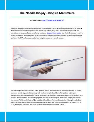
- 1. The Needle Biopsy - Biopsie Mammaire _____________________________________________________________________________________ By Mein Laon - http://imageriemedicale.fr/ A needle biopsy could be performed in most circumstances. Let's say you have a palpable lump. You can find two kinds of needle biopsies: a fine-needle aspiration (FNA) and a core needle biopsy. Both are carried out on palpable lumps as office procedures, biopsie mammaire but the techniques are not the same. In addition, different pathologists are involved: a highly trained cytopathologist reads (and might perform) the FNA, whereas a surgical pathologist reads a core needle biopsy. The advantage of an FNA is that it is the quickest way to demonstrate the presence of tumor. If tumor is shown to be existing, a definitive diagnosis has been created and days of anguished waiting are eliminated. A positive diagnosis of tumor by an FNA resolves the issue of whether you do or do not have cancer. The drawback is that a damaging FNA provides no meaningful information. Simply because of the way an FNA is carried out, a false negative is feasible. A fine-needle aspiration biopsy is carried out with a little syringe and needle-essentially like the ones utilized to provide you with a flu injection or a DPT (diphtheria, pertussis, and tetanus) shot whenever you were a little child.
- 2. The syringe and also the needle are used to suck out (aspirate) some tissue from your lump for an examination under the microscope. Often, if an unskilled individual performs the FNA, no cells appear in the aspirate. The FNA has two requirements: tissue must be obtained, and also the aspirate must be properly ready for that cytopathologist to read. In some facilities, the cytopathologist carries out the FNA and also looks at the stained tissue under the microscope, thereby giving the patient an answer almost instantly. If cells from your lump are not present or if the cells are not ready properly for that pathologist, a diagnosis can't be created and the report will examine "nondiagnostic." A damaging FNA doesn't mean anything, for cancer may be existing in spite of a negative report. This is what we have called a false negative. To become particular that there is no tumor, the surgeon needs to carry out an open biopsy, in which the whole suspicious region is surgically eliminated (an excisional biopsy). If a valid excisional biopsy proves to be damaging (that's, no tumor is found), you are able to be assured that you simply do not have tumor in that particular suspicious area of the breast area. If the FNA diagnosis is good (tumor is present), every member from the treatment team ought to be consulted-the breast surgeon, the breast area radiation oncologist, and also the breast medical oncologist. Even though you think you know regardless of whether or not you wish to preserve your breast area, you might not have the whole picture. Listen carefully to every specialist prior to making up your mind how you wish to proceed. In this instance, four heads are better than one. If a lump is palpable, a core needle biopsy or an FNA can be carried out. If you would like to know whether you have cancer, either biopsy will serve-but only when the result is good. How is a core needle
- 3. biopsy different from an FNA? Why does a different pathologist read it? At greatest, the FNA obtains only tissue. A core needle obtains a thin sample from the tissue itself and views all the cells in their proper architectural relation to other tissue in the tissue. So the core needle biopsy gives more info than the FNA. A specific cutting needle obtains a number of thin cores of tissue from the suspicious area, which are then placed in a standard "fixing" solution that prepares the tissue for that pathologist. Simply because the cores are so thin, the time in the fixing solution is much shorter than the time necessary for the much larger piece of structure from an open biopsy. Once the tissue is ready and stained, the pathologist examines it below the microscope and makes a diagnosis. What will the pathologist say? First, you will be informed regardless of whether tumor is or is not present-whether the biopsy is positive or damaging. If tumor is present in the core biopsy, the pathologist usually can get enough information from your architecture of the tissue to tell you the type of cancer that you simply have and also how orderly the tissue are (the grade). Pathologists also can give you info about whether the cancer they're seeing has special attributes such as estrogen and / or progesterone receptors, but such info is generally obtained from an open biopsy, where a larger piece of structure could be obtained. Be certain you understand that pathologists are limited to what they see. They cannot comment on structure other than the thin core they are seeing below the microscope. They diagnose only what they see. When the core needle biopsy is good, cancer is existing and your next choice is how you want your breast area tumor to be treated. It is not so simple if the core needle biopsy is negative. You might need an open biopsy to get more information. Consult with all three members of the treatment team. They could explain the issues and guide you. What if you don't have a palpable lump, but you require a biopsy because you have a suspicious mammogram? As you can see on the right side of the decision tree, a stereotactic biopsy makes it feasible to carry out a biopsy on lesions that are not palpable but visible only on a mammogram. Suppose the screening mammogram shows a cluster of suspicious microcalcifications deep in your breast. Microcalcifications are believed to be formed as a result of cells that have died in the breast duct system. The dead tissue and their debris become calcified and are visible as if they were multiple tiny white grains. Calcifications that indicate cancer could be observed either in association with a mass or long prior to a lump has been detected. So... What’s Next ? To learn more about biopsie mammaire, Click Here: http://imageriemedicale.fr/