Rotator Cuff Disorders | Shoulder Surgeon | Greater Denver Colorado

Shoulder pain is the third most common musculoskeletal symptom encountered in medical practice after back and neck pain, accounting for almost 3 million patient visits each year in the United States. A wide range of potential pathoanatomic entities can give rise to shoulder pain, from simple sprains to massive rotator cuff tears. The majority of these conditions are amenable to conservative treatment. Rotator cuff dysfunction is a particularly important entity because it occurs frequently and may necessitate surgical treatment. This report will provide a critical overview of current diagnostic and treatment techniques for rotator cuff disease. For more shoulder surgery and rotator cuff studies, visit Dr. Millett, shoulder surgeon, Greater Denver http://drmillett.com/shoulder-studies

Recomendados
Recomendados
Más contenido relacionado
La actualidad más candente
La actualidad más candente (20)
Similar a Rotator Cuff Disorders | Shoulder Surgeon | Greater Denver Colorado
Similar a Rotator Cuff Disorders | Shoulder Surgeon | Greater Denver Colorado (20)
Más de Peter Millett MD
Más de Peter Millett MD (18)
Último
Último (20)
Rotator Cuff Disorders | Shoulder Surgeon | Greater Denver Colorado
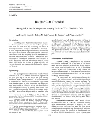
- 1. ARTHRITIS & RHEUMATISM Vol. 50, No. 12, December 2004, pp 3751–3761 DOI 10.1002/art.20668 © 2004, American College of Rheumatology REVIEW Rotator Cuff Disorders Recognition and Management Among Patients With Shoulder Pain Andreas H. Gomoll,1 Jeffrey N. Katz,1 Jon J. P. Warner,2 and Peter J. Millett1 Introduction revealed partial- and full-thickness rotator cuff tears in 4% of individuals 40 years old and in more than 50% Shoulder pain is the third most common muscu- of individuals 60 years old (5). Furthermore, autopsy loskeletal symptom encountered in medical practice studies have demonstrated a 6% prevalence of full- after back and neck pain (1), accounting for almost 3 thickness rotator cuff tears in subjects 60 years old and million patient visits each year in the United States (2). A wide range of potential pathoanatomic entities can 30% prevalence in those 60 years old (6), although it give rise to shoulder pain, from simple sprains to massive was unknown how many of these subjects had shoulder rotator cuff tears. The majority of these conditions are pain. amenable to conservative treatment. Rotator cuff dys- function is a particularly important entity because it Anatomy and pathophysiology occurs frequently and may necessitate surgical treat- ment. This report will provide a critical overview of Anatomy (Figure 1). The shoulder has the great- current diagnostic and treatment techniques for rotator est range of motion (ROM) of any joint in the human cuff disease. body. The size mismatch between the smaller glenoid and larger humeral head creates a risk of instability. Stability is provided both statically by the capsule and Epidemiology labrum, and dynamically by the rotator cuff musculature. The point prevalence of shoulder pain has been Dysfunction of any of these structures can lead to pain, estimated to be 7–25% and the incidence as 10 per 1,000 weakness, and instability. per year, peaking at 25 per 1,000 per year among The rotator cuff is a tendinous confluence of 4 individuals ages 42–46 years (3,4). The overall number muscles that initiate shoulder motion and maintain the of individuals with rotator cuff dysfunction is expected to normal relationship between the articular surfaces. The grow coincident with an aging population that is increas- supraspinatus muscle provides abduction, the infraspi- ingly active and less willing to accept functional limita- natus and teres minor muscles provide external rotation, tions. A large proportion of patients with rotator cuff and the subscapularis muscle provides internal rotation. tears remain asymptomatic. Magnetic resonance imag- In addition, the muscles of the rotator cuff balance the ing (MRI) scans of participants without shoulder pain forces of other shoulder muscles, most importantly the deltoid muscle. Contraction of the deltoid muscle in the Supported in part by the NIH (grants P60-AR-47782 and absence of supraspinatus function leads to superior K24-AR-02123 from the National Institute of Arthritis and Musculo- translocation of the humeral head, making wide abduc- skeletal and Skin Diseases). 1 Andreas H. Gomoll, MD, Jeffrey N. Katz, MD, MS, Peter J. tion difficult. Millett, MD, MSc: Brigham and Women’s Hospital and Harvard Rotator cuff dysfunction. Since Codman’s report Medical School, Boston, Massachusetts; 2Jon J. P. Warner, MD: on rotator cuff tears in 1934 (7), a continuum ranging Massachusetts General Hospital and Harvard Medical School, Boston, Massachusetts. from impingement syndrome to partial- and full- Address correspondence and reprint requests to Peter J. thickness rotator cuff tears has been described as the Millett, MD, MSc, Brigham and Women’s Hospital, 75 Francis Street, basis of rotator cuff dysfunction. Impingement syndrome Boston, MA 02115. E-mail: pmillett@partners.org. Submitted for publication March 2, 2004; accepted in revised is a chronic process that manifests as shoulder pain and form August 23, 2004. is, at least initially, reversible with rest or other conser- 3751
- 2. 3752 GOMOLL ET AL athletes who participate in overhead and throwing sports activities (10). Its anatomic correlate consists of under- surface fraying of the infraspinatus tendon, wherein it contacts the posterior glenoid as the arm is placed in maximum abduction and external rotation, such as the late cocking phase of throwing. Although this contact may often be present physiologically, the repetitive injury and eccentric loading associated with throwing can lead to labral and rotator cuff tears. Secondary impingement. Secondary, or nonoutlet, impingement is a dynamic process caused by mild gle- nohumeral instability. Subtle subluxation of the humeral head brought on by activity can severely narrow the humeroacromial interface and thus lead to impingement symptoms. Posterior capsular contractures, such as oc- cur with frozen shoulder, can cause obligate anterosu- Figure 1. Anatomy of the shoulder. Lateral view of the glenoid fossa (with humeral head removed). lig ligament. Reproduced, with perior humeral head translation with forward flexion of permission, from Turkel SJ, Panio MW, Marshall JL, Girgis FG. the humerus. This also can narrow the acromiohumeral Stabilizing mechanisms preventing anterior dislocation of the gleno- interval and result in secondary impingement humeral joint. J Bone Joint Surg Am 1981;63:1208. Intrinsic tendon degeneration. In contrast to Neer’s theory, Ogata and Uhthoff (11) attributed most vative measures. Left untreated, the pain can progress to changes in the shoulder joints to intrinsic degeneration permanent changes and eventual tearing of the rotator of the rotator cuff tendons. This degeneration was cuff, resulting in painful weakness. Impingement syn- thought to arise from relative hypoperfusion of a water- drome is classified into external, internal, and secondary shed area close to the insertion on the greater tuberos- impingement. ity, in conjunction with repetitive microtrauma. Cur- External impingement. External, or outlet, im- rently, most experts believe that both intrinsic tendon pingement, the most common form, is caused by com- degeneration and impingement are contributing factors pression of the rotator cuff tendons as they pass under- in the etiology of rotator cuff dysfunction (12). neath the coracoacromial arch (8). Narrowing of the Rotator cuff tears. The majority of rotator cuff humeroacromial motion interface, which lies between tears occur in tendons with preexisting degeneration this arch and the humeral head, causes compression of (12), which can progress to partial- and full-thickness the intervening rotator cuff tendons. Inflammation of tears, most commonly in the supraspinatus tendon. the subacromial bursa can ensue, leading to pain and Full-thickness tears also may be precipitated by acute further compression due to secondary swelling. Narrow- events; however, trauma with acute onset of weakness ing of the humeroacromial interface can occur for a has been estimated to account for only 8% of patients variety of reasons, such as acromioclavicular (AC) joint undergoing rotator cuff repair (13). osteophytes, acromial bone spurs, or malunions after Partial tears generally involve 50% of the ten- proximal humeral fractures, especially if there has been don thickness and do not lead to retraction of the muscle displacement of the greater tuberosity (9). Neer has (12). Depending on the location within the rotator cuff described several stages of external impingement, and he tendon, partial-thickness tears can be classified as intra- estimated it as the cause of 95% of rotator cuff tears in substance, bursal-sided, or articular-sided (undersur- his practice (8). Stage I affects younger patients, is fully face), the latter constituting 90% of partial tears (14). reversible, and has hemorrhage and edema as anatomic Weakness is uncommon in partial-thickness tears but correlates. Stage II is a disease affecting patients of can arise from pain, which is often greater than that in middle age, is only partially reversible, and presents as complete tears. tendon degeneration and fibrosis, also called tendinosis. In contrast, full-thickness tears represent com- Stage III occurs in elderly patients and is characterized plete discontinuity of rotator cuff fibers, resulting in by further tendon degeneration and rupture. communication between the articular and bursal spaces. Internal impingement. Internal impingement has The extent of the lesion on imaging studies is described been described more recently and occurs primarily in in both the anteroposterior (AP) and mediolateral di-
- 3. ROTATOR CUFF DISORDERS 3753 Table 1. Differential diagnoses according to affected anatomic diagnoses for the acute onset of shoulder pain include systems traumatic events such as shoulder dislocation, AC joint Articular sprains, and clavicle and proximal humerus fractures. Glenohumeral arthritis Osteoarthritis Nontraumatic etiologies of acute shoulder pain include Inflammatory arthritis calcific tendinitis, biceps tendinitis, and, much less fre- Instability quently, gout, septic arthritis, or septic bursitis. Shoulder Labral tears Periarticular pain can also be caused by the sudden exacerbation of Calcific tendinitis chronic processes such as glenohumeral and AC osteo- Adhesive capsulitis arthrosis or inflammatory arthritis. Shoulder symptoms Biceps tendinitis Fibromyalgia that occur chronically are suggestive of frozen shoulder Scapulothoracic bursitis (adhesive capsulitis), polymyalgia, or, rarely, osteomyeli- Acromioclavicular joint arthritis tis or neoplastic disorders. Referred pain from disorders Acromioclavicular joint sprains Osseous of the cervical spine is a very common source of shoulder Proximal humerus or clavicle fracture pain, and a careful clinical examination is essential to Bone cysts, infections, or tumors Referred distinguish between local and referred processes. Rarely, Cervical radiculitis and arthritis visceral radiation, as occurs with splenic, hepatic, dia- Myofascial neck pain phragmatic, and cardiac processes, may be discerned as Cardiac, splenic, hepatic, and diaphragmatic processes Neurogenic shoulder pain. Parsonage-Tuner syndrome (brachial neuritis) Carpal tunnel syndrome Evaluation and diagnosis Individuals with rotator cuff disorders can be rections. One centimeter is generally considered a small divided into 2 groups according to their presenting lesion, 1–3 cm is considered medium, 3–5 cm is consid- symptoms: 1) those with impingement-type symptoms, ered large, and 5 cm is considered massive. Tears that frequently manifested as pain at night and at rest as well involve 2 tendons can also be classified as massive, and as a painful arc of motion, which can often be success- these require more complex reconstruction (15). In fully treated by conservative measures; 2) those with larger tears, chronically retracted muscles undergo fatty symptoms of a torn rotator cuff tendon, manifested as degeneration over time that may be irreversible, and thus painful weakness and atrophy, which frequently do not may make results of direct repair unsatisfactory (16). respond fully to conservative measures alone and for Natural history. The natural history of rotator cuff dysfunction is not well understood. Yamaguchi et al which surgical intervention should be considered. demonstrated that in 50% of individuals with asymptom- History. Rotator cuff pain is frequently described atic tears pain developed within 5 years, even though as a dull ache of insidious onset, extending over the only 30% demonstrated increases in tear size (17). lateral arm and shoulder. Overhead activities exacerbate Studies investigating partial tears of the rotator cuff have the pain, and the pain frequently increases at night and demonstrated enlargement or progression to full- may awaken the individual from sleep. Weakness with thickness tears in 80% of patients over a period of 2 the inability to abduct and elevate the arm is seen in years, and these were managed with nonoperative ther- more advanced cases; patients frequently describe diffi- apy (18). Once a tear occurs, there seems to be little or culties combing hair, holding a hair dryer, and removing no evidence of spontaneous healing. A histopathologic the wallet from their back pocket. Immediate onset of study showed no signs of healing in pathologic speci- weakness, especially in association with trauma, may mens from partial-thickness tears (12). Furthermore, indicate an acute tear. although shoulder symptoms may be short-lived, persis- Clinical examination. Examination of cervical tence or recurrence of symptoms in 40–50% of individ- spine. The cervical spine is a frequent source of referred uals within 1 year after the initial presentation has been pain. Therefore, it should always be included in a clinical reported (4,19,20). examination. Differential diagnoses. Differential diagnoses to Inspection and palpation. Inspection. The shoul- consider in the evaluation of the painful shoulder can be der girdle musculature often shows evidence of muscle grouped based on the affected anatomic structures atrophy. The supra- and infraspinatus muscles typically (Table 1) or chronicity of symptoms. The differential demonstrate atrophy in advanced rotator cuff tears.
- 4. 3754 GOMOLL ET AL Figure 2. Shoulder radiographs for imaging studies of rotator cuff dysfunction. Left, Anteroposterior view. Middle, Outlet view. Right, Axillary view. Swelling over the AC joint can be a sign of traumatic or stretched. Both tests, however, have questionable value, degenerative changes. due to their relatively low sensitivity and specificity (21). Palpation. The arthritic AC joint is a frequent The cross-body adduction test reproduces pain in the source of shoulder pain and will manifest as point AC joint when the arm is adducted horizontally in front tenderness. Bicipital tendinitis can be detected with of the body. palpation over the anterior shoulder, with the arm in Functional tests. Functional tests are performed slight internal rotation. The greater tuberosity can be for each of the 3 muscle groups of the shoulder: the tender to palpation, due to the bursitis often observed in subscapularis, the infraspinatus and teres minor, and the conjunction with rotator cuff disease and calcific tendi- supraspinatus. Tests of the biceps are conducted as well, nitis, and can be palpated by extending the humerus. to determine if positive impingement test results are a Motion testing. ROM testing should first be result of biceps tendinitis. When evaluating strength, it is performed actively by the patient, and then be per- important to consider whether perceived weakness is formed passively by the examiner, with the shoulder in secondary to loss of muscle or due to voluntary or forward elevation, abduction, external rotation, and involuntary inhibition secondary to pain. Frequently, internal rotation. The contralateral shoulder can serve subacromial anesthetic and/or corticosteroid injections as a baseline referent if it is uninvolved. Comparison of are helpful to distinguish between the 2 causes. In active and passive ROM provides insight into the diag- addition to evaluating gross strength, the examiner can nosis. For example, greater passive ROM than active obtain important information by the re-creation of pain ROM, with a painful arc between 60° and 120° of with specific functional tests. abduction, is common in rotator cuff dysfunction, Gross muscle strength is tested first. More spe- whereas globally decreased active and passive ROM is cific tests of muscle function are then conducted: the typically noted in adhesive capsulitis and osteoarthrosis. subscapularis is tested with resisted internal rotation, the Impingement signs. Provocative tests of impinge- supraspinatus is tested with resisted abduction in the ment attempt to recreate shoulder pain by compressing plane of the scapula, often referred to as Jobe’s testing, the rotator cuff between the humeral head and other and the infraspinatus and teres minor are tested with bony structures, such as the acromion or coracoid pro- resisted external rotation. Side-to-side comparison is cess. The 3 most commonly used provocative maneuvers helpful. Lag signs, which are pathognomonic of rotator are Neer’s impingement sign, Hawkins’ impingement cuff tears, have been described by several authors: the sign, and Neer’s impingement test. It should be noted, lift-off and modified lift-off are tests for the subscapu- however, that the first 2 signs can be relatively nonspe- laris, the drop arm sign is used for the supraspinatus, the cific and may yield positive results in the setting of other external rotation lag is used for the infraspinatus, and pathologic entities such as AC joint arthritis or biceps Hornblower’s sign is used for teres minor dysfunction (22). tendinitis. Tests of the biceps tendon and AC joint can Imaging studies. Radiographs (Figure 2). In the be used to assess the pathologic condition of these AP radiographic view, joint space narrowing and osteo- structures. Speed’s test and Yergason’s test elicit pain in phyte formation may indicate arthritis of the glenohu- the bicipital groove when an inflamed biceps tendon is meral or AC joints. Calcium deposits from calcific
- 5. ROTATOR CUFF DISORDERS 3755 tendinitis usually occur just proximal to the rotator cuff insertion. Elevation of the humeral head on AP radio- graphs, especially when the subacromial space is de- creased to 5–7 mm, has been associated with large rotator cuff tears (23). The axillary view is essential to exclude the possibility of a dislocation. This view also shows the joint space and helps identify the rare, but occasionally symptomatic, os acromiale, which is a per- sistent and nonunited ossification center at the end of the acromion (24). Finally, the supraspinatus outlet view allows visualization of the bony structures of the scapu- lohumeral motion interface and shows acromial spurs or calcification of the coracoacromial ligament that might compress the underlying rotator cuff. Arthrography. Arthrography has been largely re- placed by other imaging techniques, such as MRI and ultrasound. Although relatively inexpensive, arthrogra- phy is invasive and less accurate than MRI, especially for the diagnosis of partial-thickness tears, but remains of value in patients with contraindications to MRI. Ultrasound. Ultrasound is noninvasive, readily available, and inexpensive. Recent studies utilizing ar- Figure 3. Oblique coronal T2 magnetic resonance image depicting a throscopy or MRI for validation of ultrasound have full-thickness rotator cuff tear (arrowhead). demonstrated sensitivities of 58–100% and specificities of 78–100% for full-thickness tears (25,26). It is less accurate in the detection of partial-thickness tears, with For the purposes of this review, we searched Cochrane’s sensitivities ranging from 25% to 94% (26–28). Database of Systematic Reviews and Medline to identify MRI and MR arthrography (Figure 3). MRI has relevant articles (see Tables 2–4), utilizing the keywords sensitivities close to 100% for full-thickness tears, and rotator cuff tear, rotator cuff tendinitis, and shoulder has all but replaced arthrography for the diagnosis of pain, with special consideration of publication types rotator cuff disease (29). Moreover, the additional quan- designated as randomized controlled trials and reviews. titative and qualitative information gleaned from this Nonoperative treatment. Nonoperative treat- cross-sectional study aids in the surgical planning and ment for shoulder pain due to rotator cuff impingement prognosis. The combination of MRI and gadolinium- and tears generally includes appropriate physical ther- enhanced arthrography further improves sensitivity, es- apy, antiinflammatory medication, corticosteroid injec- pecially for the detection of partial tears, to more than tions, and other approaches. Pain relief and restoration 90% (30), and in the detection of labral disease, the of function have been observed in 62–74% of patients sensitivity is improved to more than 80% (31). Impor- with symptomatic, radiologically proven rotator cuff tant concerns regarding MRI include the associated cost tears. Predictors of good outcomes are greater preoper- and high frequency of false-positive results. Up to 30% ative muscle strength, such as the ability to lift the arm of asymptomatic volunteer subjects have findings of above the level of the shoulder, and duration of symp- rotator cuff anomalies, and up to 50% show labral toms 6–12 months (33,34). anomalies (32). Medical management. Options for the medical management of rotator cuff disease include systemic and local approaches. Nonsteroidal antiinflammatory drugs Management options and outcome (NSAIDs) decrease symptoms of cuff irritation and The ultimate goal of any therapeutic intervention inflammation, and should be prescribed around-the- for shoulder pain is the restoration of pain-free function. clock to maximize the antiinflammatory effects. This can Specific patient factors, such as age, preinjury functional be augmented by subacromial injections of local anes- level, demand, and general health, guide the physician in thetic agents and corticosteroids. Injections should not the selection of attainable goals and choice of therapy. be instilled directly into the tendon substance, because
- 6. 3756 GOMOLL ET AL Table 2. Overview of outcomes of medical interventions* First author, Condition year (ref.) treated Treatment arms Study design Outcome measure Results Blair, 1996 Impingement Subacromial steroid RCT (n 40), Pain, ROM Significant pain reduction for treatment (67) syndrome injection, subacromial FU at 33 weeks group, from 2.4 to 1.2 (0–4 scale). local anesthetic Control group showed reduction injection from 2.3 to 2.0. At FU, 15 of 19 treatment patients were without impingement sign vs. 4 of 21 in control group. Petri, 1987 Shoulder pain Subacromial steroid RCT (n 100) Pain, ROM, function Both treatments significantly superior (68) injection, oral to placebo. Responders: 4% and 8% naproxen, naproxen with placebo, 12% and 20% with injection, placebo naproxen, 8% and 28% with steroid, 20% and 28% for the combination, at 2 weeks and 4 weeks, respectively. van der Windt, Painful, stiff Intraarticular steroid RCT (n 109), Pain, ROM, function Statistically significant difference with 1998 (69) shoulder injection, PT FU at 12 months 77% (injection) vs. 46% (PT) of patients improved at 7 weeks. Trend toward smaller difference between groups with increasing FU time. Winters, 1997 Shoulder pain PT, manipulation, steroid RCT (n 114), Pain, ROM At 5 weeks, 75% (injection), 40% (70) injection (intraarticular, FU at 11 weeks (manipulation), and 20% (PT) of subacromial, and AC patients rated themselves as “cured.” joint) Hay, 2003 (71) Shoulder pain PT, subacromial steroid RCT (n 207), Pain, ROM, function Improvements (defined as minimum of injection FU at 6 months 50% drop in disability scores) in 60% of patients in PT group and in 53% in injection group. No statistically significant difference between treatment arms at 6 weeks and 3 months. Adebajo, 1990 Rotator cuff Diclofenac steroid RCT (n 60), Pain, ROM, function Average improvements in VAS pain (72) tendinitis injection, placebo FU at 4 weeks score (% responders) of 1.35 (0%) steroid injection, with placebo, 3.6 (30%) with placebo placebo diclofenac, and 4.95 (70%) with injection triamcinolone injections. Both treatment groups significantly improved, but no difference between the 2 groups. White, 1986 Rotator cuff Indomethacin, RCT (n 40), Pain, ROM No statistically significant difference (73) tendinitis subacromial steroid FU at 6 weeks between groups. Improvement of injection 60% (injection) vs. 66% (NSAID). * RCT randomized controlled trial; ROM range of motion; FU followup; PT physical therapy; AC acromioclavicular; VAS visual analog scale; NSAID nonsteroidal antiinflammatory drug. this and multiple injections within a short period of time initial trial of oral NSAIDs and physical therapy, with increase the risk of associated tendon rupture (35). injections reserved for patients with persistent pain or Several studies (Table 2) have investigated the efficacy severe pain at the time of the initial presentation. of oral NSAIDs and injectable steroids. Overall, results Physical therapy. Physical therapy and rehabilita- demonstrate significant improvements with either form tion for rotator cuff signs and symptoms are conducted of treatment, although somewhat faster and greater pain in 3 phases (34). Phase 1 consists of activity modification relief is achieved with injections. in addition to pain control with NSAIDs and injections. Several studies have investigated the accuracy of In phase 2, gentle ROM exercises are initiated to injections, with results ranging in accuracy from 29% to prevent adhesions (39). Only after restoration of full 87% (36–38), underscoring the technique-dependence ROM should physical therapy transition to phase 3, of injections. These studies also demonstrated a signifi- which consists of a strengthening program for the rota- cant difference in outcome, with far better results for tor cuff and scapular stabilizers. Very little data com- accurate injections than for missed injections. Given the paring physical therapy with no treatment are available invasiveness of injections, many practitioners prefer an (Table 3). One retrospective study investigating the
- 7. ROTATOR CUFF DISORDERS 3757 Table 3. Overview of outcomes of physical management techniques* First author, year Condition (ref.) treated Treatment arms Study design Outcome measure Results Ginn, 1997 Shoulder pain PT for 4 weeks, no RCT (n 66), FU at Pain, ROM, function At 4 weeks, 11% of patients in the treatment (74) of mechanical treatment 1 month group scored worse for ROM and disability origin each, while in the control group 32% of patients had decreased ROM and 50% had worse disability scores. Goldberg, Full-thickness PT home exercise Prospective (n 46), Pain, function After 1 year, 59% demonstrated improvement, 2001 (41) rotator cuff program FU at 12 months 30% worsened, and 11% showed no change. tears Bang, 2000 Impingement PT alone, PT with RCT (n 52), FU at Pain, strength, function Both groups had significant improvements. (75) syndrome manual PT 2 months Pain reduction significantly better in PT with manual therapy group, with a decrease in pain scores from 575.8 to 174.4, while PT alone reduced pain from a pretreatment mean of 557.1 to a posttreatment mean of 360.6 (VAS 0–1,000). Morrison, Impingement PT NSAIDs Retrospective (n 616), Pain, ROM, function Retrospective study. 67% of patients had 1997 (40) syndrome FU at 27 months satisfactory result. 28% without improvement proceeded with SAD, 5% without improvement declined surgery. 18% of patients with initially satisfactory outcome had recurrence and were treated nonoperatively. Downing, Supraspinatus PT NSAIDs RCT (n 20), FU at Pain, ROM, function Both groups improved from a moderate-to- 1986 (76) tendinitis, with US, PT 4 weeks severe pain rating to mild-to-moderate bursitis, NSAIDs with rating without statistically significant adhesive placebo US differences between the groups. Trial limited capsulitis by small sample size. Vecchio, Rotator cuff Low-level laser RCT (n 35), FU at Pain, ROM, function Improvement in VAS pain scores of 2.2 (4 1993 (77) tendinitis therapy, placebo 8 weeks weeks) and 3.9 (8 weeks) from 6 at baseline laser therapy for laser treatment, 1.4 and 2.2, respectively, for control. Improvement in VAS functional scores of 2.9 (4 weeks) and 3.6 (8 weeks) with laser, 2 and 2.9, respectively for control. All patients improved over time. No statistically significant differences between groups. England, Rotator cuff Low-level laser RCT (n 30), FU at Pain, ROM, function Active laser therapy significantly better than 1989 (78) tendinitis therapy, placebo 2 weeks either placebo laser or NSAIDs. NSAIDs laser therapy, better than placebo laser therapy. NSAIDs Schmitt, Rotator cuff Extracorporeal RCT (n 40), FU at Pain, function Improvement in pain from 5.4 (0–10 VAS) to 2001 (79) tendinitis shock wave 12 weeks 3.2 in the control group, from 5.4 to 2.3 in therapy, placebo treatment group. No statistically significant treatment difference between groups. Speed, 2002 Rotator cuff Extracorporeal RCT (n 74), FU at Pain, ROM disability Mean change in SPADI of 16.1 in the (80) tendinitis shock wave 6 months index (SPADI) treatment group and 24.3 in the placebo therapy, placebo group at 3 months. At 6 months the mean treatment changes were 28.4 and 30.4, respectively. No significant differences between groups. * SAD subacromial decompression; US ultrasound; SPADI Shoulder Pain and Disability Index (see Table 2 for other definitions). concomitant use of physical therapy and NSAIDs ob- Several studies (Table 3) have investigated the tained satisfactory results in 67% of patients with im- use of adjuvant therapies such as ultrasound, electro- pingement symptoms (40). Another study demonstrated therapy, or laser therapy, but these studies were unable improvement of symptoms in 59% of patients treated to demonstrate a significant improvement over placebo. conservatively for full-thickness tears, whereas the symp- Extracorporeal shock wave therapy has been used suc- toms worsened in 30% of patients (41). cessfully for the treatment of calcific rotator cuff tendi-
- 8. 3758 GOMOLL ET AL Table 4. Overview of outcomes of surgical interventions* First author, year (ref.) Condition treated Treatment arms Study design Outcome measure Results Brox, 1999 Stage II impingement Supervised PT for 3–6 RCT (n 125), Functional score Successful outcome, defined as Neer (43) months, arthroscopic FU at 2.5 years score 80, in 68% (SAD), 61% SAD, placebo laser (PT), and 16% (placebo) of therapy patients. 22% and 50% of patients in PT and placebo groups, respectively, underwent SAD within 30 months. Rahme, 1998 Impingement syndrome Open SAD, supervised PT RCT (n 39), Pain score Success, defined as 50% reduction (81) FU at 1 year in VAS pain score, in 57% of surgical patients vs. 33% of PT group at 6 months. Success after 1 year in 76% of surgical patients, while 13 of 18 initially conservatively treated patients had gone on to surgical intervention. Miller, 2002 Partial-thickness Tear 50% debridement, Retrospective ´ Functional score Unsatisfactory results in 26% of (47) rotator cuff tears tear 50% repair (n 39) debridement group vs. 12.5% of ´ repair group. Cofield, 2001 Full-thickness rotator Repair Prospective Functional score Successful outcome in 80%. (53) cuff tears (n 105), Recurrent tear in 5 of 105 FU at mean shoulders. 13 years * See Tables 2 and 3 for definitions. nitis; however, several studies investigating its use in Impingement syndrome. Impingement-type symp- noncalcific tendinitis were unable to demonstrate its toms of pain brought on by overhead activities, with efficacy (Table 3). preserved strength and ROM, can be treated by arthro- Operative treatment (Table 4). Most shoulder scopic or mini-open subacromial decompression that pain secondary to rotator cuff disease responds well to involves removal of the thickened bursa, thus alleviating nonoperative, conservative measures. The dilemma for the compression that is the cause of chronic irritation the practitioner is when to forego conservative treat- and inflammation. In more advanced cases with associ- ment in favor of surgical intervention, especially in light ated partial- or full-thickness rotator cuff tears, a tendon of reports demonstrating more favorable outcomes with debridement or repair is performed during the same ´ early surgical repair (42). Surgical decision-making procedure. This provides substantial pain relief and should take into consideration the functional demands functional improvement in 75–86% of patients (44,45). and comorbidities of the individual patient. The focus in Predictors of good outcome include shorter duration of younger patients should be on restoring anatomy and symptoms and a positive result on the preoperative maximizing strength and function, whereas in older and impingement test (45). lower-demand patients the goal is to minimize surgical Partial-thickness rotator cuff tears. The choice of risk and achieve pain relief, albeit with the realization that there will be more limited gains in strength and treatment for partial-thickness rotator cuff tears remains function. In general, absolute indications for surgical controversial. For many years, experts recommended repair are the onset of acute, posttraumatic weakness in repair of tears extending across more than 50% of the physiologically younger, active individuals without pre- tendon substance, and simple debridement of lesser ´ existing rotator cuff dysfunction. Relative indications for tears (46). However, recent studies comparing the out- surgery are pain or weakness that has been refractory to comes of debridement versus repair of partial-thickness ´ an appropriate course of nonoperative management, tears demonstrated significantly better results with re- which is usually considered a period of 3–6 months. pair (47,48). Furthermore, the long-term results of de- ´ Although there is an abundance of evidence supporting bridement alone seem to deteriorate with longer fol- various types of procedures, there are, unfortunately, lowup (49). This explains the current trend for repair of few prospective randomized trials that compare surgical substantial partial-thickness tears unresponsive to med- with nonsurgical interventions (43). ical treatment (47).
- 9. ROTATOR CUFF DISORDERS 3759 Full-thickness rotator cuff tears. The type of treat- those after primary repair, with persistent weakness in ment for full-thickness tears depends on the extent of more than 70% of cases. In spite of this, pain relief was the tear, the tear pattern, and the appearance of the achieved in the majority of patients (60,61). musculature on MRI. Although, traditionally, rotator Complications. Postoperative stiffness, defined by cuff tears were repaired with open surgery, most tears some authors as decreased ROM to 80% of that in the can now be repaired arthroscopically (50), which de- contralateral shoulder (62), occurs in 4% of cases (63). creases morbidity for the patients. Some larger or more Perioperative antibiotic prophylaxis has decreased the complex tears still require open procedures. Between rate of surgical wound infections to 1%. Deltoid muscle 77% and 98% of patients are satisfied with their out- dysfunction due to intraoperative avulsions of the mus- come after rotator cuff repair, with excellent pain relief cle insertion on the acromion or postoperative disrup- and functional improvement in more than 80% of tion of a repair occurs in 0.5%, and nerve damage occurs patients (51–53). in 1% of cases (58). Massive rotator cuff tears. Tears larger than 5 cm Postoperative course. The postoperative course or tears affecting 2 tendons (usually the supra- and after rotator cuff repair depends on the location and infraspinatus) are considered massive tears. These tears extent of the tear and the strength of the repair. are often associated with retraction and fatty degenera- Partial-thickness tears are immobilized for a period of tion of the torn muscle. When there is significant 1–2 weeks postoperatively, followed by a physical ther- atrophy of the muscle and fatty replacement, the tears apy regimen with quick progression from passive ROM are considered, by some authors, to be irreparable. to active-assisted ROM and then to active ROM exer- When such tears are repaired, there may be some pain cises. After restoration of relatively pain-free ROM, relief, but often ROM and strength are not fully restored gentle strengthening exercises can be started. Overall, (functionally irreparable tear). In addition, the poor patients can expect further improvement in pain and surgical results, with rerupture rates of more than 50% function over a course of 6 months. Larger tears have a (15), have led some experts to recommend only simple less predictable clinical course. In most repairs, the debridement for the treatment of massive cuff tears, ´ patient will be restricted to passive ROM for 6 weeks to which leads to satisfactory outcomes in 83% of cases allow for tendon healing while preventing stiffness. After (54). New techniques, however, allow for the reconstruc- 6 weeks, therapy will progress to active and active- tion of massive rotator cuff tears that were previously assisted ROM with strengthening at 10–12 weeks post- believed to be irreparable. These techniques utilize operatively. Although the exact timing of healing is advanced mobilization of retracted tendons, as well as unknown and is influenced by a variety of biologic and the transfer of adjacent muscle-tendon units such as the mechanical factors, animal studies have demonstrated teres major, pectoralis major, or latissimus dorsi sufficient repair strength at 3 months (64). Complete (15,55,56). Most outcome studies of advanced tech- recovery usually is achieved by 6–8 months postopera- niques are limited by small sample size, but many have tively. shown promising results that indicate significant im- provements in pain and function in these previously Summary untreatable conditions (55,57). Revision rotator cuff repair. Primary rotator cuff Rotator cuff disease is a frequent cause of shoul- repair is highly successful for the relief of pain and der pain and encompasses a spectrum of pathologic restoration of function. Persistent or recurrent pain and changes, ranging from tendinosis to subacromial im- weakness are largely attributable to failure to heal or to pingement to partial- and full-thickness tears. Most tear recurrence. The incidence of revision for recurrent rotator cuff injuries can be adequately diagnosed on the rupture closely correlates with initial tear size. It has basis of a careful history review and physical examina- been estimated that 5–6% of primary repairs of small- tion, and respond well to conservative measures. The to-large tears are complicated by recurrent rupture subset of individuals who experience acute onset of (53,58). Interestingly, although long-term results in pa- weakness, especially in the setting of trauma in younger tients with recurrent ruptures are worse than those in patients, requires early diagnostic investigation to ex- patients with intact repairs, there is still a significant clude the possibility of a significant rotator cuff tear. improvement when compared with the patients’ preop- These patients should be referred to a shoulder special- erative function and pain (59). Not unexpectedly, out- ist early on for potential surgical intervention. comes after revision rotator cuff repair are worse than Overall, studies have found satisfactory results of
- 10. 3760 GOMOLL ET AL nonoperative treatment in more than 50% of patients 18. Yamanaka K, Matsumoto T. The joint side tear of the rotator cuff: a followup study by arthrography. Clin Orthop 1994;304:68–73. with full-thickness tears and in more than 70% of 19. Croft P, Pope D, Silman A, and the Primary Care Rheumatology patients with impingement syndrome (40). Failures in Society Shoulder Study Group. The clinical course of shoulder the treatment of full-thickness tears were generally due pain: prospective cohort study in primary care. BMJ 1996;313: to persistent weakness even in the face of substantial 601–2. 20. Van der Windt DA, Koes BW, Boeke AJ, Deville W, de Jong BA, improvements in pain and motion (65,66). Prognostic Bouter LM. Shoulder disorders in general practice: prognostic factors for poor outcome are a tear size 3 cm, and indicators of outcome. Br J Gen Pract 1996;46:519–23. duration of symptoms for longer than 6–12 months. 21. Holtby R, Razmjou H. Accuracy of the Speed’s and Yergason’s tests in detecting biceps pathology and SLAP lesions: comparison Among those patients undergoing surgical repair, 85% with arthroscopic findings. Arthroscopy 2004;20:231–6. can expect substantial pain relief and at least partial 22. Hertel R, Ballmer F, Lombert S, Gerber C. Lag signs in the restoration of strength (65). diagnosis of rotator cuff rupture. J Shoulder Elbow Surg 1996;5: 307–13. 23. Weiner DS, Macnab I. Superior migration of the humeral head: a radiological aid in the diagnosis of tears of the rotator cuff. J Bone REFERENCES Joint Surg Br 1970;52:524–7. 1. Rekola KE, Keinanen-Kiukaanniemi S, Takala J. Use of primary 24. Warner JJ, Beim GM, Higgins L. The treatment of symptomatic os health services in sparsely populated country districts by patients acromiale. J Bone Joint Surg Am 1998;80:1320–6. with musculoskeletal symptoms: consultations with a physician. J 25. Teefey S, Hasan S, Middleton W, Patel M, Wright R, Yamaguchi Epidemiol Community Health 1993;47:153–7. K. Ultrasonography of the rotator cuff: a comparison of ultrasono- 2. Praemer A, Furner S, Rice D. Musculoskeletal conditions in the graphic and arthroscopic findings in one hundred consecutive United States. 2nd ed. Rosemont, IL: American Academy of cases. J Bone Joint Surg Am 2000;82:498–504. Orthopaedic Surgeons; 1999. 26. Dinnes J, Loveman E, McIntyre L, Waugh N. The effectiveness of 3. Bjelle A. Epidemiology of shoulder problems. Baillieres Clin diagnostic tests for the assessment of shoulder pain due to soft Rheumatol 1989;3:437–51. tissue disorders: a systematic review. Health Technol Assess 4. Chard MD, Hazleman R, Hazleman BL, King RH, Reiss BB. 2003;7:1–166. Shoulder disorders in the elderly: a community survey. Arthritis 27. Brenneke S, Morgan C. Evaluation of ultrasonography as a Rheum 1991;34:766–9. diagnostic technique in the assessment of rotator cuff tendon tears. 5. Sher JS, Uribe JW, Posada A, Murphy BJ, Zlatkin MB. Abnormal Am J Sports Med 1992;20:287–9. findings on magnetic resonance images of asymptomatic shoul- 28. Van Holsbeeck M, Kolowich P, Eyler W, Craig J, Shirazi K, Habra ders. J Bone Joint Surg Am 1995;77:10–5. G, et al. US depiction of partial-thickness tear of the rotator cuff. 6. Lehman C, Cuomo F, Kummer FJ, Zuckerman JD. The incidence Radiology 1995;197:443–6. of full thickness rotator cuff tears in a large cadaveric population. 29. Iannotti J, Zlatkin M, Esterhai J, Kressel H, Dalinka M, Spindler Bull Hosp Jt Dis 1995;54:30–1. K. Magnetic resonance imaging of the shoulder: sensitivity, spec- 7. Codman EA. The shoulder: rupture of the supraspinatus tendon ificity, and predictive value. J Bone Joint Surg Am 1991;73:17–29. and other lesions in or about the subacromial bursa. Boston: 30. Meister K, Thesing J, Montgomery WJ, Indelicato PA, Walczak S, Thomas Todd Company; 1934. Fontenot W. MR arthrography of partial thickness tears of the 8. Neer C. Impingement lesions. Clin Orthop 1983;173:70–7. undersurface of the rotator cuff: an arthroscopic correlation. 9. Beredjiklian PK, Iannotti JP, Norris TR, Williams GR. Operative Skeletal Radiol 2003;33:136–41. treatment of malunion of a fracture of the proximal aspect of the 31. Schulte-Altedorneburg G, Gebhard M, Wohlgemuth W, Fischer humerus. J Bone Joint Surg Am 1998;80:1484–97. W, Zentner J, Wegener R, et al. MR arthrography: pharmacology, 10. Walch G, Liotard J, Boileau P, Noel E. Postero-superior glenoid efficacy and safety in clinical trials. Skeletal Radiol 2003;32:1–12. impingement: another impingement of the shoulder. J Radiol 32. Chandnani V, Ho C, Gerharter J, Neumann C, Kursunoglu- 1993;74:47–50. In French. Brahme S, Sartoris D, et al. MR findings in asymptomatic shoul- 11. Ogata S, Uhthoff HK. Acromial enthesopathy and rotator cuff ders: a blind analysis using symptomatic shoulders as controls. Clin tear: a radiologic and histologic postmortem investigation of the Imaging 1992;16:25–30. coracoacromial arch. Clin Orthop 1990;254:39–48. 33. Bokor DJ, Hawkins RJ, Huckell GH, Angelo RL, Schickendantz 12. Fukuda H. The management of partial-thickness tears of the MS. Results of nonoperative management of full-thickness tears of rotator cuff. J Bone Joint Surg Br 2003;85:3–11. the rotator cuff. Clin Orthop 1993:103–10. 13. Cofield RH. Rotator cuff disease of the shoulder. J Bone Joint 34. Wirth MA, Basamania C, Rockwood CA Jr. Nonoperative man- Surg Am 1985;67:974–9. agement of full-thickness tears of the rotator cuff. Orthop Clin 14. Payne LZ, Altchek DW, Craig EV, Warren RF. Arthroscopic North Am 1997;28:59–67. treatment of partial rotator cuff tears in young athletes: a prelim- 35. Speed CA. Fortnightly review: corticosteroid injections in tendon inary report. Am J Sports Med 1997;25:299–305. lesions. BMJ 2001;323:382–6. 15. Gerber C, Fuchs B, Hodler J. The results of repair of massive tears 36. Yamakado K. The targeting accuracy of subacromial injection to of the rotator cuff. J Bone Joint Surg Am 2000;82:505–15. the shoulder: an arthrographic evaluation. Arthroscopy 2002;18: 16. Goutallier D, Postel JM, Gleyze P, Leguilloux P, van Driessche S. 887–91. Influence of cuff muscle fatty degeneration on anatomic and 37. Esenyel CZ, Esenyel M, Yesiltepe R, Ayanoglu S, Bulbul M, functional outcomes after simple suture of full-thickness tears. J Sirvanci M, et al. The correlation between the accuracy of steroid Shoulder Elbow Surg 2003;12:550–4. injections and subsequent shoulder pain and function in subacro- 17. Yamaguchi K, Tetro A, Blam O, Evanoff B, Teefey S, Middleton mial impingement syndrome. Acta Orthop Traumatol Turc 2003; W. Natural history of asymptomatic rotator cuff tears: a longitu- 37:41–5. In Turkish. dinal analysis of asymptomatic tears detected sonographically. J 38. Eustace JA, Brophy DP, Gibney RP, Bresnihan B, FitzGerald O. Shoulder Elbow Surg 2001;10:199–203. Comparison of the accuracy of steroid placement with clinical
- 11. ROTATOR CUFF DISORDERS 3761 outcome in patients with shoulder symptoms. Ann Rheum Dis 61. Djurasovic M, Marra G, Arroyo JS, Pollock RG, Flatow EL, 1997;56:59–63. Bigliani LU. Revision rotator cuff repair: factors influencing 39. Goldberg BA, Scarlat MM, Harryman DT II. Management of the results. J Bone Joint Surg Am 2001;83:1849–55. stiff shoulder. J Orthop Sci 1999;4:462–71. 62. Warner JJ, Allen A, Marks PH, Wong P. Arthroscopic release for 40. Morrison DS, Frogameni AD, Woodworth P. Non-operative treat- chronic, refractory adhesive capsulitis of the shoulder. J Bone ment of subacromial impingement syndrome. J Bone Joint Surg Joint Surg Am 1996;78:1808–16. Am 1997;79:732–7. 63. Warner JJ, Greis PE. The treatment of stiffness of the shoulder 41. Goldberg BA, Nowinski RJ, Matsen FA III. Outcome of nonop- after repair of the rotator cuff. Instr Course Lect 1998;47:67–75. erative management of full-thickness rotator cuff tears. Clin 64. Gerber C, Schneeberger AG, Perren SM, Nyffeler RW. Experi- Orthop 2001;382:99–107. mental rotator cuff repair: a preliminary study. J Bone Joint Surg 42. Bassett RW, Cofield RH. Acute tears of the rotator cuff: the Am 1999;81:1281–90. timing of surgical repair. Clin Orthop 1983;175:18–24. 65. Ruotolo C, Nottage W. Surgical and nonsurgical management of 43. Brox JI, Gjengedal E, Uppheim G, Bohmer AS, Brevik JI, rotator cuff tears. Arthroscopy 2002;18:527–31. Ljunggren AE, et al. Arthroscopic surgery versus supervised 66. Hawkins RH, Dunlop R. Nonoperative treatment of rotator cuff exercises in patients with rotator cuff disease (stage II impinge- tears. Clin Orthop 1995;321:178–88. ment syndrome): a prospective, randomized, controlled study in 67. Blair B, Rokito AS, Cuomo F, Jarolem K, Zuckerman JD. Efficacy 125 patients with a 2 1/2-year follow-up. J Shoulder Elbow Surg of injections of corticosteroids for subacromial impingement syn- 1999;8:102–11. drome. J Bone Joint Surg Am 1996;78:1685–9. 44. Hawkins RJ, Plancher KD, Saddemi SR, Brezenoff LS, Moor JT. 68. Petri M, Dobrow R, Neiman R, Whiting-O’Keefe Q, Seaman WE. Arthroscopic subacromial decompression. J Shoulder Elbow Surg Randomized, double-blind, placebo-controlled study of the treat- 2001;10:225–30. ment of the painful shoulder. Arthritis Rheum 1987;30:1040–5. 45. Patel VR, Singh D, Calvert PT, Bayley JI. Arthroscopic subacro- 69. Van der Windt DA, Koes BW, Deville W, Boeke AJ, de Jong BA, mial decompression: results and factors affecting outcome. J Bouter LM. Effectiveness of corticosteroid injections versus phys- Shoulder Elbow Surg 1999;8:231–7. iotherapy for treatment of painful stiff shoulder in primary care: 46. Cordasco FA, Backer M, Craig EV, Klein D, Warren RF. The randomised trial. BMJ 1998;317:1292–6. partial-thickness rotator cuff tear: is acromioplasty without repair 70. Winters JC, Sobel JS, Groenier KH, Arendzen HJ, Meyboom-de sufficient? Am J Sports Med 2002;30:257–60. Jong B. Comparison of physiotherapy, manipulation, and cortico- 47. Miller SL, Hazrati Y, Cornwall R, Hayes P, Gothelf T, Gladstone steroid injection for treating shoulder complaints in general prac- JL, et al. Failed surgical management of partial thickness rotator tice: randomised, single blind study. BMJ 1997;314:1320–5. cuff tears. Orthopedics 2002;25:1255–7. 71. Hay EM, Thomas E, Paterson SM, Dziedzic K, Croft PR. A 48. Weber SC. Arthroscopic debridement and acromioplasty versus pragmatic randomised controlled trial of local corticosteroid injection mini-open repair in the management of significant partial-thick- and physiotherapy for the treatment of new episodes of unilateral ness tears of the rotator cuff. Orthop Clin North Am 1997;28: shoulder pain in primary care. Ann Rheum Dis 2003;62:394–9. 79–82. 72. Adebajo AO, Nash P, Hazleman BL. A prospective double blind 49. Melillo AS, Savoie FH III, Field LD. Massive rotator cuff tears: dummy placebo controlled study comparing triamcinolone hexace- debridement versus repair. Orthop Clin North Am 1997;28: tonide injection with oral diclofenac 50 mg TDS in patients with 117–24. rotator cuff tendinitis. J Rheumatol 1990;17:1207–10. 50. Jones CK, Savoie FH III. Arthroscopic repair of large and massive 73. White RH, Paull DM, Fleming KW. Rotator cuff tendinitis: rotator cuff tears. Arthroscopy 2003;19:564–71. comparison of subacromial injection of a long acting corticosteroid 51. Hawkins RJ, Misamore GW, Hobeika PE. Surgery for full- versus oral indomethacin therapy. J Rheumatol 1986;13:608–13. thickness rotator-cuff tears. J Bone Joint Surg Am 1985;67: 74. Ginn KA, Herbert RD, Khouw W, Lee R. A randomized, con- 1349–55. trolled clinical trial of a treatment for shoulder pain. Phys Ther 52. Ellman H, Hanker G, Bayer M. Repair of the rotator cuff: 1997;77:802–11. end-result study of factors influencing reconstruction. J Bone Joint 75. Bang MD, Deyle GD. Comparison of supervised exercise with and Surg Am 1986;68:1136–44. without manual physical therapy for patients with shoulder im- 53. Cofield RH, Parvizi J, Hoffmeyer PJ, Lanzer WL, Ilstrup DM, pingement syndrome. J Orthop Sports Phys Ther 2000;30:126–37. Rowland CM. Surgical repair of chronic rotator cuff tears: a 76. Downing DS, Weinstein A. Ultrasound therapy of subacromial prospective long-term study. J Bone Joint Surg Am 2001;83:71–7. bursitis: a double blind trial. Phys Ther 1986;66:194–9. 54. Rockwood CA Jr, Williams GR Jr, Burkhead WZ Jr. Debridement 77. Vecchio P, Cave M, King V, Adebajo AO, Smith M, Hazleman BL. of degenerative, irreparable lesions of the rotator cuff. J Bone A double-blind study of the effectiveness of low level laser treatment Joint Surg Am 1995;77:857–66. of rotator cuff tendinitis. Br J Rheumatol 1993;32:740–2. 55. Warner JJ. Management of massive irreparable rotator cuff tears: 78. England S, Farrell AJ, Coppock JS, Struthers G, Bacon PA. Low the role of tendon transfer. Instr Course Lect 2001;50:63–71. power laser therapy of shoulder tendonitis. Scand J Rheumatol 56. Miniaci A, MacLeod M. Transfer of the latissimus dorsi muscle 1989;18:427–31. after failed repair of a massive tear of the rotator cuff: a two to 79. Schmitt J, Haake M, Tosch A, Hildebrand R, Deike B, Griss P. five-year review. J Bone Joint Surg Am 1999;81:1120–7. Low-energy extracorporeal shock-wave treatment (ESWT) for 57. Jost B, Puskas GJ, Lustenberger A, Gerber C. Outcome of tendinitis of the supraspinatus: a prospective, randomised study. pectoralis major transfer for the treatment of irreparable subscap- J Bone Joint Surg Br 2001;83:873–6. ularis tears. J Bone Joint Surg Am 2003;85:1944–51. 80. Speed CA, Richards C, Nichols D, Burnet S, Wies JT, Humphreys 58. Mansat P, Cofield RH, Kersten TE, Rowland CM. Complications H, et al. Extracorporeal shock-wave therapy for tendonitis of the of rotator cuff repair. Orthop Clin North Am 1997;28:205–13. rotator cuff: a double-blind, randomised, controlled trial. J Bone 59. Jost B, Pfirrmann CW, Gerber C, Switzerland Z. Clinical outcome Joint Surg Br 2002;84:509–12. after structural failure of rotator cuff repairs. J Bone Joint Surg 81. Rahme H, Solem-Bertoft E, Westerberg CE, Lundberg E, So- Am 2000;82:304–14. rensen S, Hilding S. The subacromial impingement syndrome: a 60. Bigliani LU, Cordasco FA, McIlveen SJ, Musso ES. Operative study of results of treatment with special emphasis on predictive treatment of failed repairs of the rotator cuff. J Bone Joint Surg factors and pain-generating mechanisms. Scand J Rehabil Med Am 1992;74:1505–15. 1998;30:253–62.