Pneumonia / Community Acquired Pneumonia
•Descargar como PPTX, PDF•
3 recomendaciones•459 vistas

1. Pneumonia is an inflammatory lung condition caused by infection, usually bacterial, that is characterized by consolidation of the lung tissue. 2. Community-acquired pneumonia is defined as pneumonia acquired outside of a hospital setting, within 14 days of symptoms. Healthcare-associated pneumonia refers to pneumonia acquired in other healthcare settings such as nursing homes. 3. Hospital-acquired pneumonia refers to pneumonia that develops 48 hours or more after admission to the hospital. Risk factors include mechanical ventilation, underlying diseases, and antibiotic resistance of hospital-acquired pathogens.

Recomendados
Más contenido relacionado
La actualidad más candente
La actualidad más candente (20)
Similar a Pneumonia / Community Acquired Pneumonia
Similar a Pneumonia / Community Acquired Pneumonia (20)
Más de Dr. Pawan Kumar B
Más de Dr. Pawan Kumar B (12)
Último
Último (20)
Pneumonia / Community Acquired Pneumonia
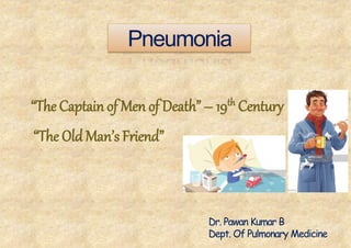
- 1. Pneumonia “The Captain of Men of Death” – 19th Century “The Old Man’s Friend” Dr. Pawan Kumar B Dept. Of Pulmonary Medicine
- 2. Whatispneumonia? • Lung parenchyma/ alveolarinflammation and (abnormal) alveolar filling with fluid. Syndrome caused by acute infection , usually bacterial, characterized by clinical and/or radiological signs of consolidation of a part or parts of one or both lungs
- 3. Lobes
- 6. Pneumonia Community acquired pneumonia Nosocomial pneumonia Hospital‐acquired pneumonia(HAP) Ventilator‐associated pneumonia(VAP) Healthcare‐associated pneumonia(HCAP) 1. Bacterial 2. Viral 3. Fungal 4. Parasitic 5. eosinophilic 1. Lobarpneumonia 2. Bronchopneumonia 3. Interstitialpneumonia 4. Segmentalpneumonia 12th November – World Pneumonia Day
- 9. Communityacquiredpneumonia • Definition: – An acute infection of the pulmonary parenchyma that is associated with at least some symptoms of acute infection, accompanied by the presence of an acute infiltrate on a chest radiograph, orauscultatory findings consistent with pneumonia, in a patient not hospitalized or residing in a long term carefacilityfor >14days before onset of symptoms.
- 10. Communityacquiredpneumonia • CAP can be defined both on clinical and radiographic findings. In the absence of chest radiograph, CAP is defined as: • (a) symptoms of an acute lower respiratory tract illness (cough with or without expectoration, shortness of breath, pleuritic chest pain) for less than 1 week; and • (b) at least one systemic feature (temperature >37.7°C, chills, and rigors, and/or severe malaise); and • (c) new focal chest signs on examination (bronchial breath sounds and/or crackles); with • (d) no other explanation for the illness
- 11. • When a chest radiograph is available, CAP is defined as symptoms and signs as above with new radiographic shadowing for which there is no other explanation (not due to pulmonary edema or infarction).
- 12. • Streptococcus pneumoniae (20% to 60%of CAP cases) • Haemophilus influenzae (3% to 10%of CAP cases) • L. pneumophila (1% to 5% of adult pneumonias) (2% to 8% of CAP cases) • Klebsiella, Pseudomonas, Escherichia coli, Staphylococcus aureus (3% to 5% of CAP cases) • Atypical organisms such as M. pneumoniae, C. pneumoniae, and L. pneumophila implicated in up to 40% of cases of CAP • Pneumococcal infection responsible for 50% to 75% of CAPs. Influenza infection is one of the important predisposing factors to S. pneumoniae and S. aureus pneumonia; • gram‐negative organisms cause .80% of nosocomial pneumonias
- 13. Riskfactors 1. Viral infections (damage cilia and produce serous exudates) 2. Age (Children & elderly‐ defect in swallowing, ↓ immunity) 3. Alcoholism (depress coughing and epiglottis function) 4. Smoking (damage epithelial cells and impair cilia functions) 5. Asthma/COPD 6. Immunosuppression (AIDS, transplant pt, cancer chemo)
- 14. 7. Dementia 8. Diabetes Mellitus‐ defective neutrophil function, ↓ CMI 9. Renal Failure‐ ↓ humural response, ↓ leukocyte chemotaxis, complement depletion 10.Chronic lung diseases 11.Cold Weather (dry mucous membrane & person to person spread)‐ common in winter 12.Heart Disease‐ Impaired lymphatics & alv macrophage function, edema promotes bacterial growth Riskfactors
- 15. Pathogenesis Reduces or suppress cough Impairs mucociliary activity Reduces the effective phagocytic activity of alveolar macrophages and neutrophils Reduces immunoglobuin production Predisposed by Condition that,
- 16. Pathogenesis • Primaryinhalation:whenorganismsbypassnormal respiratorydefensemechanismsorwhenthePt inhales aerobicGNorganismsthatcolonizetheupper respiratory tractorrespiratorysupportequipment. • Aspiration:occurswhenthePtaspiratescolonized upper respiratorytractsecretions • Hematogenous:originatefromadistantsourceand reach thelungsviathebloodstream(IVcannula,chronic hemodialysis).
- 17. z Stages Of Pneumonia i. Congestion / Consolidation ii. Red Hepatization iii. Grey Hepatization iv. Resolution
- 18. SymptomsandSigns
- 19. Typical pneumonia • Sudden/subacute onset • Fever with chills, rigors • Productive cough, Mucopurulent sputum • Tachypnea and tachycardia • breathlessness • Pleuritic chest pain • Breath sound: crackles and rales • CXR: air‐bronchogram, consolidation Usually bacteria Clinical presentation
- 21. Atypicalpneumonia: Clinicalpresentation Atypical – Gradualonset – Afebrile – Drycough – Breathsound:Rales – Uni/bilateralpatchy,infiltrates – WBC:usualnormalorslighthigh – Sorethroat,myalgia,fatigue,diarrhea – Commonetiology • Mycoplasmapneumoniae • Chlamydiapneumoniae • Legionellapneumophilla • Mycobactria • Virus,Others
- 22. Differentialdiagnosis • Pulmonary edema • Pulmonary infarction • Acute respiratory distress syndrome • Pulmonary hemorrhage • Lung cancer or metastatic cancer • Atelectasis • Radiation pneumonitis • Drug reactions involving the lung
- 23. DIAGNOSI S
- 24. LaboratoryTests: Microbiologicaltests SputumGramstain Sputumforculture SputumforZiehlNeelsenstain Sputumcytology Routine bloodinvestigations CBC with differential BUN/Cr, electrolytes Glucose, liverenzymes Blood culture Imaging studies X‐RaychestP/A&lateralview Computetomography Pulse oximetry Arterial oxygensaturation Serological test Pneumococcalantigentest Legionellaantigen
- 25. Sputum gram stain and culture – Theyieldofsputumculturesvariesfrom34to86%. – Aninitial sputumGramstainandculture(oraninvasive respiratorysample asappropriate)shouldbeobtainedinall hospitalized patients withCAP – Sputum quality should beensured • PMN’s>25/LPF • Fewepithelial cells<10/LPF • Single predominant organism
- 26. Chestradiography Postero‐ant erior and lateral view‐ important • Establish the diagnosis • Delineate the extent of consolidation • Indicate the presence of underlying disorders • Identify complications (pleural effusion, multilobar disease, lung abscess) • To prognosticate the disease
- 27. Chest X Ray (mostlyfivepatterns) 1. Lobar‐ S.Pneumoniae 2. Patchy pattern‐ VirusAtypicals,Mycoplasma, Chlamydia,Legionella 3. Interstitial‐Influenza,CMV ,PCP ,MilliaryTB 4. Lung abscess‐ S.Aureus,anerobes 5. Nodular‐Fungalinfection(Histoplasmosis, Coccidiomycosis,cryptococosis) ‘Bulging fissure’sign ofKlebsiella, Pleuraleffusion‐ Streptococcus,anaerobes.
- 28. • 32Y/Omale • Coughfor1wk • Feverfor2days • CreptsoverLLL
- 33. Why do we needtotreat • Itspotentiallyfatal • Eradicatethecausativeorganismfromthesite and reversetheinflammatoryprocess • Preventcomplications • Preventmortality TREATMENT
- 34. Principles ofmanagement • Prompt initiation of antibiotictherapy • Pathogen directed antimicrobialtherapy whenever possible • Rational use of microbiologylaboratory • Decision to hospitalize based onprognostic criteria
- 35. How to proceed >>>>>>?
- 37. Criteria for risk stratification(CURB‐65) Confusion / Altered mental state Urea ≥ 7 mmol/L (Low U/O <20ml/hr) Respiratory rate ≥30/min Low blood pressure (DBP ≤60 or SBP ≤90 mm Hg) Age ≥65 years
- 38. Managementoptions • Outpatient (Emperically) • Inpatient • ICU management
- 39. Factorsforconsideration (IP) • Risk of death from the pneumonia, • Disease severity • Presence of comorbid conditions, • Need for advanced diagnostics, • Inability to take oral medications • Lack of of social support
- 40. Inpatientmanagement • S.pneumoniae • M. pneumoniae • C.pneumoniae • H.influenzae • Legionellaspecies • Aspiration • Respiratoryvirusesa Pathogen directed treatment/Emperic Amoxycillin +clavulanic acid +Macrolide 3rdgeneration cephalosporins Fluoroquinolone Important: 1. Injectable drugs areusedinitially 2. Combinations of drugs arepreferred 3. In elderly, diabetics, alcoholism, those with structural lung disease, cephalosporins are preferred
- 41. Criteria for ICUadmission Major criteria Minor criteria 1. Invasivemechanicalventilation 2. Septicshockwithneedfor vasopressors 1. Respiratoryrate≥30breaths/min 2. PaO2/FIO2ratio<250 3. Multilobarradiographicinvolvement 4. Confusionordisorientation 5. Uremia(BUNlevel>20mg/dL) 6. Leukopenia(WBCcount<4000cells/dL)or Leucocytosis>30000c/dl 7. Thrombocytopenia(plateletcount< 100,000cells/dL) 8. Hypothermia(coretemperature<36°C) 9. Hypotensionrequiringaggressivefluid resuscitation
- 42. ICUmanagement • S.pneumoniae • Staphylococcus aureus • Legionellaspecies • Gram‐negativebacilli • H.influenzae Pathogen directedtreatment Amoxycillin +clavulanic acid +Macrolide 3rd & 4th generationcephalosporins Fluoroquinolone Aminoglycosides Carbapenems Vancomycin/Teicoplanin Metronidazole/clindamycin
- 43. Criteria for clinicalstability • Temperature≤ 37.8°C • Heartrate≤ 100beats/minute • Systolicbloodpressure≥ 90mmHg • Respiratoryrate≤ 24breaths/minute • Oxyhemoglobinsaturation≥90%orPO2≥60mmHg on preadmissionlevelofoxygensupplementation. 4outof5criterianeedtobefulfilledinstabilitycriteria • Generally7‐10daysofantibioticsare sufficient • Oncethestabilitycriteriaaremet,patientcan beswitchedtooral antibiotics(samegroup)
- 45. Definition HAP is an inflammatory condition of the lung parenchyma, caused by infectious agents, neither present nor incubating at the time of hospital admission. It is defined as pneumonia developing 48h after admission to the hospital. Divided into ICU HAP or non‐ICU HAP depending upon whether this infection is acquired in the intensive care unit (ICU) or in other clinical areas (e.g. wards)
- 46. “Nosocomial”Pneumonia Hospital‐acquired pneumonia (HAP) • Occurs 48 hours or more after admission, which was not incubating at the time of admission Ventilator‐associated pneumonia (VAP) • Arises more than 48‐72 hours after endotracheal intubation Healthcare‐associated pneumonia (HCAP) • Patients who were hospitalized in an acute care hospital for two or more days within 90 days of the infection; resided in a nursing home; received recent IV, chemotherapy, or wound care within the past 30 days of the current infection; or attended a hospital or hemodialysis clinic
- 47. • Early‐onset HAP (and VAP) is defined as pneumonia occurring within the first 4 days of hospitalization (or endotracheal intubation. It usually carries a better prognosis and is more likely to be caused by antibiotic‐sensitive bacteria. • Late‐onset HAP and VAP (day 5 or thereafter) are more likely to be caused by MDR pathogens, and are associated with higher morbidity and mortality.
- 48. Burdenofdisease HAP is the second most common nosocomial infection HAP accounts for up to 25% of all ICU infections and more than 50% of the entire antibiotic prescriptions. The crude mortality rate for HAP may be as high as 30–70% The risk of HAP/VAP is the highest early in the course of hospital stay
- 50. Organism profile inIndia Aerobic Gram‐negative bacilli (mostcommon) • P .aeruginosa • E.coli • K.pneumoniae • Acinetobacter species. • Staph. aureus (more common in diabetes,head trauma and in ICU admittedpatients) HAP/VAP can be clinically defined using modified CDCcriteria
- 51. Modified CDC criteriafor diagnosisof HAP/ VAP Chestradiographicopacities(new,progressive,orpersistent infiltrateor cavitation)andatleasttwoofthefollowing Fever >38°C or>100.4°F Leukopenia (<4000 WBC/μL) orleukocytosis (≥12,000 WBC/μL) Altered mental statuswith noother recognized cause in theelderly New onset ofpurulent sputum, orchange in character ofsputum, or increased respiratory secretions, orincreased suctioning requirements Worsening gas exchange (e.g.desaturations, increased oxygen requirements, orincreased ventilator demand) New onset orworsening cough, ordyspnea, or tachypnea Rales orbronchial breath sounds
- 53. • Oneormorelowerrespiratorytractsamplesand bloodshouldbe sentforculturespriortoinstitution ofantibiotics • Good‐qualitysputummicrobiology • Appropriatemanagementshouldnotbedelayedin clinically unstablepatientsforthepurposeof performingdiagnostic sampling. • Quantitativeandorsemi‐quantitativecultures usingvarious samplingtechniqueslikeETA, bronchoscopicornon‐bronchoscopic BALareequallyusefulforestablishingthe diagnosisofHAP/VAP. Management
- 56. Basicprinciples • Start antibiotics as early as possible • The exact choice of antibiotic to be started is based on local availability, antibiotic resistance patterns, preferred routes of delivery, other complicating factors, and cost. • The initial combination therapy should be converted to appropriate monotherapy once culture reports are available • The strategy for de‐escalation of antibiotics is strongly recommended
- 58. • Among patients with suspected VAP in whom an alternate cause for pulmonary infiltrates is identified, it is recommended that antibiotics should be stopped . • If cultures are sent after initiation of antibiotics and there is clinical improvement with subsequent cultures being sterile, antibiotics should be continued for 7 days followed by assessment. • Empiric antifungal therapy (on day 3) should not be used as a routine in all patients if cultures are sterile and there is clinical worsening
- 59. • In patients with VAPdue to Pseudomonas, Acinetobacter, and MRSA, a longer duration (14 days) of antibiotic course is recommended. • In other patients with VAPwho are clinically improving, a 7‐day course of antibiotics is recommended. Supportive Treatment Respiratory Support Fluid and Electrolyte replacement TPN Pleuritic Pain Physiotherapy Corticosteroids Inotropic agents
- 61. Thankyou
- 62. z