Glioma.pptx
•Descargar como PPTX, PDF•
0 recomendaciones•12 vistas

Presented By Mr. Pradeepsingh N B Asst.Professor HOD of Medical Surgical Nursing

Recomendados
Más contenido relacionado
Similar a Glioma.pptx
Similar a Glioma.pptx (20)
Más de pradeepsingh855
Más de pradeepsingh855 (20)
Último
Último (20)
Glioma.pptx
- 1. Glioma
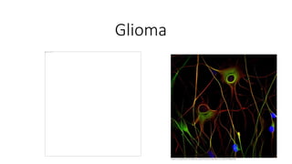
- 2. Definition • Glioma is a type of tumor that occurs in the brain and spinal cord. Gliomas begin in the gluey supportive cells (glial cells) that surround nerve cells and help them function.
- 3. Risk factors • Gliomas are most common in adults between ages 45 and 65 years old. • Certain types of gliomas, such as ependymomas and pilocytic astrocytomas, are more common in children and young adults. • Exposure to radiation:People who have been exposed to a type of radiation called ionizing radiation have an increased risk of brain tumor. Examples of ionizing radiation include radiation therapy used to treat cancer and radiation exposure caused by atomic bombs.
- 4. • Family history of glioma. It's rare for glioma to run in families. But having a family history of glioma can double the risk of developing it. Some genes have been weakly associated with glioma, but more study is needed to confirm a link between these genetic variations and brain tumors.
- 5. Types of glioma • Astrocytomas, including astrocytoma, anaplastic astrocytoma and glioblastoma • Ependymomas, including anaplastic ependymoma, myxopapillary ependymoma and subependymoma • Oligodendrogliomas, including oligodendroglioma, anaplastic oligodendroglioma and anaplastic oligoastrocytoma
- 6. Symptoms • The symptoms of glioma vary by tumor type as well as the tumor's size, location and rate of growth. • Common signs and symptoms of gliomas include: • Headache • Nausea or vomiting • Confusion or a decline in brain function • Memory loss • Personality changes or irritability • Difficulty with balance • Urinary incontinence • Vision problems, such as blurred vision, double vision or loss of peripheral vision • Speech difficulties • Seizures, especially in someone without a history of seizures
- 7. Diagnosis • Neurology consultation • A neurological exam. During a neurological exam, The doctor may check your vision, hearing, balance, coordination, strength and reflexes. Problems in one or more of these areas may provide clues about the part of the brain that could be affected by a brain tumor. • Imaging tests. Magnetic resonance imaging (MRI) is often used to help diagnose brain tumors. In some cases, a dye (contrast material) may be injected through a vein in the arm during MRI study to help show differences in brain tissue. • computerized tomography (CT) scan and positron emission tomography (PET).
- 8. • Tests to find cancer in other parts of your body. To rule out other types of brain tumors that may have spread from other parts of the body, the doctor may recommend tests and procedures to determine where the cancer originated. Gliomas originate within the brain and are not the result of cancer that has spread (metastasized) from elsewhere. • Collecting and testing a sample of abnormal tissue (biopsy). Depending on the location of the glioma, a biopsy may be performed with a needle before treatment or as part of an operation to remove the brain tumor. • A stereotactic needle biopsy may be done for gliomas in hard-to-reach areas or very sensitive areas within the brain that might be damaged by a more extensive operation. During a stereotactic needle biopsy, the neurosurgeon drills a small hole into the skull. A thin needle is then inserted through the hole. Tissue is removed through the needle, which is frequently guided by CT or MRI scanning.
- 9. Treatment • Treatment for glioma depends on the type, size, grade and location of the tumor, as well as your age, overall health and preferences. • In addition to actions to remove the tumor itself, treatment for glioma may also require using drugs to reduce the signs and symptoms of tumor.
- 10. Surgery • Surgery to remove as much of the tumor as possible is usually the first step in treating most types of gliomas. • In some cases, neuropathologists may analyze tissue samples removed by a surgeon and report the results while surgery is underway. This information helps the surgeon decide how much tissue to remove.
- 12. • A variety of surgical technologies and techniques may be used to assist the neurosurgeon in protecting as much healthy brain tissue as possible while removing the tumor, including computer-assisted brain surgery, intraoperative MRI, awake brain surgery and lasers. For example, during awake brain surgery, may be asked to perform a task with the goal of ensuring the area of the brain controlling that function is not damaged.
- 14. • Radiation therapy usually follows surgery in treatment of glioma, especially high-grade gliomas. Radiation uses high-energy beams, such as X-rays or protons, to kill tumor cells. Radiation therapy for glioma comes from a machine outside the body (external beam radiation). • Radiation therapy options include: 1. Using computers to pinpoint delivery of radiation treatment to the exact location of the brain tumor. Techniques include intensity- modulated radiation therapy and 3D conformal radiation therapy. 2. Using protons — the positive parts of atoms — rather than X-rays as the source of radiation. This technique, called conformal proton beam therapy, delivers radiation only once proton beams reach the tumor, causing less damage than X-rays to surrounding tissue.
- 15. • Using multiple beams of radiation to give a highly focused form of radiation treatment. While this technique is called stereotactic radiation therapy (radiosurgery), it doesn't actually involve surgery in the traditional sense. Each beam of radiation isn't particularly powerful, but the point where all the beams meet — at the brain tumor — receives a very large dose of radiation to kill the tumor cells in a very small area.
- 17. Chemotherapy • Chemotherapy uses drugs to kill tumor cells. Chemotherapy drugs can be taken in pill form (orally) or injected into a vein (intravenously). • Chemotherapy is usually used in combination with radiation therapy to treat gliomas. • The chemotherapy drug used most often to treat gliomas is temozolomide (Temodar), which is taken as a pill. • Side effects of chemotherapy depend on the type and dose of drugs receive. Common side effects include nausea and vomiting, headache, hair loss, fever, and weakness. Some side effects may be managed with medication.
- 18. Targeted drug therapy • Targeted drug treatments focus on specific abnormalities present within cancer cells. By blocking these abnormalities, targeted drug treatments can cause cancer cells to die. • One targeted drug therapy used to treat a type of brain cancer called glioblastoma is bevacizumab (Avastin). This drug, given through a vein (intravenously), stops the formation of new blood vessels, cutting off blood supply to a tumor and killing the tumor cells.
- 19. Rehabilitation • Physical therapy can help the regain lost motor skills or muscle strength. • Occupational therapy, which can help get back to the normal daily activities, including work, after a brain tumor or other illness • Speech therapy with specialists in speech difficulties (speech pathologists), which can help if patient have difficulty speaking • Tutoring for school-age children, which can help kids cope with changes in memory and thinking after a brain tumor