SlideShare una empresa de Scribd logo
1 de 19
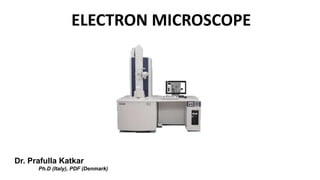
ELECTRON MICROSCOPE
Dr. Prafulla Katkar
Ph.D (Italy), PDF (Denmark)
In the 1920s it was discovered that accelerated electrons
(parts of the atom) behave in vacuum just like light. They
travel in straight lines and have a wavelength which is about
100 000 times smaller than that of light. Furthermore, it was
found that electric and magnetic fields have the same effect
on electrons as glass lenses and mirrors have on visible light.
Why use electrons instead of light ?
The electron microscope uses a beam of electrons to create an
image of the specimen. It is capable of much higher
magnifications and has a greater resolving power than a light
microscope, allowing it to see much smaller objects in finer
detail. They are large, expensive pieces of equipment,
generally standing alone in a small, specially designed room
and requiring trained personnel to operate them.
What is Electron Microscope…?
Dr. Ernst Ruska at the University of Berlin and Max Knoll
combined built the first electron microscope in 1931.
For this and subsequent work on the subject, Ernst Ruska
was awarded the Nobel Prize for Physics in 1986.
Max Knoll Ernst Ruska
History of Electron Microscope
Type of Electron Microscope
 Transmission Electron Microscope (TEM)
 Scanning Electron Microscope (SEM)
Types of Electron Microscope
Components
Transmission Electron Microscope-TEM
(a) Electron gun:
It consists of a cathode that emits electrons on receiving
high voltage electric current (50,000-100,000 volts).
(c) Condense lens: It is the electromagnetic coil which
focuses the electron beam in the plane of the specimen.
(d) Objective lens: It is the electromagnetic coil which
produces the first magnified image formed by the objective lens
and produces the final image.
(e) Projector lens: It is also an electromagnetic coil
which further magnifies the first image formed by the
objective lens and produces the final image.
(f) Fluorescent Screen or Photographic Film: Since
unaided human eye cannot observe electrons, therefore, a
fluorescent screen is used for viewing the final image of the
specimen
Components
• A heated tungsten filament in the electron gun generates a
beam of electrons that is then focused on the specimen by the
condenser.
• Since electrons cannot pass through a glass lens, magnetic
lenses are used to focus the beam.
• lenses and specimen must be under high vacuum to obtain a
clear image because electrons are deflected by collisions with air
molecules.
• The specimen scatters electron passing through it, and the beam
is focused by magnetic lenses to form an enlarged , visible image
of the specimen on a fluorescent screen.
• A denser region in the specimen scatters more electron and
therefore appears darker in the image.
• In contrast, electron-transparent regions are brighter.
Scanning Electron Microscope-SEM
 SEM image of sample by scanning it with a high energy beam of electrons.
 The electrons interaction with sample atoms produce signals that contain
information about the sample’s surface topography, composition, and other
properties.
Scanning Electron Microscope
 Electron gun consisting of cathode and anode
 The condenser lens controls the amount of electrons
travelling down the column.
 The objective lens focuses the beam into a spot on the
sample.
 Deflection coil helps to deflect the electron beam.
 SED attracts the secondary electrons.
 Additional sensors detect backscattered electrons
and x-rays.
Components of SEM
 Beam of electrons is generated by a suitable
source, typically a tungsten filament or a field
electron gun.
 The electron beam is accelerated through a
high voltage and pass through a system of
electromagnetic lenses to produce a thin
beam of electrons.
 Then the beam scans the surface of the
specimen, secondary electrons are emitted
from the specimen by the action of the
scanning beam and collected by a suitably
positioned detector.
Working Mechanism
Application
 It gives detailed 3d and topographical imaging and the
versatile information.
 This works very fast.
 Modern SEMs allow for the generation data in digital
form.
 Most SEM samples require minimal preparation actions.
 Enable us to view without thinning dehydrating, fixing
the sample
Electron microscope (SEM and TEM)

Más contenido relacionado

La actualidad más candente

Confocal microscopy Likhith K
Confocal microscopy Likhith KConfocal microscopy Likhith K
Confocal microscopy Likhith K
LIKHITHK1
 
Electron microscope
Electron microscopeElectron microscope
Electron microscope
shabeel pn
 

La actualidad más candente (20)

Sem (2)
Sem (2)Sem (2)
Sem (2)
 
Transmission electron microscope (TEM) Likhith K
Transmission electron microscope  (TEM) Likhith KTransmission electron microscope  (TEM) Likhith K
Transmission electron microscope (TEM) Likhith K
 
Generation and application of attosecond laser pulse
Generation and application of attosecond laser pulseGeneration and application of attosecond laser pulse
Generation and application of attosecond laser pulse
 
Transmission electron microscopy (1) (1)
Transmission electron microscopy (1) (1)Transmission electron microscopy (1) (1)
Transmission electron microscopy (1) (1)
 
Tem ppt
Tem ppt Tem ppt
Tem ppt
 
2018 HM-Transmission electron microscope
2018 HM-Transmission electron microscope2018 HM-Transmission electron microscope
2018 HM-Transmission electron microscope
 
Tem ppt
Tem pptTem ppt
Tem ppt
 
Confocal microscope presentation pt
Confocal microscope presentation  ptConfocal microscope presentation  pt
Confocal microscope presentation pt
 
Scanning Electron microscopy
Scanning Electron microscopy Scanning Electron microscopy
Scanning Electron microscopy
 
Transmission electron
Transmission        electronTransmission        electron
Transmission electron
 
Atomic Force Microscopy
Atomic Force MicroscopyAtomic Force Microscopy
Atomic Force Microscopy
 
Confocal microscopy Likhith K
Confocal microscopy Likhith KConfocal microscopy Likhith K
Confocal microscopy Likhith K
 
Scanning electron microscope
Scanning electron microscope  Scanning electron microscope
Scanning electron microscope
 
Cofocal microscope
Cofocal microscopeCofocal microscope
Cofocal microscope
 
Principles of Confocal Microscopy
Principles of Confocal MicroscopyPrinciples of Confocal Microscopy
Principles of Confocal Microscopy
 
Transmission electron microscope, high resolution tem and selected area elect...
Transmission electron microscope, high resolution tem and selected area elect...Transmission electron microscope, high resolution tem and selected area elect...
Transmission electron microscope, high resolution tem and selected area elect...
 
Electron microscope
Electron microscopeElectron microscope
Electron microscope
 
Electron microscopy
Electron microscopyElectron microscopy
Electron microscopy
 
Scanning electron microscopy
Scanning electron microscopyScanning electron microscopy
Scanning electron microscopy
 
Electron Microscope
Electron MicroscopeElectron Microscope
Electron Microscope
 

Similar a Electron microscope (SEM and TEM)

Scanning electron microscope
Scanning electron microscopeScanning electron microscope
Scanning electron microscope
SuganyaPaulraj
 
Scanning electron microscopy mubbu
Scanning electron microscopy mubbuScanning electron microscopy mubbu
Scanning electron microscopy mubbu
Mubashshir Arif
 

Similar a Electron microscope (SEM and TEM) (20)

Electron microscopy, M. Sc. Zoology, University of Mumbai
Electron microscopy, M. Sc. Zoology, University of MumbaiElectron microscopy, M. Sc. Zoology, University of Mumbai
Electron microscopy, M. Sc. Zoology, University of Mumbai
 
Sem md ppt
Sem md pptSem md ppt
Sem md ppt
 
Chapter-3. Electron Microscopy.pptx
Chapter-3. Electron Microscopy.pptxChapter-3. Electron Microscopy.pptx
Chapter-3. Electron Microscopy.pptx
 
ELECTRON MICROSCOPY-Scanning Electron MicroscopySEM.pptx
ELECTRON MICROSCOPY-Scanning Electron MicroscopySEM.pptxELECTRON MICROSCOPY-Scanning Electron MicroscopySEM.pptx
ELECTRON MICROSCOPY-Scanning Electron MicroscopySEM.pptx
 
electron microscopy
electron microscopyelectron microscopy
electron microscopy
 
Sem
SemSem
Sem
 
Electron Microscopy
Electron MicroscopyElectron Microscopy
Electron Microscopy
 
Lecture_3_conventional-Microscope.pdf
Lecture_3_conventional-Microscope.pdfLecture_3_conventional-Microscope.pdf
Lecture_3_conventional-Microscope.pdf
 
Scanning Electron Microscope
Scanning Electron Microscope Scanning Electron Microscope
Scanning Electron Microscope
 
Electron microscopy
Electron microscopyElectron microscopy
Electron microscopy
 
Scanning electron microscope
Scanning electron microscopeScanning electron microscope
Scanning electron microscope
 
SEM & FESEM.pptx
SEM & FESEM.pptxSEM & FESEM.pptx
SEM & FESEM.pptx
 
Scanning electron microscopy (sem)
Scanning electron microscopy (sem)Scanning electron microscopy (sem)
Scanning electron microscopy (sem)
 
Scanning electron microscopy
Scanning electron microscopyScanning electron microscopy
Scanning electron microscopy
 
Presentation forensic micoscopy SEM microscope.pptx
Presentation forensic micoscopy SEM microscope.pptxPresentation forensic micoscopy SEM microscope.pptx
Presentation forensic micoscopy SEM microscope.pptx
 
Electron microscope
Electron microscopeElectron microscope
Electron microscope
 
Electron Microscopy By Rimsha Khan.pptx
Electron Microscopy By Rimsha Khan.pptxElectron Microscopy By Rimsha Khan.pptx
Electron Microscopy By Rimsha Khan.pptx
 
Scanning electron microscopy mubbu
Scanning electron microscopy mubbuScanning electron microscopy mubbu
Scanning electron microscopy mubbu
 
Scanning and Transmission Electron Microscopy
Scanning and Transmission Electron MicroscopyScanning and Transmission Electron Microscopy
Scanning and Transmission Electron Microscopy
 
ELECTRON MICROSCOPE
ELECTRON MICROSCOPEELECTRON MICROSCOPE
ELECTRON MICROSCOPE
 

Más de Guru Nanak College of Science, Chandrapur MS india (6)

Dark field and Phase Contrast Microscope
Dark field and Phase Contrast MicroscopeDark field and Phase Contrast Microscope
Dark field and Phase Contrast Microscope
 
DNA sequencing
DNA  sequencingDNA  sequencing
DNA sequencing
 
Dna fingerprinting
Dna fingerprintingDna fingerprinting
Dna fingerprinting
 
Topoisomerase inhibiton by Gold III
Topoisomerase inhibiton by Gold IIITopoisomerase inhibiton by Gold III
Topoisomerase inhibiton by Gold III
 
Topoisomerase inhibtion by thaspine
Topoisomerase inhibtion by thaspineTopoisomerase inhibtion by thaspine
Topoisomerase inhibtion by thaspine
 
Topoisomerase inhibitor
Topoisomerase inhibitor Topoisomerase inhibitor
Topoisomerase inhibitor
 

Último

Jual Obat Aborsi Hongkong ( Asli No.1 ) 085657271886 Obat Penggugur Kandungan...
Jual Obat Aborsi Hongkong ( Asli No.1 ) 085657271886 Obat Penggugur Kandungan...Jual Obat Aborsi Hongkong ( Asli No.1 ) 085657271886 Obat Penggugur Kandungan...
Jual Obat Aborsi Hongkong ( Asli No.1 ) 085657271886 Obat Penggugur Kandungan...
ZurliaSoop
 

Último (20)

Accessible Digital Futures project (20/03/2024)
Accessible Digital Futures project (20/03/2024)Accessible Digital Futures project (20/03/2024)
Accessible Digital Futures project (20/03/2024)
 
HMCS Max Bernays Pre-Deployment Brief (May 2024).pptx
HMCS Max Bernays Pre-Deployment Brief (May 2024).pptxHMCS Max Bernays Pre-Deployment Brief (May 2024).pptx
HMCS Max Bernays Pre-Deployment Brief (May 2024).pptx
 
Jamworks pilot and AI at Jisc (20/03/2024)
Jamworks pilot and AI at Jisc (20/03/2024)Jamworks pilot and AI at Jisc (20/03/2024)
Jamworks pilot and AI at Jisc (20/03/2024)
 
HMCS Vancouver Pre-Deployment Brief - May 2024 (Web Version).pptx
HMCS Vancouver Pre-Deployment Brief - May 2024 (Web Version).pptxHMCS Vancouver Pre-Deployment Brief - May 2024 (Web Version).pptx
HMCS Vancouver Pre-Deployment Brief - May 2024 (Web Version).pptx
 
This PowerPoint helps students to consider the concept of infinity.
This PowerPoint helps students to consider the concept of infinity.This PowerPoint helps students to consider the concept of infinity.
This PowerPoint helps students to consider the concept of infinity.
 
Google Gemini An AI Revolution in Education.pptx
Google Gemini An AI Revolution in Education.pptxGoogle Gemini An AI Revolution in Education.pptx
Google Gemini An AI Revolution in Education.pptx
 
Micro-Scholarship, What it is, How can it help me.pdf
Micro-Scholarship, What it is, How can it help me.pdfMicro-Scholarship, What it is, How can it help me.pdf
Micro-Scholarship, What it is, How can it help me.pdf
 
How to Manage Global Discount in Odoo 17 POS
How to Manage Global Discount in Odoo 17 POSHow to Manage Global Discount in Odoo 17 POS
How to Manage Global Discount in Odoo 17 POS
 
How to Add New Custom Addons Path in Odoo 17
How to Add New Custom Addons Path in Odoo 17How to Add New Custom Addons Path in Odoo 17
How to Add New Custom Addons Path in Odoo 17
 
Plant propagation: Sexual and Asexual propapagation.pptx
Plant propagation: Sexual and Asexual propapagation.pptxPlant propagation: Sexual and Asexual propapagation.pptx
Plant propagation: Sexual and Asexual propapagation.pptx
 
Python Notes for mca i year students osmania university.docx
Python Notes for mca i year students osmania university.docxPython Notes for mca i year students osmania university.docx
Python Notes for mca i year students osmania university.docx
 
How to Give a Domain for a Field in Odoo 17
How to Give a Domain for a Field in Odoo 17How to Give a Domain for a Field in Odoo 17
How to Give a Domain for a Field in Odoo 17
 
Sociology 101 Demonstration of Learning Exhibit
Sociology 101 Demonstration of Learning ExhibitSociology 101 Demonstration of Learning Exhibit
Sociology 101 Demonstration of Learning Exhibit
 
Holdier Curriculum Vitae (April 2024).pdf
Holdier Curriculum Vitae (April 2024).pdfHoldier Curriculum Vitae (April 2024).pdf
Holdier Curriculum Vitae (April 2024).pdf
 
TỔNG ÔN TẬP THI VÀO LỚP 10 MÔN TIẾNG ANH NĂM HỌC 2023 - 2024 CÓ ĐÁP ÁN (NGỮ Â...
TỔNG ÔN TẬP THI VÀO LỚP 10 MÔN TIẾNG ANH NĂM HỌC 2023 - 2024 CÓ ĐÁP ÁN (NGỮ Â...TỔNG ÔN TẬP THI VÀO LỚP 10 MÔN TIẾNG ANH NĂM HỌC 2023 - 2024 CÓ ĐÁP ÁN (NGỮ Â...
TỔNG ÔN TẬP THI VÀO LỚP 10 MÔN TIẾNG ANH NĂM HỌC 2023 - 2024 CÓ ĐÁP ÁN (NGỮ Â...
 
SOC 101 Demonstration of Learning Presentation
SOC 101 Demonstration of Learning PresentationSOC 101 Demonstration of Learning Presentation
SOC 101 Demonstration of Learning Presentation
 
Jual Obat Aborsi Hongkong ( Asli No.1 ) 085657271886 Obat Penggugur Kandungan...
Jual Obat Aborsi Hongkong ( Asli No.1 ) 085657271886 Obat Penggugur Kandungan...Jual Obat Aborsi Hongkong ( Asli No.1 ) 085657271886 Obat Penggugur Kandungan...
Jual Obat Aborsi Hongkong ( Asli No.1 ) 085657271886 Obat Penggugur Kandungan...
 
Sensory_Experience_and_Emotional_Resonance_in_Gabriel_Okaras_The_Piano_and_Th...
Sensory_Experience_and_Emotional_Resonance_in_Gabriel_Okaras_The_Piano_and_Th...Sensory_Experience_and_Emotional_Resonance_in_Gabriel_Okaras_The_Piano_and_Th...
Sensory_Experience_and_Emotional_Resonance_in_Gabriel_Okaras_The_Piano_and_Th...
 
Graduate Outcomes Presentation Slides - English
Graduate Outcomes Presentation Slides - EnglishGraduate Outcomes Presentation Slides - English
Graduate Outcomes Presentation Slides - English
 
Key note speaker Neum_Admir Softic_ENG.pdf
Key note speaker Neum_Admir Softic_ENG.pdfKey note speaker Neum_Admir Softic_ENG.pdf
Key note speaker Neum_Admir Softic_ENG.pdf
 

Electron microscope (SEM and TEM)

  • 1. ELECTRON MICROSCOPE Dr. Prafulla Katkar Ph.D (Italy), PDF (Denmark)
  • 2. In the 1920s it was discovered that accelerated electrons (parts of the atom) behave in vacuum just like light. They travel in straight lines and have a wavelength which is about 100 000 times smaller than that of light. Furthermore, it was found that electric and magnetic fields have the same effect on electrons as glass lenses and mirrors have on visible light. Why use electrons instead of light ?
  • 3. The electron microscope uses a beam of electrons to create an image of the specimen. It is capable of much higher magnifications and has a greater resolving power than a light microscope, allowing it to see much smaller objects in finer detail. They are large, expensive pieces of equipment, generally standing alone in a small, specially designed room and requiring trained personnel to operate them. What is Electron Microscope…?
  • 4.
  • 5.
  • 6. Dr. Ernst Ruska at the University of Berlin and Max Knoll combined built the first electron microscope in 1931. For this and subsequent work on the subject, Ernst Ruska was awarded the Nobel Prize for Physics in 1986. Max Knoll Ernst Ruska History of Electron Microscope
  • 7. Type of Electron Microscope  Transmission Electron Microscope (TEM)  Scanning Electron Microscope (SEM) Types of Electron Microscope
  • 9. (a) Electron gun: It consists of a cathode that emits electrons on receiving high voltage electric current (50,000-100,000 volts). (c) Condense lens: It is the electromagnetic coil which focuses the electron beam in the plane of the specimen. (d) Objective lens: It is the electromagnetic coil which produces the first magnified image formed by the objective lens and produces the final image. (e) Projector lens: It is also an electromagnetic coil which further magnifies the first image formed by the objective lens and produces the final image. (f) Fluorescent Screen or Photographic Film: Since unaided human eye cannot observe electrons, therefore, a fluorescent screen is used for viewing the final image of the specimen Components
  • 10. • A heated tungsten filament in the electron gun generates a beam of electrons that is then focused on the specimen by the condenser. • Since electrons cannot pass through a glass lens, magnetic lenses are used to focus the beam. • lenses and specimen must be under high vacuum to obtain a clear image because electrons are deflected by collisions with air molecules. • The specimen scatters electron passing through it, and the beam is focused by magnetic lenses to form an enlarged , visible image of the specimen on a fluorescent screen. • A denser region in the specimen scatters more electron and therefore appears darker in the image. • In contrast, electron-transparent regions are brighter.
  • 11.
  • 12.
  • 13. Scanning Electron Microscope-SEM  SEM image of sample by scanning it with a high energy beam of electrons.  The electrons interaction with sample atoms produce signals that contain information about the sample’s surface topography, composition, and other properties.
  • 14. Scanning Electron Microscope  Electron gun consisting of cathode and anode  The condenser lens controls the amount of electrons travelling down the column.  The objective lens focuses the beam into a spot on the sample.  Deflection coil helps to deflect the electron beam.  SED attracts the secondary electrons.  Additional sensors detect backscattered electrons and x-rays. Components of SEM
  • 15.  Beam of electrons is generated by a suitable source, typically a tungsten filament or a field electron gun.  The electron beam is accelerated through a high voltage and pass through a system of electromagnetic lenses to produce a thin beam of electrons.  Then the beam scans the surface of the specimen, secondary electrons are emitted from the specimen by the action of the scanning beam and collected by a suitably positioned detector. Working Mechanism
  • 16.
  • 17.
  • 18. Application  It gives detailed 3d and topographical imaging and the versatile information.  This works very fast.  Modern SEMs allow for the generation data in digital form.  Most SEM samples require minimal preparation actions.  Enable us to view without thinning dehydrating, fixing the sample