Brow elevation post frontotemporal craniotomy
•
1 recomendación•757 vistas

Dr Patrick Treacy shares some of his most challenging cases. This month he talks about brow elevation post frontotemporal craniotomy

Recomendados
Más contenido relacionado
La actualidad más candente
La actualidad más candente (20)
Destacado
Similar a Brow elevation post frontotemporal craniotomy
Similar a Brow elevation post frontotemporal craniotomy (20)
Más de Dr. Patrick J. Treacy
Más de Dr. Patrick J. Treacy (20)
Último
Último (20)
Brow elevation post frontotemporal craniotomy
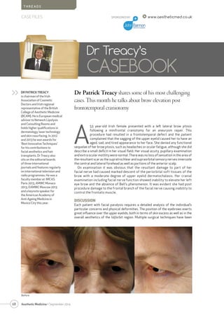
- 1. 68 Aesthetic Medicine • September 2014 SPONSORED BY www.aestheticmed.co.uk T H R E A D S CASE FILES Dr Patrick Treacy shares some of his most challenging cases. This month he talks about brow elevation post frontotemporal craniotomy Dr Treacy’s CASEBOOK DR PATRICK TREACY is chairman of the Irish Association of Cosmetic Doctors and Irish regional representative of the British College of Aesthetic Medicine (BCAM). He is European medical advisor to Network Lipolysis and Consulting Rooms and holds higher qualifications in dermatology, laser technology and skin resurfacing. In 2012 and 2013 he won awards for ‘Best Innovative Techniques’ for his contributions to facial aesthetics and hair transplants. Dr Treacy also sits on the editorial boards of three international journals and features regularly on international television and radio programmes. He was a faculty member at IMCAS Paris 2013, AMWC Monaco 2013, EAMWC Moscow 2013 and a keynote speaker for the American Academy of Anti-Ageing Medicine in Mexico City this year. >> A 53 year-old Irish female presented with a left lateral brow ptosis following a minifrontal craniotomy for an aneurysm repair. This procedure had resulted in a frontotemporal defect and the patient complained that the sagging of the upper eyelid caused her to have an aged, sad, and tired appearance to her face. She denied any functional sequelae of her brow ptosis, such as headaches or ocular fatigue, although she did describe a small deficit in her visual field. Her visual acuity, pupillary examination and extra ocular motility were normal. There was no loss of sensation in the area of the resultant scar as the supratrochlear and supraorbital sensory nerves innervate the central and lateral forehead as well as portions of the anterior scalp. On examination it was obvious that the resultant damage to part of her facial nerve had caused marked descent of the periorbital soft tissues of the brow with a moderate degree of upper eyelid dermatochalasis. Her cranial examination including facial nerve function showed inability to elevate her left eye brow and the absence of Bell’s phenomenon. It was evident she had post procedure damage to the frontal branch of the facial nerve causing inability to control the frontalis muscle. DISCUSSION Each patient with facial paralysis requires a detailed analysis of the individual’s particular concerns and physical deformities. The position of the eyebrows exerts great influence over the upper eyelids, both in terms of skin excess as well as in the overall aesthetics of the lid/orbit region. Multiple surgical techniques have been Before
- 2. describedandadvocatedforfacialreanimationafterfacial nerve paralysis. It was decided here to use some contour threads as the procedure is quite simple to perform under a local anaesthetic. Ideal candidates for thread lifts include people with minimal signs of ageingwhoneedjustasmalllift.Usually, these are women between 35 and 45. The threads are indicated because these patients have begun to see more prominence of the jaw, a relaxed (or minimally sagging) mid-facial appearance or slight bags under the eyes or on the neck. 1 Since the invention of the first barbed (short) suture by Sulamanidze in the late 1990s, different techniques have been described including Woffles (long) thread lifting, Waptos suture lifting, Isse unidirectional barbed-threads lifting, and silhouette lifting. However, essentially, there are two types of barbed threads which are available. These are: A. Bi-directional threads, with no anchoring points, insertedwithinahollowneedleandplacedinsuchamanner that the thread cannot move either way because of the two-way direction of barbs fixing it nicely. Examples are the APTOS® threads B. Uni-directional barbed threads, which are anchored at a higher level fixation point. Examples are the Contour® and Silhouette® threads2 Barbs along the thread act as cogs to grasp lift and suspend a relaxed facial area. The barbs open like an umbrella to form a support structure that lifts the sagging tissue. This creates tension in the thread, and the tension lifts the skin tissue. Collagen formation occurs around the threads and their cogs or barbs, producing an increasing effect ANATOMY Understanding the temporal and forehead anatomy is important to any successful browlift surgery. The frontal branch of the facial nerve is locatedinthesuperficialtemporal fascia and innervates the muscles of the forehead (frontalis, corrugators, depressor supercilii, and procerus). The blood supply to the forehead scalp is from the internal (supra-trochlear, supraorbital) and external carotid (superficial temporal) arteries. Hair follicles are located in the subcutaneous layer. Injury to the follicles results in temporary or permanent alopecia. The scalp is composed of five layers (skin, connective tissue, galea aponeurotica, loose areolar connective tissue, and periosteum). > 69 CASE FILES Aesthetic Medicine • September 2014 T H R E A D S www.aestheticmed.co.uk SPONSORED BY On examination it was obvious that the resultant damage to part of her facial nerve had cause marked descent of the periorbital soft tissues of the brow with a moderate degree of upper eyelid dermatochalasis The procedure
- 3. 70 Aesthetic Medicine • September 2014 S U R G I C A L CASE FILES SPONSORED BY METHOD The procedure was performed with the patient under local anaesthesia and no sedation was required. The patient’s face was marked preoperatively to determine the appropriate vector of the thread and its end fixation points. The presence of prominent dynamic and static rythides in the patient’s forehead was noted and influenced incision placement. The location of the hairline was noted. The superior border of the thread was placed above the hairline and exited at the level of the lateral brow. The sutures were trimmed, and the proximal ends were secured on the deep temporal fascia and reinforced with Vicryl interrupted sutures. PHYSICAL EVALUATION A thorough past medical and facial surgical history was obtained. The patient’s visual acuity, hairline position and brow symmetry were noted. We also noted skin quality and rhytiddepthinthemedialandlateralforehead.Theresidual motor function was noted prior to the procedure. REFERENCES 1. DeLorenziC.Barbedsutures:Rationaleandtechnique.AesthetSurgJ. 2006;26:223–9. 2. WuWT.Barbedsuturesinfacialrejuvenation.AesthetSurgJ.2004;24:582–7] 3. PaulMD.Complicationsofbarbedsutures.AesthetPlastSurg.2008;32:149 4. AestheticPlastSurg.2014Feb;38(1):69-74.Facialrejuvenationwithfine- barbedthreads:thesimpleMizlift.ParkTH1,SeoSW,WhangKW. 5 TranspalpebralOrbitofrontalCraniotomy:AMinimallyInvasiveApproachto AnteriorCranialVaultLesionsKofiD.OwusuBoahene,M.D.,MichaelLim, M.D.,EugeneChu,M.D.andAlfredoQuinones-Hinojosa,M.D www.aestheticmed.co.uk CONCLUSION Despite improvements in microsurgical techniques and intraoperativefacialnervemonitoring,itisoftenimpossible to preserve normal facial nerve function during craniotomy, especially when performing emergency intracranial surgeryorremovingtumourswithfacialnerveinvolvement. Although the facial nerve is anatomically preserved in most cases,functionmaybepartiallyorcompletelylost,resulting in some level of paralysis4 . In these circumstances, full facial paralysis is often physiologically and psychologically devastating to the patient. Associated problems include painful corneal irritation, visual loss, difficulties in eating and speaking and in the worse circumstance, self-imposed social isolation 5 . AM ADDENDUM ON BROW PTOSIS The main etiologic factors in brow ptosis are gravity and age. The ageing face undergoes a loss of tone from a diminution in the amount of elastin and collagen in the skin. Because the lateral brow has fewer attachments to the periosteum and has no underlying frontalis muscle, it usually descends more than the medial brow. Brow ptosis can happen secondary to paralysis of the frontalis muscle as in this case, but also because of Bell’s palsy, acoustic neuroma or even birth trauma. Medical causes include conditions such as myasthenia gravis, myotonic and oculopharyngeal dystrophy After treatment Despite improvements in microsurgical techniques and intraoperative facial nerve monitoring, it is often impossible to preserve normal facial nerve function during craniotomy,involvement