

Recomendados
Más contenido relacionado
La actualidad más candente
La actualidad más candente (20)
Destacado
Destacado (18)
Similar a 23205001
Similar a 23205001 (20)
Último
Último (20)
23205001
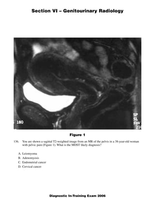
- 1. Section VI – Genitourinary Radiology Figure 1 136. You are shown a sagittal T2-weighted image from an MR of the pelvis in a 38-year-old woman with pelvic pain (Figure 1). What is the MOST likely diagnosis? A. Leiomyoma B. Adenomyosis C. Endometrial cancer D. Cervical cancer 1 Diagnostic In-Training Exam 2006
- 2. Section VI – Genitourinary Radiology Question #136 Rationales: A. Incorrect. Although leiomyomas typically have low intensity on T2 weighted images, they would be expected to be round and well defined. B. Correct. Adenomyosis results from the presence of heterotopic endometrial glands and stroma in the myometrium with adjacent myometrial hyperplasia. It is frequently associated with symptoms of pelvic pain, hypermenorrhea, and uterine enlargement. The diffuse thickening of the low intensity junctional zone is typical of diffuse adenomyosis of the uterus (junctional zone thickness ? 12 mm is generally considered diagnostic), and other imaging findings include poor definitions of the bor- ders of the junctional zone, or the presence of high-signal foci on T2- or T1-weighted images. This case demonstrates diffuse adenomyosis; focal adenomyosis may also be seen. C. Incorrect. For endometrial cancer, one would expect the high intensity endometrial stripe to be thickened, as well as inhomogeneous. D. Incorrect. For cervical cancer, one would expect an isointense mass in the area of the cervix, which may deform the endocervical canal or disrupt the low-signal-intensity fibrous stroma, and one may see tumor extension towards the vagina and/or parametrium. In this case, the endocervical canal and cervical region appears normal. 2 American College of Radiology
- 3. Section VI – Genitourinary Radiology Figure 2A 137. You are shown two ultrasound images of the scrotum in a 44-year-old man (Figures 2A and 2B). What is the MOST LIKELY diagnosis? A. Testicular torsion B. Seminoma C. Epididymo-orchitis D. Lymphoma 3 American College of Radiology
- 4. Section VI – Genitourinary Radiology Figure 2B 4 Diagnostic In-Training Exam 2006
- 5. Section VI – Genitourinary Radiology Question #137 Rationales: A. Incorrect. Testicular torsion typically presents as acute testicular pain with or without testicular enlargement. Testicular echogenicity is typically homogeneous, with normal testicular echogenicity initially, becoming hypoechoic with ongoing torsion and infarct. B. Correct. The images show an intratesticular mass. Seminoma is the most common solid intratestic- ular neoplasm. C. Incorrect. While epididymo-orchitis can cause enlargement of the testicle as well as hypoechoic areas within the testicle, it is not typically painless. D. Incorrect. While lymphoma could present as a testicular mass, it is less common than germ cell tumors such as seminoma. 5 American College of Radiology
- 6. Section VI – Genitourinary Radiology Figure 3A Figure 3B 6 American College of Radiology
- 7. Section VI – Genitourinary Radiology Figure 3C 138. You are shown a pelvic ultrasound (Figure 3A) and T1-weighted (Figure 3B) and fat-saturated T1-weighted (Figure 3C) MR images in a pregnant patient. What is the MOST likely diagnosis? A. Ectopic pregnancy B. Ovarian teratoma C. Ovarian serous cystadenoma D. Ovarian fibroma 7 Diagnostic In-Training Exam 2006
- 8. Section VI – Genitourinary Radiology Question #138 Rationales: A. Incorrect. An ectopic pregnancy can present as a complex mass by ultrasound, although it typically will not have the classic features of a teratoma described in the discussion for the correct answer in “A”. More importantly, a heterotopic pregnancy (concurrent intra-uterine and ectopic pregnancy) incidence is estimated at 1 out of 30,000 pregnancies. The finding of an intrauterine pregnancy effectively excludes an ectopic pregnancy in a patient except for those with high risk factors (ovula- tion induction, etc.) B. Correct. The ovarian teratoma (dermoid) is the most common ovarian neoplasm and occur most commonly during the reproductive years of a woman’s life. The ultrasound exam demonstrates a complex right adnexal mass that has two features highly sug- gestive of an ovarian teratoma. The first is the highly echogenic, non-shadowing nodule along the caudal wall of the mass. This is most consistent with a Rokitansky protuberance in a teratoma. The second is the hyperechoic lines and dots within the cystic portion of the mass that is caused by hair within the teratoma. The MRI confirms the diagnosis by showing high signal intensity fat within a portion of the mass on T1 images that “saturates” or loses signal intensity on T1 images with fat suppression technique. This is diagnostic of an ovarian teratoma containing fat. C. Incorrect. Serous cystadenoma is the most common epithelial neoplasm of the ovary and can occur in a young, pregnant female. However, the sonographic appearance is typically of an anechoic, unilocular cyst or minimally complex cyst with a few internal septations. Additionally, there would be no evidence for fat within the mass as is seen with the teratoma in this case. D. Incorrect. The ovarian fibroma is an uncommon neoplasm of the ovary in the stromal tumor catego- ry. It comprises only 4% of ovarian neoplasms. The sonographic appearance is typically of a solid, hypoechoic or mixed echogenicity mass that may attenuate sound posteriorly much like a peduncu- lated leiomyoma. Additionally, no fat would be present within this neoplasm. 8 American College of Radiology
- 9. Section VI – Genitourinary Radiology Figure 4 139. You are shown a delayed contrast-enhanced CT image (Figure 4) in a 60-year-old with hematuria. What is the MOST LIKELY diagnosis? A. Schistosomiasis B. Blood clots C. Cystitis cystica D. Transitional cell carcinoma 9 American College of Radiology
- 10. Section VI – Genitourinary Radiology Question #139 Rationales: A. Incorrect. Not a typical appearance. No calcifications. B. Incorrect. Should not be adherent to the wall and enhancing. C. Incorrect. Not cystic. D. Correct. Often multifocal. Classically enhance. Rarely calcify. 10 Diagnostic In-Training Exam 2006
- 11. Section VI – Genitourinary Radiology Figure 5 140. You are shown an image from a hysterosalpingogram on a 32-year-old woman (Figure 5). What is the MOST LIKELY diagnosis? A. Uterine hypoplasia B. Unicornuate uterus C. Fundal fibroid D. Asherman’s syndrome 11 American College of Radiology
- 12. Section VI – Genitourinary Radiology Question #140 Rationales: A. Incorrect. No contrast has entered the endometrial cavity. Only the endocervix contains contrast. Thus, you cannot comment on the size of the uterus. B. Incorrect. No contrast has entered the endometrial cavity. Thus, there is no evidence that only one uterine horn exists. C. Incorrect. No contrast has entered the endometrial cavity. In addition, HSG doesn’t allow the specif- ic diagnosis of filling defects which might be seen within the endometrial cavity. A differential diagnosis must be given, including polyp, fibroid, synechia, and cancer. D. Correct. Contrast fills only the endocervix, despite multiple attempts to fill the endometrial cavity. These women usually report having very short and light menstrual days and give a history of a prior D&C (common) or prior complications from pregnancy (uncommon). 12 Diagnostic In-Training Exam 2006
- 13. Section VI – Genitourinary Radiology Figure 6A Figure 6B 13 American College of Radiology
- 14. Section VI – Genitourinary Radiology Figure 6C 141. You are shown three images (Figures 6A through 6C) from a contrast-enhanced MR exam on a man with renal cell carcinoma. Based on these images, what is the stage by Robson classification? A. Stage II B. Stage IIIA C. Stage IIIB D. Stage IV 14 Diagnostic In-Training Exam 2006
- 15. Section VI – Genitourinary Radiology Question #141 Rationales: A. Incorrect. Stage II extends beyond the renal capsule but not through Gerota’s fascia or into the renal veins or local lymph nodes. Stage II includes involvement of the ipsilateral adrenal gland, which this patient does have on the left. B. Correct. Stage IIIA extends into the renal vein and may progress into the inferior vena cava. C. Incorrect. Stage IIIB involves regional lymph nodes but no extension into the renal veins or IVC. D. Incorrect. Stage IV includes distant metastases. 15 American College of Radiology
- 16. Section VI – Genitourinary Radiology 142. Which of the following is associated with testicular microlithiasis? A. Testicular torsion B. Epididymo-orchitis C. Right-sided varicocele D. Testicular neoplasm Question #142 Rationales: A. Incorrect. Microlithiasis is not typically seen in testicular torsion. B. Incorrect. While the calcifications may be the result of prior infection, it does not have an increased association with infection. C. Incorrect. There is no increased incidence of varicocele with testicular microlithiasis.. D. Correct. While testicular microlithiasis is often incidental, there is an increased incidence of testic- ular neoplasm, most of which are germ cell tumors. 16 American College of Radiology
- 17. Section VI – Genitourinary Radiology Concerning renal medullary carcinoma, which one is TRUE? 143. A. Usually peripheral in location B. Commonly seen in diabetic females C. Common in patients with sickle trait D. Often very small at presentation Question #143 Rationales: A. Incorrect. They are usually central. B. Incorrect. Commonly seen in African American patients with sickle trait; more commonly male. There is no association with diabetes. C. Correct. Renal medullary carcinoma typically is seen as an infiltrative mass in patients with sickle trait. D. Incorrect. They are usually large at presentation. 17 Diagnostic In-Training Exam 2006
- 18. Section VI – Genitourinary Radiology Concerning bladder rupture, which one is TRUE? 144. A. There is equal incidence between intraperitoneal and extraperitoneal rupture. B. Extraperitoneal bladder rupture causes elevated serum creatinine. C. Delayed images after a contrast-enhanced CT scan are sufficient to exclude it. D. Intraperitoneal bladder rupture requires surgical repair. Question #144 Rationales: A. Incorrect. Most bladder ruptures, two-thirds, are extraperitoneal, caused by trauma and pelvic frac- tures. B. Incorrect. The uremia and elevated creatinine occur because of INTRAPERITONEAL ruptures, not extraperitoneal ruptures. C. Incorrect. CT cystograms require active distention of the bladder with contrast. Passive filling of the bladder, such as seen in delayed images through the bladder after a normal contrast exam, may miss intraperitoneal ruptures which occur high in the dome, and thus may be higher than the level of the contrast-opacified urine seen during passive filling of the bladder. A CT cystogram, with active fill- ing, overcomes this obstacle, by filling the entire bladder lumen with contrast. In addition, CT cys- tograms do not require post-void images. D. Correct. Intraperitoneal bladder rupture generally require surgical closure. They do not close on their own. If uncorrected, they cause uremia and elevated creatinine as noted above. 18 American College of Radiology
- 19. Section VI – Genitourinary Radiology Concerning prostate carcinoma, which one of the following is CORRECT? 145. A. 30% of prostate cancers arise from the peripheral zone of the prostate. B. T1-weighted images provide the best contrast for detecting most prostate carcinomas. C. Most prostate cancers demonstrate increased enhancement on immediate post- gadoliniumfat-saturated T1 images. D. Prostate cancer metastasizes early along the gonadal vein/lymphatic pathway to the periaortic and pericaval region near the level of the kidneys. Question #145 Rationales: A. Incorrect. 70% of prostate cancers arise from the peripheral zone, the remainder from the transition- al and central zones. B. Incorrect. Prostate carcinomas in the peripheral zone are generally isointense to surrounding prostate tissue on T1-weighted images. T2-weighted images demonstrate prostate cancers as low signal intensity compared to the surrounding normal high signal intensity peripheral zone. C. Correct. Prostate cancer in the peripheral zone (where the majority of prostate cancers arise) demonstrates increased enhancement compared to the normal peripheral zone tissue. D. Incorrect. This metastatic pathway is characteristic of testicular neoplasms, not prostatic. Lymph node metastases from prostate carcinoma are generally first to the obturator, external and internal iliac chains. 19 Diagnostic In-Training Exam 2006
- 20. Section VI – Genitourinary Radiology Concerning gonadal vein thrombosis, which one is TRUE? 146. A. Most common on the right side in post partum women B. Best study for diagnosis is excretory urography C. Usually treated surgically D. Commonly seen in diabetic males Question #146 Rationales: A. Correct. TRUE. Gonadal vein thrombosis is in the differential for cause of fever in post partum woman. B. Incorrect. FALSE. CT or MR are most sensitive in detection of gonadal vein thrombosis. The diag- nosis may also be made with US. IVU would not be expected to be helpful in this diagnosis. C. Incorrect. FALSE. Patients are usually treated with anticoagulation and antibiotics. D. Incorrect. FALSE. There is no association with diabetes; gonadal vein thrombosis is most com- monly seen in post partum women (answer A). 20 American College of Radiology
- 21. Section VI – Genitourinary Radiology Concerning pseudodiverticula of the ureter, which one is TRUE? 147. A. They represent ulcerations within a transitional cell carcinoma lesion. B. They represent the site of premalignant lesions, similar to carcinoma in situ. C. 40% of patients have co-existing transitional cell carcinoma. D. They warrant semi-annual investigation. Question #147 Rationales: A. Incorrect. They represent intramural outpouchings from the ureter. They indicate an increased risk of transitional cell carcinoma, either in the ipsilateral ureter or in the bladder. B. Incorrect. They are benign, but indicate mural inflammation, thought to predispose the patient to developing malignancy. C. Incorrect. Up to 25% of patients with pseudodiverticula have TCC in the ipsilateral ureter or blad- der. D. Correct. Patients require immediate work-up. If, however, they do not have transitional cell carci- noma, they need semi-annual follow-up to exclude the interval development of tumor. 21 Diagnostic In-Training Exam 2006
- 22. Section VI – Genitourinary Radiology 148. Concerning cervical carcinoma, what stage is a lesion that is confined to the upper two thirds of the vagina on clinical exam and that shows right hydroureter to the level of a poorly defined cer- vical soft tissue mass on CT exam? A. Stage II A B. Stage II B C. Stage III A D. Stage III B Question #148 Rationales: A. Incorrect. At stage II A the tumor has spread beyond the cervix but has no obvious parametrial involvement, is confined to the upper two thirds of the vagina and no invasion of the ureter or blad- der. B. Incorrect. Stage II B has obvious parametrial involvement but does not extend to the pelvic side wall. C. Incorrect. Stage III A extends to the lower third of the vagina but not the pelvic sidewall and does not obstruct the ureters or invade adjacent organs. D. Correct. Stage III B tumors extend to pelvic sidewall and/or causes hydronephrosis or non-func- tioning kidney. 22 American College of Radiology
- 23. Section VI – Genitourinary Radiology Concerning renal lymphoma, which one is TRUE? 149. A. Multiple or solitary focal nodular masses are the most common form. B. It demonstrates uniform, hyperintense enhancement after IV gadolinium. C. Direct extension to and involvement of the psoas muscle is more characteristic of primary renal cell carcinoma than of renal lymphoma. D. Tumor thrombus commonly occurs in renal lymphoma. Question #149 Rationales: A. Correct. There are 3 basic patterns of renal involvement by lymphoma: 1) direct invasion by adja- cent nodal disease, 2) focal masses that may be solitary or multiple (most common), and 3) diffuse infiltration. B. Incorrect. Renal lymphoma typically enhances minimally to a mildly heterogenous pattern. C. Incorrect. Renal lymphoma can commonly extend to and involve the adjacent psoas muscle. This feature is rare in primary renal carcinoma. D. Incorrect. Renal lymphoma rarely causes tumor thrombus. This is a common feature of renal carci- noma. 23 Diagnostic In-Training Exam 2006
- 24. Section VI – Genitourinary Radiology Which one would result in a pelvic CT image that is abnormally noisy? 150. A. Higher-than-normal tube voltage (kVp) B. Thicker-than-normal slice thickness C. Smoothing reconstruction algorithm D. Lower-than-normal tube current Question #150 Rationales: A. Incorrect. Higher kVp yields lower image noise. B. Incorrect. Increasing slice thickness decreases image noise. C. Incorrect. Normally smoothing algorithms decreases image noise. D. Correct. Lower tube current means fewer x-ray photons, therefore increased image noise. 24 American College of Radiology
- 25. Section VI – Genitourinary Radiology Concerning adrenal cortical carcinoma, which one is TRUE? 151. A. It is the most common cause of an adrenal mass. B. It most often displays areas of macroscopic fat. C. It usually presents with <10 H.U. on non contrast CT. D. It usually presents as a large heterogeneous soft tissue mass. Question #151 Rationales: A. Incorrect. Adrenal adenoma and metastatic disease are much more common than primary adrenal cortical carcinoma. B. Incorrect. While fat can rarely be seen in these tumors, macroscopic fat in an adrenal lesion is almost always in a myelolipoma. C. Incorrect. Adrenal adenomas are more likely to present with the above characteristics. D. Correct. Most adrenal cortical carcinomas are > 6 cm and often have central necrosis. Calcification is seen in approximately 30% of these lesions. 25 Diagnostic In-Training Exam 2006
- 26. Section VI – Genitourinary Radiology Concerning post-transplantation lymphoproliferative disorder, which one is TRUE? 152. A. Epstein Barr virus infection is associated with the disorder. B. T1- and T2-weighted MR images reveal hyperintense areas of soft tissue centrally within the kidney. C. It affects 12% of patients with solid organ transplants. D. Radiation may be necessary if chemotherapy fails to involute the tumor. Question #152 Rationales: A. Correct. Despite advances in antiviral therapy, Epstein-Barr virus-induced posttransplant lympho- proliferative disease (EBV-PTLD) continues to be a major complication after solid organ transplan- tation. B. Incorrect. MR imaging reveals hypointense tissue on both T1- and T2-weighted images. The tissue shows little enhancement with gadolinium. C. Incorrect. It affects 2% of patients with solid organ transplants. D. Incorrect. First line of therapy is reduction in the level of immunosuppression. If that fails, chemotherapy is warranted. 26 American College of Radiology
- 27. Section VI – Genitourinary Radiology Which one of the following findings on IVU is MOST sensitive in detecting mild, acute ureteral 153. obstruction? A. Delayed, increasingly dense nephrogram B. Demonstration of medullary rays in the nephrogram C. Delayed opacification of the calyces and collecting system D. Blunting of the calyceal fornices Question #153 Rationales: A. Incorrect. The classic “obstructive nephrogram” is often absent in mild, acute obstruction. B. Incorrect. Medullary rays or faint striations may be seen in acute obstruction of moderate severity, but may be absent in cases of mild obstruction. C. Incorrect. Delayed opacification of the collecting system is a consequence of more severe obstruc- tion and secondary oliguria. D. Correct. Calyceal blunting is an excellent sign of mild obstruction. Visualizing sharp fornices virtu- ally excludes mild obstruction. 27 Diagnostic In-Training Exam 2006
- 28. Section VI – Genitourinary Radiology Concerning adrenal adenomas, which one is TRUE? 154. A. They are a more common cause of adrenal mass than is metastatic disease. B. They typically have non-IV contrast density of more than 20 Hounsfield units. C. They typically show no decrease in signal intensity on opposed phase MR imaging. D. They typically retain 90% of their immediate contrast value on 10-minute delayed CT exam. Question #154 Rationales: A. Correct. Adenomas are the most common adrenal mass and occur in 2 - 8 % of the population. B. Incorrect. Adenomas most often have non contrast density of less than 10 H.U. C. Incorrect. Adenomas typically decrease in signal intensity on the out of phase portion of opposed phase imaging. D. Incorrect. Adenomas typically washout 50% of their initial enhancement levels on a 10 minute delayed CT exam. 28 American College of Radiology
- 29. Section VI – Genitourinary Radiology Concerning urothelial malignancy, which one is TRUE? 155. A. Adenocarcinoma is the second most common urothelial malignancy. B. Leukoplakia is associated with transitional cell carcinoma of the bladder at the urachal remant. C. Transitional cell carcinoma of the upper collecting system is less common than within the bladder. D. 50% of primary renal malignancies develop from the urothelium. Question #155 Rationales: A. Incorrect. Transitional cell carcinoma is the most common urothelial malignancy followed by squa- mous cell carcinoma. Adenocarcinoma is distinctly rare. B. Incorrect. Leukoplakia is associated with squamous cell carcinoma of the bladder, not transitional cell carcinoma. The urachus is a site where transitional epithelium can undergo metaplasia to glan- dular epithelium and result in adenocarcinoma. C. Correct. The entire urothelium is at risk, but transitional cell carcinomas are more common in the bladder than in the upper tracts. D. Incorrect. 8% of renal malignancies develop from the urothelium. 29 Diagnostic In-Training Exam 2006
- 30. Section VI – Genitourinary Radiology Concerning renal cystic disease, which one is TRUE? 156. A. Autosomal recessive polycystic disease typically presents as multiple bilateral cysts in adult- hood. B. Autosomal dominant polycystic disease typically presents as enlarged hyperechoic kidneys in the neonatal period. C. Acquired cystic renal disease in chronic renal failure patients on dialysis is indistinguishable from autosomal dominant polycystic disease. D. Autosomal dominant polycystic disease has a higher incidence of associated hepatic cysts than does autosomal recessive polycystic disease. Question #156 Rationales: A. Incorrect. Autosomal dominant polycystic disease usually presents with multiple bilateral simple renal cysts between ages 20-39 years. Autosomal recessive polycystic disease has a spectrum of presentation ages but is typically seen from the neonatal through childhood periods rather than adulthood. B. Incorrect. This description is more typical of the appearance of the infantile form of ARPKD. C. Incorrect. The kidneys are typically small and atrophic with multiple cysts in acquired cystic renal disease of dialysis as compared to markedly enlarged kidneys in ADPCD. D. Correct. ADPCD typically has multiple hepatic cysts in over 50% of cases. Autosomal recessive polycystic disease is associated with hepatic fibrosis particularly in the juvenile onset form. 30 American College of Radiology
- 31. Section VI – Genitourinary Radiology Concerning renal angiomyolipomas, which one finding is MOST diagnostic? 157. A. Fluid/fluid levels B. Fat C. Homogeneous soft tissue D. Large irregular calcification Question #157 Rationales: A. Incorrect. These lesions may occasionally hemorrhage but are usually incidental masses with mixed amounts of soft tissue and macroscopic fat. B. Correct. While other lesions such as renal cell carcinoma, oncocytoma, Wilm’s and metastasis have also been reported with areas of fat within these tumors, these cases are rare. C. Incorrect. Angiomyolipomas have varying amounts of fat and soft tissue. Some have no fat visible by CT and a solid soft tissue renal mass in such a case is indistinguishable from renal cell carcino- ma and should be treated as such. D. Incorrect. Calcification in angiomyolipomas is unusual but may occur if there has been prior hem- orrhage. 31 Diagnostic In-Training Exam 2006
- 32. Section VI – Genitourinary Radiology 158. What characterizes Type II posterior urethral injury? A. The membranous urethra is disrupted with extension of injury into the proximal bulbous urethra and/or disruption of the urogenital diaphragm (UGD). B. On urethrography, contrast material extravasates into the perineum. C. Disruption of the urethra above the urogenital diaphragm. D. The posterior urethra is stretched but intact. Question #158 Rationales: A. Incorrect. This is the definition of Type III posterior urethral injury. B. Incorrect. Extravasation of contrast below the urogenital diaphragm (UGD) into the perineum on urethrography indicates disruption of the UGD and signifies Type III injury. C. Correct. This is the definition of Type II posterior urethral injury. D. Incorrect. This is the definition of Type I posterior urethral injury. 32 American College of Radiology
- 33. Section VI – Genitourinary Radiology Which one of the following conditions is MOST closely associated with female pelvic 159. inflammatory disease (PID)? A. Intramural pseudodiverticulosis B. Salpingitis isthmica nodosa C. Adenomyosis D. Leukoplakia Question #159 Rationales: A. Incorrect. Intramural pseudodiverticuli are prominent submucosal esophageal glands associated with gastroesophageal reflux and Candida.. B. Correct. Salpingitis isthmica nodosa manifests as tiny diverticula arising from the fallopian tubes secondary to chronic inflammation. They is most easily seen during hysterosalpingography. C. Incorrect. Adenomyosis is the presence of endometrial glands and supporting tissue in the myometrium. Increased incidence due to childbirth, Cesarean section, trauma, and tubal ligation. D. Incorrect. Leukoplakia is an inflammatory condition of the ureter or bladder, associated with chron- ic urinary tract infection and not PID per se. 33 Diagnostic In-Training Exam 2006
- 34. Section VI – Genitourinary Radiology 160. A lateral abdominal radiograph is taken of a pregnant woman with a transmission path length of 30 cm. If the entrance dose is 10 mGy (1 rad), and the half-value layer thickness for the x-ray beam is 3 cm of tissue, what is the approximate dose to the center of the uterus from the primary radiation? A. 0.3 mGy B. 1 mGy C. 2 mGy D. 5 mGy Question #160 Rationales: A. Correct. The middle of the uterus would be midline in the patient, at a depth of 15cm. Since the HVL equals 3 cm of tissue, the radiation must pass through 5 HVL’s of tissue to reach the uterus. The primary radiation will then be reduced by (1/2)^5 or 1/32nd of the incident intensity. B. Incorrect. See correct answer C. Incorrect. See correct answer D. Incorrect. See correct answer 34 American College of Radiology
- 35. Section VI – Genitourinary Radiology Concerning epididymo-orchitis, which one is TRUE? 161. A. Physical exam shows increasing testicular pain when the scrotum is raised above the level of the symphysis pubis. B. Hypervascularity in the epididymis and adjacent testicle supports the diagnosis. C. Testicular involvement is seen in 80% of cases of epididymitis. D. Treatment requires antibiotic therapy for 10 days to 2 weeks. Question #161 Rationales: A. Incorrect. Raising the scrotum above the level of the symphysis pubis DECREASES the scrotal pain. This maneuver, known as the Prehn sign, helps to differentiate between epididymo-orchitis and testicular torsion. B. Correct. Hypervascularity of the epididymis and adjacent testicle are typically seen in epididymo- orchitis. Studies have shown that males with epididymo-orchitis have resistive indices below 0.5 in 50% of cases. A peak systolic velocity higher than 15 cm/sec yields sensitivity for epididymo-orchi- tis of 90-93%. C. Incorrect. Orchitis is seen in 20-40% of cases of epididymo-orchitis. D. Incorrect. The testicle is a sanctuary zone. Thus, antibiotic therapy is recommended for 4-6 weeks to exclude recurrence of infection. 35 Diagnostic In-Training Exam 2006