SlideShare una empresa de Scribd logo
1 de 54
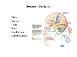
Sensory Systems
• Vision
• Hearing
• Taste
• Smell
• Equilibrium
• Somatic Senses
Sensory Systems
• Somatic sensory
• General – transmit impulses from skin, skeletal muscles,
and joints
• Special senses - hearing, balance, vision
• Visceral sensory
• Transmit impulses from visceral organs
• Special senses - olfaction (smell), gustation (taste)
• Stimulus - energy source
• Internal
• External
• Receptors
• Sense organs - structures specialized to respond to
stimuli
• Transducers - stimulus energy converted into
action potentials
• Conduction
• Afferent pathway
• Nerve impulses to the CNS
• Translation
• CNS integration and information processing
• Sensation and perception – your reality
Properties of Sensory Systems
Sensory Pathways
• Stimulus as physical energy  sensory receptor acts as a transducer
• Stimulus > threshold  action potential to CNS
• Integration in CNS  cerebral cortex or acted on subconsciously
Classification by Function (Stimuli)
• Mechanoreceptors – respond to touch, pressure, vibration, stretch, and itch
• Thermoreceptors – sensitive to changes in temperature
• Photoreceptors – respond to light energy (e.g., retina)
• Chemoreceptors – respond to chemicals (e.g., smell, taste, changes in
blood chemistry)
• Nociceptors – sensitive to pain-causing stimuli
• Osmoreceptors – detect changes in concentration of solutes, osmotic
activity
• Baroreceptors – detect changes in fluid pressure
Classification by Location
• Exteroceptors – sensitive to stimuli arising from outside the body
• Located at or near body surfaces
• Include receptors for touch, pressure, pain, and temperature
• Interoceptors – (visceroceptors) receive stimuli from internal viscera
• Monitor a variety of stimuli
• Proprioceptors – monitor degree of stretch
• Located in musculoskeletal organs
Classification by Structure
• General somatic – include touch,
pain, vibration, pressure, temperature
• Proprioceptive – detect stretch in
tendons and muscle provide
information on body position,
orientation and movement of body in
space
Somatic Senses
Somatic Receptors
• Divided into two groups
• Free or Unencapsulated nerve endings
• Encapsulated nerve endings - consist of one or more neural
end fibers enclosed in connective tissue
Free Nerve Endings
• Abundant in epithelia and underlying connective tissue
• Nociceptors - respond to pain
• Thermoreceptors - respond to temperature
• Two specialized types of free nerve endings
• Merkel discs – lie in the epidermis, slowly adapting receptors for light touch
• Hair follicle receptors – Rapidly adapting receptors that wrap around hair follicles
Encapsulated Nerve Endings
• Meissner’s corpuscles
• Spiraling nerve ending surrounded by Schwann cells
• Occur in the dermal papillae of hairless areas of the skin
• Rapidly adapting receptors for discriminative touch
• Pacinian corpuscles
• Single nerve ending surrounded by layers of flattened Schwann cells
• Occur in the hypodermis
• Sensitive to deep pressure – rapidly adapting receptors
• Ruffini’s corpuscles
• Located in the dermis and respond to pressure
• Monitor continuous pressure on the skin – adapt slowly
Encapsulated Nerve Endings - Proprioceptors
• Monitor stretch in locomotory organs
• Three types of proprioceptors
• Muscle spindles – monitors the changing length of a muscle, imbedded in the
perimysium between muscle fascicles
• Golgi tendon organs – located near the muscle-tendon junction, monitor tension
within tendons
• Joint kinesthetic receptors - sensory nerve endings within the joint capsules, sense
pressure and position
Muscle Spindle & Golgi Tendon Organ
Special Senses
Figure 10-4: Sensory pathways
• Taste, smell, sight,
hearing, and balance
• Localized – confined to
the head region
• Receptors are not free
endings of sensory
neurons but specialized
receptor cells
Anatomy of the Eyeball
• Function of the eyeball
• Protect and support the photoreceptors
• Gather, focus, and process light into precise images
• External walls – composed of three tunics (layers)
• Internal cavity – contains fluids (humors)
The Fibrous Layer
• Most external layer of the eyeball
• Cornea
• Anterior one-sixth of the fibrous tunic
• Composed of stratified Squamous externally, simple squamous internally
• Refracts (bends) light
• Sclera
• Posterior five-sixths of the tunic
• White, opaque region composed of dense irregular connective tissue Provides
shape and an anchor for eye muscles,
• Scleral venous sinus – allows aqueous humor to drain
The Vascular Layer
• Middle layer consists of choroid, ciliary body, and iris
• Iris and Pupil
• Composed of smooth muscle, melanocytes, and blood vessels that forms the colored portion of
the eye.
• Function: It regulates the amount of light entering the eye through the pupil.
• It is attached to the ciliary body.
• Pupil is the opening in center of iris through which light enters the eye
• Ciliary body
• Composed of a ring of muscle called ciliary muscle and ciliary processes which are folds located
at the posterior surface of ciliary bodies
• Suspensory ligaments attach to these processes
• Function: secretes the aqueous humor
• The suspensory ligaments position the lens so that light passing through the pupil passes through
the center of the lens of the eye.
The Vascular Layer
• Choroid - vascular layer in the wall of the eye.
• Dark brown (pigmented) membrane with melanocytes that lines most of
the internal surface of the sclera. Has lots of blood vessels
• Lines most of the interior of the sclera.
• Extends from the ciliary body to the lens.
• Corresponds to arachnoid and pia mater
• Functions:
• Delivers oxygen and nutrients to the retina.
• Absorb light rays so that the light rays are not reflected within the eye
The Inner Layer (Retina)
• Retina is the innermost layer of the eye lining the posterior cavity
• The retina contains 2 layers:
• Pigmented layer made of a single layer of melanocytes, absorbs light after it
passes through the neural layer
• Neural layer – sheet of nervous tissue, contains three main types of neurons
• Photoreceptor cells
• Bipolar cells
• Ganglion cells
Photoreceptors
• Two main types
• Rod cells
• More sensitive to light
• Allow vision in dim light
• In periphery
• Cone cells
• Operate best in bright light
• High-acuity
• Color vision – blue, green, red cones
• Concentrated in fovea
Regional Specializations of the Retina
• Ora serrata retinae
• Neural layer ends at the posterior margin of the ciliary body
• Pigmented layer covers ciliary body and posterior surface of the iris
• Macula lutea – contains mostly cones
• Fovea centralis – contains only cones
• Region of highest visual acuity
• Optic disc – blind spot
The Lens
• A thick, transparent, biconvex disc
• Held in place by its ciliary zonule
• Lens epithelium – covers anterior surface of the
lens
The Eye as an Optical Device
• Structures in the eye bend light rays
• Light rays converge on the retina at a single focal point
• Light bending structures (refractory media)
• The lens, cornea, and humors
• Accommodation – curvature of the lens is adjustable
• Allows for focusing on nearby objects
Internal Chambers and Fluids
Figure 16.8
Internal Chambers and Fluids
• Anterior segment
• Divided into anterior and posterior chambers
• Anterior chamber – between the cornea and iris
• Posterior chamber – between the iris and lens
• Filled with aqueous humor
• Renewed continuously
• Formed as a blood filtrate
• Supplies nutrients to the lens and cornea
Internal Chambers and Fluids
• The lens and ciliary zonules divide the eye
• Posterior segment (cavity)
• Filled with vitreous humor - clear, jelly-like substance
• Transmits light
• Supports the posterior surface of the lens
• Helps maintain intraocular pressure
Accessory Structures of the Eye
• Eyebrows – coarse hairs on
the superciliary arches
• Eyelids (palpebrae)
• Separated by the palpebral
fissure
• Meet at the medial and
lateral angles (canthi)
• Conjunctiva – transparent
mucous membrane
• Palpebral conjunctiva
• Bulbar (ocular) conjunctiva
• Conjunctival sac
• Moistens the eye
Figure 16.5a
Accessory Structures of the Eye
• Lacrimal apparatus –
keeps the surface of the
eye moist
• Lacrimal gland –
produces lacrimal fluid
• Lacrimal sac – fluid
empties into nasal
cavity
Figure 16.5b
Extrinsic Eye Muscles
Figure 16.6a, b
• Six muscles that control movement of the eye
• Originate in the walls of the orbit
• Insert on outer surface of the eyeball
Visual Pathways to the Cerebral Cortex
• Pathway begins at the retina
• Light activates photoreceptors
• Photoreceptors signal bipolar cells
• Bipolar cells signal ganglion cells
• Axons of ganglion cells exit eye as the optic nerve
• Optic nerve
• Optic chiasm
• Optic tract
• Thalamus
• Visual cortex
• Other pathways
include the
midbrain and
diencephalon
Vision Integration / Pathway
Figure 10-29b, c: Neural pathways for vision and the papillary reflex
The Ear: Hearing and Equilibrium
• The ear – receptor organ for hearing and
equilibrium
• Composed of three main regions
• Outer ear – functions in hearing
• Middle ear – functions in hearing
• Inner ear – functions in both hearing and
equilibrium
The Outer (External) Ear
• Auricle (pinna) - helps direct sounds
• External acoustic meatus
• Lined with skin
• Contains hairs, sebaceous glands, and ceruminous glands
• Tympanic membrane
• Forms the boundary between the external and middle ear
The Middle Ear
• The tympanic cavity
• A small, air-filled space
• Located within the petrous portion of the temporal bone
• Medial wall is penetrated by
• Oval window
• Round window
• Pharyngotympanic tube (auditory or eustachian tube) - Links the middle ear
and pharynx
Figure 16.17
The Middle Ear
• Ear ossicles – smallest
bones in the body
• Malleus – attaches to
the eardrum
• Incus – between the
malleus and stapes
• Stapes – vibrates
against the oval
window
The Inner (Internal) Ear
• Inner ear – also called the
labyrinth
• Bony labyrinth – a cavity
consisting of three parts
• Semicircular canals
• Vestibule
• Cochlea
• Bony labyrinth is filled
with perilymph
The Membranous Labyrinth
Figure 16.18
• Membranous labyrinth - series of membrane-walled sacs and ducts
• Fit within the bony labyrinth
• Consists of three main parts
• Semicircular ducts
• Utricle and saccule
• Cochlear duct
• Filled with a clear fluid – endolymph
The Cochlea
• A spiraling chamber in the bony labyrinth
• Coils around a pillar of bone – the modiolus
• Spiral lamina – a spiral of bone in the modiolus
• The cochlear nerve runs through the core of the modiolus
The Cochlea
• The cochlear duct (scala media) – contains receptors for hearing
• Lies between two chambers
• The scala vestibuli
• The scala tympani
• The vestibular membrane – the roof of the cochlear duct
• The basilar membrane – the floor of the cochlear duct
The Cochlea
• The cochlear duct (scala media) – contains receptors for
hearing
• Organ of Corti – the receptor epithelium for hearing
• Consists of hair cells (receptor cells)
The Role of the Cochlea in Hearing
Figure 16.20
Auditory Pathway from the Organ of Corti
• The ascending
auditory pathway
• Transmits
information from
cochlear receptors
to the cerebral
cortex
Figure 16.23
The Vestibule
• Utricle and saccule – suspended in perilymph
• Two egg-shaped parts of the membranous labyrinth
• House the macula – a spot of sensory epithelium
• Macula – contains receptor cells
• Monitor the position of the head when the head is still
• Contains columnar supporting cells
• Receptor cells – called hair cells
• Synapse with the vestibular nerve
Anatomy and Function of the Maculae
Figure 16.21b
The Semicircular Canals
• Lie posterior and lateral to the vestibule
• Anterior and posterior semicircular canals lie in the vertical plane at
right angles
• Lateral semicircular canal lies in the horizontal plane
The Semicircular Canals
• Semicircular duct – snakes through each semicircular canal
• Membranous ampulla – located within bony ampulla
• Houses a structure called a crista ampullaris
• Cristae contain receptor cells of rotational acceleration
• Epithelium contains supporting cells and receptor hair cells
Structure and Function of the Crista
Ampullaris
Figure 16.22b
The Chemical Senses: Taste and Smell
• Taste – gustation
• Smell – olfaction
• Receptors – classified as chemoreceptors
• Respond to chemicals
Taste – Gustation
• Taste receptors
• Occur in taste buds
• Most are found on the surface of the
tongue
• Located within tongue papillae
• Two types of papillae (with taste buds)
• Fungiform papillae
• Circumvallate papillae
Taste Buds
• Collection of 50 –100 epithelial cells
• Contain three major cell types (similar
in all special senses)
• Supporting cells
• Gustatory cells
• Basal cells
• Contain long microvilli – extend
through a taste pore
Taste Sensation and the Gustatory Pathway
• Four basic qualities of taste
• Sweet, sour, salty, and bitter
• A fifth taste – umami, “deliciousness”
• No structural difference among taste buds
Gustatory Pathway from Taste Buds
Figure 16.2
• Taste information reaches the
cerebral cortex
• Primarily through the facial
(VII) and glossopharyngeal
(IX) nerves
• Some taste information
through the vagus nerve (X)
• Sensory neurons synapse in
the medulla
• Located in the solitary
nucleus
• Olfactory epithelium with olfactory receptors, supporting cells, basal cells
• Olfactory receptors are modified neurons
• Surfaces are coated with secretions from olfactory glands
• Olfactory reception involves detecting dissolved chemicals as they interact with
odorant binding proteins
Smell (Olfaction)
Olfactory Receptors
• Bipolar sensory neurons located within olfactory epithelium
• Dendrite projects into nasal cavity, terminates in cilia
• Axon projects directly up into olfactory bulb of cerebrum
• Olfactory bulb projects to olfactory cortex, hippocampus, and amygdaloid
nuclei

Más contenido relacionado

La actualidad más candente

La actualidad más candente (20)

Sensory organs eyes
Sensory organs eyesSensory organs eyes
Sensory organs eyes
 
Sense organs
Sense organsSense organs
Sense organs
 
Introduction to the nervous system
Introduction to the nervous systemIntroduction to the nervous system
Introduction to the nervous system
 
Nervous system
Nervous systemNervous system
Nervous system
 
Endocrine system
Endocrine systemEndocrine system
Endocrine system
 
Central nervous system
Central nervous systemCentral nervous system
Central nervous system
 
Sense organs
Sense organsSense organs
Sense organs
 
Anatomy and physiology of nervous system
Anatomy and physiology of nervous systemAnatomy and physiology of nervous system
Anatomy and physiology of nervous system
 
Sense of smell
Sense of smellSense of smell
Sense of smell
 
ear anatomy, structures, functions
ear anatomy, structures, functions ear anatomy, structures, functions
ear anatomy, structures, functions
 
Nervous system
Nervous systemNervous system
Nervous system
 
special sense organs (anatomy and physiology) - a brief discussion
special sense organs (anatomy and physiology) - a brief discussion special sense organs (anatomy and physiology) - a brief discussion
special sense organs (anatomy and physiology) - a brief discussion
 
Nose
NoseNose
Nose
 
Nervous System
Nervous SystemNervous System
Nervous System
 
Nervous System
Nervous SystemNervous System
Nervous System
 
anatomy of central nervous system
anatomy of central nervous systemanatomy of central nervous system
anatomy of central nervous system
 
Nervous system
Nervous systemNervous system
Nervous system
 
Introduction to nervous system
Introduction to nervous systemIntroduction to nervous system
Introduction to nervous system
 
The nervous system anatomy
The nervous system anatomyThe nervous system anatomy
The nervous system anatomy
 
ppt of Sensory organ
ppt of Sensory organppt of Sensory organ
ppt of Sensory organ
 

Destacado

Destacado (6)

Sensory organs
Sensory organsSensory organs
Sensory organs
 
SENSES
SENSES SENSES
SENSES
 
Skin Structure
Skin StructureSkin Structure
Skin Structure
 
Anatomy of skin
Anatomy of skinAnatomy of skin
Anatomy of skin
 
Anatomy Of The Skin Lecture
Anatomy Of The Skin LectureAnatomy Of The Skin Lecture
Anatomy Of The Skin Lecture
 
The integumentary system
The integumentary systemThe integumentary system
The integumentary system
 

Similar a Sensory organs

160456 water-template-16x9
160456 water-template-16x9160456 water-template-16x9
160456 water-template-16x9bennardnicholas1
 
SPECIAL SENSES - eye , ear, taste, touch WPS Office.pptx
SPECIAL SENSES - eye , ear, taste, touch WPS Office.pptxSPECIAL SENSES - eye , ear, taste, touch WPS Office.pptx
SPECIAL SENSES - eye , ear, taste, touch WPS Office.pptxnandithapradeep92
 
SENSE_ORGANS_physiology[1].pptx
SENSE_ORGANS_physiology[1].pptxSENSE_ORGANS_physiology[1].pptx
SENSE_ORGANS_physiology[1].pptxObsa2
 
Anatomy and physilogy of eye,nose and throat
Anatomy and physilogy of eye,nose and throatAnatomy and physilogy of eye,nose and throat
Anatomy and physilogy of eye,nose and throatDeeps Gupta
 
Special senses 👀.pptxuiiiiiiiiihjjjjiiiii
Special senses 👀.pptxuiiiiiiiiihjjjjiiiiiSpecial senses 👀.pptxuiiiiiiiiihjjjjiiiii
Special senses 👀.pptxuiiiiiiiiihjjjjiiiiiRawalRafiqLeghari
 
Anatomy and Physiology of Special senses
Anatomy and Physiology of Special senses Anatomy and Physiology of Special senses
Anatomy and Physiology of Special senses DR .PALLAVI PATHANIA
 
ANATOMY & PHYSIOLOGY OF EYE.pptx
ANATOMY  & PHYSIOLOGY OF EYE.pptxANATOMY  & PHYSIOLOGY OF EYE.pptx
ANATOMY & PHYSIOLOGY OF EYE.pptxSaili Gaude
 
ANATOMY OF EYE.pptx
ANATOMY OF EYE.pptxANATOMY OF EYE.pptx
ANATOMY OF EYE.pptxSaili Gaude
 
The Special Senses.pptx
The Special Senses.pptxThe Special Senses.pptx
The Special Senses.pptxchriskimeu103
 
The Special Senses.pptx
The Special Senses.pptxThe Special Senses.pptx
The Special Senses.pptxJohnmvula3
 
Eye Anatomy & Physiology
Eye Anatomy & Physiology Eye Anatomy & Physiology
Eye Anatomy & Physiology Pari Pratik
 
The eye (sense of sight organ)
The eye (sense of sight organ)The eye (sense of sight organ)
The eye (sense of sight organ)Jordan Mwelwa
 
1 GNM anatomy Unit - 12 - sense organs.pptx
1 GNM anatomy Unit - 12 - sense organs.pptx1 GNM anatomy Unit - 12 - sense organs.pptx
1 GNM anatomy Unit - 12 - sense organs.pptxthiru murugan
 
The Eye notes physiology notes for medical students
The Eye notes physiology notes for medical studentsThe Eye notes physiology notes for medical students
The Eye notes physiology notes for medical studentskenosewe1
 

Similar a Sensory organs (20)

160456 water-template-16x9
160456 water-template-16x9160456 water-template-16x9
160456 water-template-16x9
 
SPECIAL SENSES - eye , ear, taste, touch WPS Office.pptx
SPECIAL SENSES - eye , ear, taste, touch WPS Office.pptxSPECIAL SENSES - eye , ear, taste, touch WPS Office.pptx
SPECIAL SENSES - eye , ear, taste, touch WPS Office.pptx
 
SENSE_ORGANS_physiology[1].pptx
SENSE_ORGANS_physiology[1].pptxSENSE_ORGANS_physiology[1].pptx
SENSE_ORGANS_physiology[1].pptx
 
Anatomy and physilogy of eye,nose and throat
Anatomy and physilogy of eye,nose and throatAnatomy and physilogy of eye,nose and throat
Anatomy and physilogy of eye,nose and throat
 
Vision.ppt
Vision.pptVision.ppt
Vision.ppt
 
Sense organs
Sense organsSense organs
Sense organs
 
Special senses 👀.pptxuiiiiiiiiihjjjjiiiii
Special senses 👀.pptxuiiiiiiiiihjjjjiiiiiSpecial senses 👀.pptxuiiiiiiiiihjjjjiiiii
Special senses 👀.pptxuiiiiiiiiihjjjjiiiii
 
Special senses
Special sensesSpecial senses
Special senses
 
Anatomy and Physiology of Special senses
Anatomy and Physiology of Special senses Anatomy and Physiology of Special senses
Anatomy and Physiology of Special senses
 
ANATOMY & PHYSIOLOGY OF EYE.pptx
ANATOMY  & PHYSIOLOGY OF EYE.pptxANATOMY  & PHYSIOLOGY OF EYE.pptx
ANATOMY & PHYSIOLOGY OF EYE.pptx
 
ANATOMY OF EYE.pptx
ANATOMY OF EYE.pptxANATOMY OF EYE.pptx
ANATOMY OF EYE.pptx
 
The Special Senses.pptx
The Special Senses.pptxThe Special Senses.pptx
The Special Senses.pptx
 
The Special Senses.pptx
The Special Senses.pptxThe Special Senses.pptx
The Special Senses.pptx
 
Eye Anatomy & Physiology
Eye Anatomy & Physiology Eye Anatomy & Physiology
Eye Anatomy & Physiology
 
The eye (sense of sight organ)
The eye (sense of sight organ)The eye (sense of sight organ)
The eye (sense of sight organ)
 
1 GNM anatomy Unit - 12 - sense organs.pptx
1 GNM anatomy Unit - 12 - sense organs.pptx1 GNM anatomy Unit - 12 - sense organs.pptx
1 GNM anatomy Unit - 12 - sense organs.pptx
 
The Eye notes physiology notes for medical students
The Eye notes physiology notes for medical studentsThe Eye notes physiology notes for medical students
The Eye notes physiology notes for medical students
 
The Eye.pdf
The Eye.pdfThe Eye.pdf
The Eye.pdf
 
The vascular coat of the eye
The vascular coat of the eyeThe vascular coat of the eye
The vascular coat of the eye
 
Anatomy Of The Eye
Anatomy Of The EyeAnatomy Of The Eye
Anatomy Of The Eye
 

Más de rajender arutla

ACTH and Corticosteroids.ppt
ACTH and Corticosteroids.pptACTH and Corticosteroids.ppt
ACTH and Corticosteroids.pptrajender arutla
 
Antiplatelets_and_fibrinolytics_1_.ppt
Antiplatelets_and_fibrinolytics_1_.pptAntiplatelets_and_fibrinolytics_1_.ppt
Antiplatelets_and_fibrinolytics_1_.pptrajender arutla
 
Biogeographical classification of india
Biogeographical classification of indiaBiogeographical classification of india
Biogeographical classification of indiarajender arutla
 
Chemical digestion in the small intestine
Chemical digestion in the small intestineChemical digestion in the small intestine
Chemical digestion in the small intestinerajender arutla
 

Más de rajender arutla (8)

ACTH and Corticosteroids.ppt
ACTH and Corticosteroids.pptACTH and Corticosteroids.ppt
ACTH and Corticosteroids.ppt
 
Antiplatelets_and_fibrinolytics_1_.ppt
Antiplatelets_and_fibrinolytics_1_.pptAntiplatelets_and_fibrinolytics_1_.ppt
Antiplatelets_and_fibrinolytics_1_.ppt
 
muscular ppt.pptx
muscular ppt.pptxmuscular ppt.pptx
muscular ppt.pptx
 
Micromeritics
MicromeriticsMicromeritics
Micromeritics
 
Biogeographical classification of india
Biogeographical classification of indiaBiogeographical classification of india
Biogeographical classification of india
 
Chemical digestion in the small intestine
Chemical digestion in the small intestineChemical digestion in the small intestine
Chemical digestion in the small intestine
 
Vision
VisionVision
Vision
 
The digestive system
The digestive systemThe digestive system
The digestive system
 

Último

Dyslexia AI Workshop for Slideshare.pptx
Dyslexia AI Workshop for Slideshare.pptxDyslexia AI Workshop for Slideshare.pptx
Dyslexia AI Workshop for Slideshare.pptxcallscotland1987
 
Making communications land - Are they received and understood as intended? we...
Making communications land - Are they received and understood as intended? we...Making communications land - Are they received and understood as intended? we...
Making communications land - Are they received and understood as intended? we...Association for Project Management
 
Vishram Singh - Textbook of Anatomy Upper Limb and Thorax.. Volume 1 (1).pdf
Vishram Singh - Textbook of Anatomy  Upper Limb and Thorax.. Volume 1 (1).pdfVishram Singh - Textbook of Anatomy  Upper Limb and Thorax.. Volume 1 (1).pdf
Vishram Singh - Textbook of Anatomy Upper Limb and Thorax.. Volume 1 (1).pdfssuserdda66b
 
Python Notes for mca i year students osmania university.docx
Python Notes for mca i year students osmania university.docxPython Notes for mca i year students osmania university.docx
Python Notes for mca i year students osmania university.docxRamakrishna Reddy Bijjam
 
How to Manage Global Discount in Odoo 17 POS
How to Manage Global Discount in Odoo 17 POSHow to Manage Global Discount in Odoo 17 POS
How to Manage Global Discount in Odoo 17 POSCeline George
 
Single or Multiple melodic lines structure
Single or Multiple melodic lines structureSingle or Multiple melodic lines structure
Single or Multiple melodic lines structuredhanjurrannsibayan2
 
ICT role in 21st century education and it's challenges.
ICT role in 21st century education and it's challenges.ICT role in 21st century education and it's challenges.
ICT role in 21st century education and it's challenges.MaryamAhmad92
 
SOC 101 Demonstration of Learning Presentation
SOC 101 Demonstration of Learning PresentationSOC 101 Demonstration of Learning Presentation
SOC 101 Demonstration of Learning Presentationcamerronhm
 
Understanding Accommodations and Modifications
Understanding  Accommodations and ModificationsUnderstanding  Accommodations and Modifications
Understanding Accommodations and ModificationsMJDuyan
 
Introduction to Nonprofit Accounting: The Basics
Introduction to Nonprofit Accounting: The BasicsIntroduction to Nonprofit Accounting: The Basics
Introduction to Nonprofit Accounting: The BasicsTechSoup
 
ComPTIA Overview | Comptia Security+ Book SY0-701
ComPTIA Overview | Comptia Security+ Book SY0-701ComPTIA Overview | Comptia Security+ Book SY0-701
ComPTIA Overview | Comptia Security+ Book SY0-701bronxfugly43
 
How to Create and Manage Wizard in Odoo 17
How to Create and Manage Wizard in Odoo 17How to Create and Manage Wizard in Odoo 17
How to Create and Manage Wizard in Odoo 17Celine George
 
Jual Obat Aborsi Hongkong ( Asli No.1 ) 085657271886 Obat Penggugur Kandungan...
Jual Obat Aborsi Hongkong ( Asli No.1 ) 085657271886 Obat Penggugur Kandungan...Jual Obat Aborsi Hongkong ( Asli No.1 ) 085657271886 Obat Penggugur Kandungan...
Jual Obat Aborsi Hongkong ( Asli No.1 ) 085657271886 Obat Penggugur Kandungan...ZurliaSoop
 
1029 - Danh muc Sach Giao Khoa 10 . pdf
1029 -  Danh muc Sach Giao Khoa 10 . pdf1029 -  Danh muc Sach Giao Khoa 10 . pdf
1029 - Danh muc Sach Giao Khoa 10 . pdfQucHHunhnh
 
General Principles of Intellectual Property: Concepts of Intellectual Proper...
General Principles of Intellectual Property: Concepts of Intellectual  Proper...General Principles of Intellectual Property: Concepts of Intellectual  Proper...
General Principles of Intellectual Property: Concepts of Intellectual Proper...Poonam Aher Patil
 
ICT Role in 21st Century Education & its Challenges.pptx
ICT Role in 21st Century Education & its Challenges.pptxICT Role in 21st Century Education & its Challenges.pptx
ICT Role in 21st Century Education & its Challenges.pptxAreebaZafar22
 
Google Gemini An AI Revolution in Education.pptx
Google Gemini An AI Revolution in Education.pptxGoogle Gemini An AI Revolution in Education.pptx
Google Gemini An AI Revolution in Education.pptxDr. Sarita Anand
 
Salient Features of India constitution especially power and functions
Salient Features of India constitution especially power and functionsSalient Features of India constitution especially power and functions
Salient Features of India constitution especially power and functionsKarakKing
 
Unit-IV- Pharma. Marketing Channels.pptx
Unit-IV- Pharma. Marketing Channels.pptxUnit-IV- Pharma. Marketing Channels.pptx
Unit-IV- Pharma. Marketing Channels.pptxVishalSingh1417
 
How to Give a Domain for a Field in Odoo 17
How to Give a Domain for a Field in Odoo 17How to Give a Domain for a Field in Odoo 17
How to Give a Domain for a Field in Odoo 17Celine George
 

Último (20)

Dyslexia AI Workshop for Slideshare.pptx
Dyslexia AI Workshop for Slideshare.pptxDyslexia AI Workshop for Slideshare.pptx
Dyslexia AI Workshop for Slideshare.pptx
 
Making communications land - Are they received and understood as intended? we...
Making communications land - Are they received and understood as intended? we...Making communications land - Are they received and understood as intended? we...
Making communications land - Are they received and understood as intended? we...
 
Vishram Singh - Textbook of Anatomy Upper Limb and Thorax.. Volume 1 (1).pdf
Vishram Singh - Textbook of Anatomy  Upper Limb and Thorax.. Volume 1 (1).pdfVishram Singh - Textbook of Anatomy  Upper Limb and Thorax.. Volume 1 (1).pdf
Vishram Singh - Textbook of Anatomy Upper Limb and Thorax.. Volume 1 (1).pdf
 
Python Notes for mca i year students osmania university.docx
Python Notes for mca i year students osmania university.docxPython Notes for mca i year students osmania university.docx
Python Notes for mca i year students osmania university.docx
 
How to Manage Global Discount in Odoo 17 POS
How to Manage Global Discount in Odoo 17 POSHow to Manage Global Discount in Odoo 17 POS
How to Manage Global Discount in Odoo 17 POS
 
Single or Multiple melodic lines structure
Single or Multiple melodic lines structureSingle or Multiple melodic lines structure
Single or Multiple melodic lines structure
 
ICT role in 21st century education and it's challenges.
ICT role in 21st century education and it's challenges.ICT role in 21st century education and it's challenges.
ICT role in 21st century education and it's challenges.
 
SOC 101 Demonstration of Learning Presentation
SOC 101 Demonstration of Learning PresentationSOC 101 Demonstration of Learning Presentation
SOC 101 Demonstration of Learning Presentation
 
Understanding Accommodations and Modifications
Understanding  Accommodations and ModificationsUnderstanding  Accommodations and Modifications
Understanding Accommodations and Modifications
 
Introduction to Nonprofit Accounting: The Basics
Introduction to Nonprofit Accounting: The BasicsIntroduction to Nonprofit Accounting: The Basics
Introduction to Nonprofit Accounting: The Basics
 
ComPTIA Overview | Comptia Security+ Book SY0-701
ComPTIA Overview | Comptia Security+ Book SY0-701ComPTIA Overview | Comptia Security+ Book SY0-701
ComPTIA Overview | Comptia Security+ Book SY0-701
 
How to Create and Manage Wizard in Odoo 17
How to Create and Manage Wizard in Odoo 17How to Create and Manage Wizard in Odoo 17
How to Create and Manage Wizard in Odoo 17
 
Jual Obat Aborsi Hongkong ( Asli No.1 ) 085657271886 Obat Penggugur Kandungan...
Jual Obat Aborsi Hongkong ( Asli No.1 ) 085657271886 Obat Penggugur Kandungan...Jual Obat Aborsi Hongkong ( Asli No.1 ) 085657271886 Obat Penggugur Kandungan...
Jual Obat Aborsi Hongkong ( Asli No.1 ) 085657271886 Obat Penggugur Kandungan...
 
1029 - Danh muc Sach Giao Khoa 10 . pdf
1029 -  Danh muc Sach Giao Khoa 10 . pdf1029 -  Danh muc Sach Giao Khoa 10 . pdf
1029 - Danh muc Sach Giao Khoa 10 . pdf
 
General Principles of Intellectual Property: Concepts of Intellectual Proper...
General Principles of Intellectual Property: Concepts of Intellectual  Proper...General Principles of Intellectual Property: Concepts of Intellectual  Proper...
General Principles of Intellectual Property: Concepts of Intellectual Proper...
 
ICT Role in 21st Century Education & its Challenges.pptx
ICT Role in 21st Century Education & its Challenges.pptxICT Role in 21st Century Education & its Challenges.pptx
ICT Role in 21st Century Education & its Challenges.pptx
 
Google Gemini An AI Revolution in Education.pptx
Google Gemini An AI Revolution in Education.pptxGoogle Gemini An AI Revolution in Education.pptx
Google Gemini An AI Revolution in Education.pptx
 
Salient Features of India constitution especially power and functions
Salient Features of India constitution especially power and functionsSalient Features of India constitution especially power and functions
Salient Features of India constitution especially power and functions
 
Unit-IV- Pharma. Marketing Channels.pptx
Unit-IV- Pharma. Marketing Channels.pptxUnit-IV- Pharma. Marketing Channels.pptx
Unit-IV- Pharma. Marketing Channels.pptx
 
How to Give a Domain for a Field in Odoo 17
How to Give a Domain for a Field in Odoo 17How to Give a Domain for a Field in Odoo 17
How to Give a Domain for a Field in Odoo 17
 

Sensory organs

  • 1. Sensory Systems • Vision • Hearing • Taste • Smell • Equilibrium • Somatic Senses
  • 2. Sensory Systems • Somatic sensory • General – transmit impulses from skin, skeletal muscles, and joints • Special senses - hearing, balance, vision • Visceral sensory • Transmit impulses from visceral organs • Special senses - olfaction (smell), gustation (taste)
  • 3. • Stimulus - energy source • Internal • External • Receptors • Sense organs - structures specialized to respond to stimuli • Transducers - stimulus energy converted into action potentials • Conduction • Afferent pathway • Nerve impulses to the CNS • Translation • CNS integration and information processing • Sensation and perception – your reality Properties of Sensory Systems
  • 4. Sensory Pathways • Stimulus as physical energy  sensory receptor acts as a transducer • Stimulus > threshold  action potential to CNS • Integration in CNS  cerebral cortex or acted on subconsciously
  • 5. Classification by Function (Stimuli) • Mechanoreceptors – respond to touch, pressure, vibration, stretch, and itch • Thermoreceptors – sensitive to changes in temperature • Photoreceptors – respond to light energy (e.g., retina) • Chemoreceptors – respond to chemicals (e.g., smell, taste, changes in blood chemistry) • Nociceptors – sensitive to pain-causing stimuli • Osmoreceptors – detect changes in concentration of solutes, osmotic activity • Baroreceptors – detect changes in fluid pressure
  • 6. Classification by Location • Exteroceptors – sensitive to stimuli arising from outside the body • Located at or near body surfaces • Include receptors for touch, pressure, pain, and temperature • Interoceptors – (visceroceptors) receive stimuli from internal viscera • Monitor a variety of stimuli • Proprioceptors – monitor degree of stretch • Located in musculoskeletal organs
  • 8. • General somatic – include touch, pain, vibration, pressure, temperature • Proprioceptive – detect stretch in tendons and muscle provide information on body position, orientation and movement of body in space Somatic Senses
  • 9. Somatic Receptors • Divided into two groups • Free or Unencapsulated nerve endings • Encapsulated nerve endings - consist of one or more neural end fibers enclosed in connective tissue
  • 10. Free Nerve Endings • Abundant in epithelia and underlying connective tissue • Nociceptors - respond to pain • Thermoreceptors - respond to temperature • Two specialized types of free nerve endings • Merkel discs – lie in the epidermis, slowly adapting receptors for light touch • Hair follicle receptors – Rapidly adapting receptors that wrap around hair follicles
  • 11. Encapsulated Nerve Endings • Meissner’s corpuscles • Spiraling nerve ending surrounded by Schwann cells • Occur in the dermal papillae of hairless areas of the skin • Rapidly adapting receptors for discriminative touch • Pacinian corpuscles • Single nerve ending surrounded by layers of flattened Schwann cells • Occur in the hypodermis • Sensitive to deep pressure – rapidly adapting receptors • Ruffini’s corpuscles • Located in the dermis and respond to pressure • Monitor continuous pressure on the skin – adapt slowly
  • 12. Encapsulated Nerve Endings - Proprioceptors • Monitor stretch in locomotory organs • Three types of proprioceptors • Muscle spindles – monitors the changing length of a muscle, imbedded in the perimysium between muscle fascicles • Golgi tendon organs – located near the muscle-tendon junction, monitor tension within tendons • Joint kinesthetic receptors - sensory nerve endings within the joint capsules, sense pressure and position
  • 13. Muscle Spindle & Golgi Tendon Organ
  • 14. Special Senses Figure 10-4: Sensory pathways • Taste, smell, sight, hearing, and balance • Localized – confined to the head region • Receptors are not free endings of sensory neurons but specialized receptor cells
  • 15. Anatomy of the Eyeball • Function of the eyeball • Protect and support the photoreceptors • Gather, focus, and process light into precise images • External walls – composed of three tunics (layers) • Internal cavity – contains fluids (humors)
  • 16. The Fibrous Layer • Most external layer of the eyeball • Cornea • Anterior one-sixth of the fibrous tunic • Composed of stratified Squamous externally, simple squamous internally • Refracts (bends) light • Sclera • Posterior five-sixths of the tunic • White, opaque region composed of dense irregular connective tissue Provides shape and an anchor for eye muscles, • Scleral venous sinus – allows aqueous humor to drain
  • 17. The Vascular Layer • Middle layer consists of choroid, ciliary body, and iris • Iris and Pupil • Composed of smooth muscle, melanocytes, and blood vessels that forms the colored portion of the eye. • Function: It regulates the amount of light entering the eye through the pupil. • It is attached to the ciliary body. • Pupil is the opening in center of iris through which light enters the eye • Ciliary body • Composed of a ring of muscle called ciliary muscle and ciliary processes which are folds located at the posterior surface of ciliary bodies • Suspensory ligaments attach to these processes • Function: secretes the aqueous humor • The suspensory ligaments position the lens so that light passing through the pupil passes through the center of the lens of the eye.
  • 18. The Vascular Layer • Choroid - vascular layer in the wall of the eye. • Dark brown (pigmented) membrane with melanocytes that lines most of the internal surface of the sclera. Has lots of blood vessels • Lines most of the interior of the sclera. • Extends from the ciliary body to the lens. • Corresponds to arachnoid and pia mater • Functions: • Delivers oxygen and nutrients to the retina. • Absorb light rays so that the light rays are not reflected within the eye
  • 19. The Inner Layer (Retina) • Retina is the innermost layer of the eye lining the posterior cavity • The retina contains 2 layers: • Pigmented layer made of a single layer of melanocytes, absorbs light after it passes through the neural layer • Neural layer – sheet of nervous tissue, contains three main types of neurons • Photoreceptor cells • Bipolar cells • Ganglion cells
  • 20. Photoreceptors • Two main types • Rod cells • More sensitive to light • Allow vision in dim light • In periphery • Cone cells • Operate best in bright light • High-acuity • Color vision – blue, green, red cones • Concentrated in fovea
  • 21. Regional Specializations of the Retina • Ora serrata retinae • Neural layer ends at the posterior margin of the ciliary body • Pigmented layer covers ciliary body and posterior surface of the iris • Macula lutea – contains mostly cones • Fovea centralis – contains only cones • Region of highest visual acuity • Optic disc – blind spot
  • 22. The Lens • A thick, transparent, biconvex disc • Held in place by its ciliary zonule • Lens epithelium – covers anterior surface of the lens
  • 23. The Eye as an Optical Device • Structures in the eye bend light rays • Light rays converge on the retina at a single focal point • Light bending structures (refractory media) • The lens, cornea, and humors • Accommodation – curvature of the lens is adjustable • Allows for focusing on nearby objects
  • 24. Internal Chambers and Fluids Figure 16.8
  • 25. Internal Chambers and Fluids • Anterior segment • Divided into anterior and posterior chambers • Anterior chamber – between the cornea and iris • Posterior chamber – between the iris and lens • Filled with aqueous humor • Renewed continuously • Formed as a blood filtrate • Supplies nutrients to the lens and cornea
  • 26. Internal Chambers and Fluids • The lens and ciliary zonules divide the eye • Posterior segment (cavity) • Filled with vitreous humor - clear, jelly-like substance • Transmits light • Supports the posterior surface of the lens • Helps maintain intraocular pressure
  • 27. Accessory Structures of the Eye • Eyebrows – coarse hairs on the superciliary arches • Eyelids (palpebrae) • Separated by the palpebral fissure • Meet at the medial and lateral angles (canthi) • Conjunctiva – transparent mucous membrane • Palpebral conjunctiva • Bulbar (ocular) conjunctiva • Conjunctival sac • Moistens the eye Figure 16.5a
  • 28. Accessory Structures of the Eye • Lacrimal apparatus – keeps the surface of the eye moist • Lacrimal gland – produces lacrimal fluid • Lacrimal sac – fluid empties into nasal cavity Figure 16.5b
  • 29. Extrinsic Eye Muscles Figure 16.6a, b • Six muscles that control movement of the eye • Originate in the walls of the orbit • Insert on outer surface of the eyeball
  • 30. Visual Pathways to the Cerebral Cortex • Pathway begins at the retina • Light activates photoreceptors • Photoreceptors signal bipolar cells • Bipolar cells signal ganglion cells • Axons of ganglion cells exit eye as the optic nerve
  • 31. • Optic nerve • Optic chiasm • Optic tract • Thalamus • Visual cortex • Other pathways include the midbrain and diencephalon Vision Integration / Pathway Figure 10-29b, c: Neural pathways for vision and the papillary reflex
  • 32. The Ear: Hearing and Equilibrium • The ear – receptor organ for hearing and equilibrium • Composed of three main regions • Outer ear – functions in hearing • Middle ear – functions in hearing • Inner ear – functions in both hearing and equilibrium
  • 33. The Outer (External) Ear • Auricle (pinna) - helps direct sounds • External acoustic meatus • Lined with skin • Contains hairs, sebaceous glands, and ceruminous glands • Tympanic membrane • Forms the boundary between the external and middle ear
  • 34. The Middle Ear • The tympanic cavity • A small, air-filled space • Located within the petrous portion of the temporal bone • Medial wall is penetrated by • Oval window • Round window • Pharyngotympanic tube (auditory or eustachian tube) - Links the middle ear and pharynx
  • 35. Figure 16.17 The Middle Ear • Ear ossicles – smallest bones in the body • Malleus – attaches to the eardrum • Incus – between the malleus and stapes • Stapes – vibrates against the oval window
  • 36. The Inner (Internal) Ear • Inner ear – also called the labyrinth • Bony labyrinth – a cavity consisting of three parts • Semicircular canals • Vestibule • Cochlea • Bony labyrinth is filled with perilymph
  • 37. The Membranous Labyrinth Figure 16.18 • Membranous labyrinth - series of membrane-walled sacs and ducts • Fit within the bony labyrinth • Consists of three main parts • Semicircular ducts • Utricle and saccule • Cochlear duct • Filled with a clear fluid – endolymph
  • 38. The Cochlea • A spiraling chamber in the bony labyrinth • Coils around a pillar of bone – the modiolus • Spiral lamina – a spiral of bone in the modiolus • The cochlear nerve runs through the core of the modiolus
  • 39. The Cochlea • The cochlear duct (scala media) – contains receptors for hearing • Lies between two chambers • The scala vestibuli • The scala tympani • The vestibular membrane – the roof of the cochlear duct • The basilar membrane – the floor of the cochlear duct
  • 40. The Cochlea • The cochlear duct (scala media) – contains receptors for hearing • Organ of Corti – the receptor epithelium for hearing • Consists of hair cells (receptor cells)
  • 41. The Role of the Cochlea in Hearing Figure 16.20
  • 42. Auditory Pathway from the Organ of Corti • The ascending auditory pathway • Transmits information from cochlear receptors to the cerebral cortex Figure 16.23
  • 43. The Vestibule • Utricle and saccule – suspended in perilymph • Two egg-shaped parts of the membranous labyrinth • House the macula – a spot of sensory epithelium • Macula – contains receptor cells • Monitor the position of the head when the head is still • Contains columnar supporting cells • Receptor cells – called hair cells • Synapse with the vestibular nerve
  • 44. Anatomy and Function of the Maculae Figure 16.21b
  • 45. The Semicircular Canals • Lie posterior and lateral to the vestibule • Anterior and posterior semicircular canals lie in the vertical plane at right angles • Lateral semicircular canal lies in the horizontal plane
  • 46. The Semicircular Canals • Semicircular duct – snakes through each semicircular canal • Membranous ampulla – located within bony ampulla • Houses a structure called a crista ampullaris • Cristae contain receptor cells of rotational acceleration • Epithelium contains supporting cells and receptor hair cells
  • 47. Structure and Function of the Crista Ampullaris Figure 16.22b
  • 48. The Chemical Senses: Taste and Smell • Taste – gustation • Smell – olfaction • Receptors – classified as chemoreceptors • Respond to chemicals
  • 49. Taste – Gustation • Taste receptors • Occur in taste buds • Most are found on the surface of the tongue • Located within tongue papillae • Two types of papillae (with taste buds) • Fungiform papillae • Circumvallate papillae
  • 50. Taste Buds • Collection of 50 –100 epithelial cells • Contain three major cell types (similar in all special senses) • Supporting cells • Gustatory cells • Basal cells • Contain long microvilli – extend through a taste pore
  • 51. Taste Sensation and the Gustatory Pathway • Four basic qualities of taste • Sweet, sour, salty, and bitter • A fifth taste – umami, “deliciousness” • No structural difference among taste buds
  • 52. Gustatory Pathway from Taste Buds Figure 16.2 • Taste information reaches the cerebral cortex • Primarily through the facial (VII) and glossopharyngeal (IX) nerves • Some taste information through the vagus nerve (X) • Sensory neurons synapse in the medulla • Located in the solitary nucleus
  • 53. • Olfactory epithelium with olfactory receptors, supporting cells, basal cells • Olfactory receptors are modified neurons • Surfaces are coated with secretions from olfactory glands • Olfactory reception involves detecting dissolved chemicals as they interact with odorant binding proteins Smell (Olfaction)
  • 54. Olfactory Receptors • Bipolar sensory neurons located within olfactory epithelium • Dendrite projects into nasal cavity, terminates in cilia • Axon projects directly up into olfactory bulb of cerebrum • Olfactory bulb projects to olfactory cortex, hippocampus, and amygdaloid nuclei