The document discusses the structure and function of DNA and RNA. It describes DNA as a double-stranded helical structure composed of deoxyribonucleotides held together by phosphodiester bonds. The bases adenine, guanine, cytosine and thymine form hydrogen bonds between the strands in a complementary fashion according to Watson-Crick base pairing rules. RNA is single-stranded and exists in various types that serve different functions, such as messenger RNA, transfer RNA and ribosomal RNA, which are involved in protein synthesis. The structures of DNA and RNA allow them to carry out their roles in genetic inheritance and expression.
2. DNA -the chemical basis of heredity - carries the genetic
information
found in chromosomes, mitochondria and chloroplasts
DNA is organized into genes - fundamental units of genetic
information.
Knowledge of the structure and function of nucleic acids is
essential in understanding genetics and the genetic basis of
disease.
DR.N.SIVARANJANI
4. • DNA is a polymer of deoxyribonucleotides
• Composed of monomeric units namely
• Deoxyadenylate (dAMP)
• Deoxyguanylate (dGMP)
• Deoxycytidylate (dCMP)
• Deoxythymidylate (dTMP)
• The monomeric units held together by 3’,5’-Phosphodiester (PDE)
bonds as back bone.
DR.N.SIVARANJANI
6. Erwin Chargaff is biochemist (1905
- 2002) quantitatively analyzed
DNA from different species.
He found some crucial rule present
in the DNA.
He got Nobel prize for this at
1950 – 1954.
Chargaff’s rule
Edwin Chargaff
DR.N.SIVARANJANI
7. Purine = Pyrimidines
Single stranded DNA &
RNAs do not obey rule
Double stranded DNA &
RNA (in some viruses)
satisfies chargaff’s rule.
DR.N.SIVARANJANI
DR.N.SIVARANJA
NI
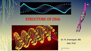
9. James Watson and Francis Crick – (1953)
Proposed - DNA as double helical structure
The informational content of DNA resides in the sequence in which
the deoxyribonucleotides are ordered / arranged.
Salient Features
1.DNA is a right handed double helix – 2 polynucleotide chain
twisted around each other on common axis
Watson-Crick Model of DNA Structure
DR.N.SIVARANJANI
10. 2. Two strands are Antiparallel - one strand runs in 5'-3' direction,
other in 3'-5' direction
3. Width (or diameter) of a double helix is 20 Å (2 nm)
4. Each turn of helix is 34 Å (3.4nm) with 10 pairs of nucleotides,
each pair placed at a distance of about 3.4 Å
5. DNA helix - Deoxyribose-PO4 – backbone – Hydrophilic,
Nitrogenous bases – inside – Hydrophobic
6. Two polynucleotide chains are not identical but complementary to
each other due to base pairing.
DR.N.SIVARANJA
NI
11. 7. The two strands are held together by Hydrogen bonds -
A = T , G = C
The hydrogen bonds are formed between a purine & pyrimidine
8. Major groove – wide PDE backbone
Minor groove – Narrow PDE backbone
9. Complementary bp – Chargaff’s rule
Adenine = Thymine
Guanine = Cytosine
10. Genetic information resides on one of the two strands - Template
strand or antisense strand or non coding
opposite strand -Sense strand / Non template / coding strand
Proteins interact with
the exposed bases
DR.N.SIVARANJANI
18. • DNA exists in 6 forms - A,B,C,D,E and Z form.
• B-form is most predominant form under physiological conditions.
• A-form – Right handed helix , 11 bp per turn, tilting of bp by 20Å
away from the central axis.
• Z-form – Left handed helix, 12 bp per turn, move in ZIG-ZAG
Conformations of DNA double helix
DR.N.SIVARANJANI
22. Unusual Structures of DNA
•Bent DNA
• Adenine base containing DNA tracts – produce bend
• Six adenosines in a row produce a bend of about 18⁰.
• Important in the binding of some proteins to DNA.
• Certain antitumor drugs (eg-cisplastin) produce bent
structure in DNA.
DR.N.SIVARANJANI
23. Triple standard of DNA
due to additional hydrogen bonds between the bases
Thymine forms two Hoogsteen hydrogen bonds to the
adenine of A-T pair to form T-A-T.
Cytosine forms two hydrogen bonds with guanine of
G-C pairs that results in C-G-C.
Triple helical structure is less stable than double
helix - increased electrostatic repulsion.
24. Four-stranded DNA
• High content of Guanine – form tetrameric
structure called G-quartets.
• These structures are planar & are connected
by Hoogsteen hydrogen bonds.
• Antiparallel four stranded DNA structures -
G-tetraplexes.
• Eukaryotic chromosomes - Telomeres are
rich in guanine - forms G-tetraplexes.
DR.N.SIVARANJANI
26. Denaturation of DNA
• ds DNA are held together by hydrogen bonds
• Disruption of hydrogen bonds (by change in pH or increase in temperature)
results in separation of strands
• The phenomenon of loss of helical structure of DNA is known as
denaturation
• Phosphodiester bonds are not broken by denaturation.
• It is measured by absorbance at 260nm.
• ss DNA has a higher relative absorbance than ds DNA
(Hyperchromatic effect)
27. Melting Temperature (Tm)
• It is defined as the temperature at which half of the helical structure of
DNA is lost.
• G-C base pairs are more stable than A-T bp.
• Tm is greater for DNAs with high content of GC.
• Formamide destabilizes hydrogen bonds of base pairs - used in rDNA
technology.
Renaturation (reannealing):
• It is a process in which the separated complementary DNA strands can
form a double helix.
• Renaturation is highly essential in the process of Replication.
29. Organization of DNA in cell
• Prokaryotic DNA:
• The DNA is organized as a single chromosome in the form of
double stranded circle.
• Packed in the form of nucleoids.
• Eukaryotic DNA:
• DNA is associated with various proteins - chromatin which then
organized into compact structures - chromosomes.
DR.N.SIVARANJANI
32. RNA
• Single stranded Polymer of ribonucleotides held together by 3’5’
phosphodiester bonds.
• Chemically less stable than DNA.
• Presence of 2’-OH makes RNA more susceptible to hydrolytic
attack (especially form Alkali)
• Prone to degradation by Ribonucleases (Rnases)
33. • RNA base composition:
• A + G ≠ U + C
Chargaff’s rule does not apply (RNA usually
prevails as single strand)
• All types of RNA are generated by nuclear processing of a
precursor molecule – Post transcriptional modification.
34. Major types of
RNA
Composition Functions
Ribosomal RNA
(rRNA)
(very abundant)
50 - 80 %
Integral part of ribosomes & act
as a machinery for synthesis of
proteins.
Transfer RNA
(tRNA)
10 - 20 % Carries activated amino acids to
ribosomes.
Messenger RNA
(mRNA)
5 – 10 % Encodes sequences of amino acids
in proteins.
35. mRNA
• The template strand of DNA is transcribed into a single stranded
mRNA by RNA polymerase enzyme.
• It carries the message to be translated to a protein
• Pre-m RNA or hnRNA on processing liberates functional mRNA
which enter cytoplasm & take part in protein synthesis.
• Shorter lifespan - quickly broken down after translation
36. 5’ Cap –
• mRNA is capped by 7 methyl GTP at 5’ terminal end attached
"backward" through a triphosphate linkage
• stabilizes the mRNA, prevent the attack of 5’ exonuclease.
• helps in recognition of mRNA for protein synthesis.
Coding region (introns) - which is translated to proteins
• Initiating codon – AUG
• Contains specific codon for different amino acids
• Terminating codon – UGA , UAA, UAG.
mRNA contains nucleotide sequence that is converted to a.a
sequence of polypeptide chain in the process of translation.
37. 3’ Poly A tail :
- Polymer of adenylate residues (20-250 nucleotides)
– maintains intracellular stability by preventing attach of 3’
exonuclease.
-Can be used to separate mRNA from other species of RNA.
AUGUUUUACGCAUGCUAG
38. tRNA
• They transfer amino acids from cytoplasm to the ribosomal
protein synthesizing machinery
• Soluble RNA molecule Varying in length from 74 – 95 nucleotides.
• At least 20 species of tRNA in every cell corresponding to each
20 a.a required for protein synthesis.
• Structure resembles clover leaf model- Robert Holley .
39. Unusual bases seen in tRNA –
Thymine,
Pseudouridine,
Dihydrouracil,
Hypoxanthine,
Methyl adenine,
Dimethyl Guanine.
tRNA serves as an "adaptor"
molecule that carries specific
amino acid to the site of protein
synthesis
40. • Acceptor arm – carriers amino acids
has 7 base pair, capped with a sequence CCA (5’-3’)
3’ OH forms ester bond with COOH of a.a
• DHU arm – dihydrouridine
3-4 base pair
serve as recognition site for enzyme which adds a.a
• Pseudouridine arm (TψC) – 5 base pair
involve in binding of tRNA to ribosome
41. • Anti codon arm – 5 base pair
recognizes the triplet nucleotide codon present in mRNA
contains anticodon that base pair with codon of mRNA.
(contains base sequences complementary to that of mRNA
codon)
responsible for the specificity of tRNA.
For ex: mRNA contains AUG UUU UAC
anticodon of tRNA UAC AAA AUG
tRNA accepts the specific a.a coded by that codon of mRNA
42. Variable arm – tRNA
divide into
class I – 75% , 3-5 bp
class II – 13-20 bp
The nucleotides of codon
has no affinity for a.a so
tRNA act as adapters
(mediates b/w mRNA & a.a)
44. rRNA
• Nucleolus - rRNA is synthesized and assembled with proteins to
form ribosome subunits.
• Ribosomes provide necessary infrastructure for the mRNA, tRNA
and amino acids to interact with each other for the translation.
• Acts as a machinery for the synthesis of proteins.
4 different rRNA – 18 S, 5.8 S, 28 S & 5 S.
• They are distributed in both 40S and 60S ribosomal subunits.
46. DR.N.SIVARANJANI
Types of RNA Functions
Heterogeneous nuclear RNA
(hnRNA)
Serves as a precursor for mRNA
Small nuclear RNA (snRNA) Involved in mRNA splicing
Small nucleolar RNA (snoRNA) Involved in rRNA processing
Small cytoplasmic RNA (scRNA) Involved in selection of proteins for export
Transfer messenger RNA
(tmRNA)
Mostly present in bacteria.
Promotes degradation of incorrectly
synthesized proteins.
Micro-RNAs (miRNAs) and Small
Interfering RNAs (siRNAs)
Inhibition of gene expression by decreasing
specific protein production
47. Ribozymes
Enzymes made up of RNA are called ribozymes
Ribozymes or RNA enzymes are catalytic RNA molecules with
sequence specific cleavage activity
Ex: Spliceosomes contain ribozymes as well as protein components
which serve to stabilize the structure of ribozymes.
RNAse-P is another ribozyme, which generates the ends of
tRNAs.
Peptidyl transferase present in ribosomes - used for protein
synthesis.
48. DNA RNA
Site Nucleus Cytoplasm
Strand Double Single
Base pair Millions of bp 100-5000 bp
Sugar Deoxy ribose Ribose
Base A, G, C, Thymine A, G, C, Uracil
Purine / pyrimidine
content
A = T , G = C .
Obeys Chargaff’s rule.
A ≠ U , G ≠ C
Types A ,B ,C ,D, E & Z m RNA, t RNA, r RNA.
Alkali hydrolysis Stable Susceptible
Importance Carriers genetic information
(Replication , Transcription)
Protein synthesis
(Translation)
Notas del editor
non-Watson-Crick pairing is called Hoogsteen pairing, after Karst Hoogsteen. 2pu,1py or 1pu 2py
Ss dna flexible. Dec in viscosity.
Cooled 5-20degree melting point
Modified bases at internal structure- 6 methyladenylates
Greek alphabet -psi
Codon and anticodon complementary to each other.
Eu Mitochondrial are smaller than cytoplasmic ribosomes.
antibiotics will inhibit bacterial protein synthesis, but will do no harm to human cells