SlideShare una empresa de Scribd logo
1 de 9
Descargar para leer sin conexión
IMMUNOLOGY                   ORIGINAL ARTICLE


Expression and regulation of interleukin-33 in human monocytes


Christopher J. Nile, Emma Barksby,              Summary
Paiboon Jitprasertwong, Philip M.
                                                Interleukin-33 (IL-33) is an IL-1 family cytokine that has a role in regu-
Preshaw and John J. Taylor
                                                lating T helper type 2 cytokines and mast cell development. Expression of
Periodontal Immunobiology Research Group,
                                                IL-33 is also associated with chronic inflammatory conditions such as
Institute of Cellular Medicine, Newcastle
University, Newcastle upon Tyne, UK             rheumatoid arthritis. However, there is little information regarding IL-33
                                                in myeloid cell immune responses, which are important in immunity and
                                                inflammation. We therefore investigated the expression, intracellular loca-
                                                tion and regulation of myeloid cell IL-33 by lipopolysaccharide (LPS)
                                                from Escherichia coli and the periodontal pathogen Porphyromonas gingi-
                                                valis. We detected IL-33 messenger RNA in the human promonocytic cell
                                                line THP-1, in monocytes derived from these cells and in primary human
                                                monocytes. However, IL-33 was not expressed in primary monocyte-
                                                derived dendritic cells. Stimulation of monocytes with E. coli LPS (Toll-
                                                like receptor 4 agonist) and LPS from P. gingivalis (Toll-like receptor 2
                                                agonist) up-regulated IL-33 at both the messenger RNA and protein levels
                                                but IL-1b and tumour necrosis factor-a had no effect. The IL-33 protein
                                                was mainly found in the cytoplasm of monocytes with no evidence of
                                                nuclear translocation in stimulated cells. Furthermore, no IL-33 secretion
                                                was detected after stimulation with LPS and/or ATP. These data indicate
                                                that the function, if any, of IL-33 in activated monocytes is primarily
                                                intracellular. Interestingly, immunofluorescence analysis indicated that
doi:10.1111/j.1365-2567.2009.03221.x            IL-33 was sequestered in the nucleus of monocytes undergoing apoptosis
Received 6 March 2009; revised 30 October       but released into the extracellular milieu by LPS-stimulated cells in which
2009; accepted 18 November 2009.
                                                necrosis had been induced by freeze–thawing. Therefore, this endorses the
Correspondence: Dr J. J. Taylor, Periodontal
Immunobiology Research Group, Institute of      view that IL-33 may function as an ‘alarmin’ and have a role in signalling
Cellular Medicine, School of Dental Sciences,   cellular damage and inflammatory disease pathogenesis through release
Newcastle University, Framlington Place,        from damaged or necrotic cells.
Newcastle upon Tyne, NE2 4BW, UK.
Email: j.j.taylor@ncl.ac.uk                     Keywords: cytokines; interleukin-1 family; monocytes; myeloid immune
Senior author: John J. Taylor                   cells


                                                                    weight of between 20 000 and 22 000 by both caspase-12,3
Introduction
                                                                    and caspase-3.3 The receptor for IL-33 has recently been
The interleukin-1 (IL-1) family of cytokines are important          identified and has a structure analogous to other IL-1
mediators of destructive inflammatory disorders such as              cytokine receptors: hence IL-33 binds to a heterodimer
rheumatoid arthritis and periodontitis and are important            comprising the IL-1 accessory protein IL-1RAcP and the
therapeutic targets, for example through the use of neu-            Toll-interleukin-1 receptor superfamily member ST2.2,4,5
tralizing monoclonal antibodies.1 The most recently dis-              Studies of complementary DNA (cDNA) libraries indi-
covered member of the IL-1 family is IL-33 (IL-F11).2               cated constitutive expression of IL-33 in bronchial and
Interleukin-33 exhibits structural similarity to IL-18 and          arterial smooth muscle cells and epithelial cells from the
is synthesized as a 30 000 molecular weight precursor               bronchus and small airways.2 Activation of primary lung
protein that lacks a signal peptide. The IL-33 precursor            or dermal fibroblasts and keratinocytes by IL-1b and
has been found to be cleaved to a form with a molecular             tumour necrosis factor-a (TNF-a) resulted in expression


172                                                                     Ó 2010 Blackwell Publishing Ltd, Immunology, 130, 172–180
IL-33 in monocytes

of IL-33.2 The IL-33 is also expressed in the central ner-    secreted from phorbol 12-myristate 13-acetate-stimulated
vous system and in particular the astrocytes of central       rat cardiac fibroblasts18 and also from adenosine triphos-
nervous system glia.2,6 Significantly, IL-33 is expressed in   phate (ATP) -stimulated mixed glial cell cultures and
high endothelial venules associated with human tonsil,        astrocyte-enriched cultures6 there are no reports of mea-
Crohn’s disease intestine and rheumatoid arthritis            surement of IL-33 secretion from other cell types and in
synovium.7 and may be involved in endothelial cell acti-      particular immune cells relevant to chronic inflammation.
vation, for example during angiogenesis.8 Other recent           Perhaps significantly, IL-33 has been reported to be
reports indicate that IL-33 is expressed at the site of       identical to nuclear factor of high endothelial venules
immune-mediated pathologies; hence IL-33 was found to         (NF-HEV), which is a peptide that is associated with
be expressed in synovial fibroblasts isolated from             chromatin and has transcriptional repressor activity
patients with rheumatoid arthritis where it is also           in vitro.7,19 These authors propose that IL-33 may be a
up-regulated by IL-1b and TNF-a9 and IL-33 was                ‘dual function’ cytokine and, like IL-1a and HMGB1,
increased in the brain tissues of mice infected with          may therefore have an intracellular function (for example
Theiler’s murine encephalomyelitis virus.6 Significantly,      in high endothelial venules, where it is highly expressed)
Moussion et al.10 reported that IL-33 is constitutively       as well as mediating pro-inflammatory responses as an
expressed in endothelial cells from both small and large      extracellular cytokine.7 Indeed, recent published evidence
blood vessels, in the fibroblastic reticular cells of lym-     demonstrates that biologically active IL-33 is released
phoid tissues as well as in a number of epithelial cells      from damaged endothelial cells suggesting that this cyto-
including epidermal keratinocytes; this further endorses      kine may function as an ‘alarmin’, providing an endo-
the potential role of IL-33 in immune–inflammatory             genous signal activating innate immunity during tissue
reactions. In their screen of a cDNA library, Schmitz         damage and infection.3
et al.2 did report modest levels of IL-33 expression in          In this study we report that intracellular IL-33 expres-
lipopolysaccharide (LPS) -activated human monocytes           sion is up-regulated by THP-1 monocytes and primary
and dendritic cells (of unknown phenotype); there is no       monocytes but not by dendritic cells in response to stimu-
other detailed information on the expression and regula-      lation with LPS from Escherichia coli and LPS from the
tion of IL-33 in myeloid immune cells. There is strong        periodontal pathogen, Porphyromonas gingivalis. However,
evidence for a role of IL-33 in regulating T helper type      IL-1b or TNF-a had no effect on IL-33 messenger RNA
2 (Th2) cytokines and in stimulating mast cell develop-       (mRNA) expression in monocytes. Our data suggest that
ment and associated pathologies.2,11–16 Hence, ST2 is         in monocytes, IL-33 is primarily an intracellular protein:
strongly expressed on Th2 cells and IL-33 stimulates the      we have not observed secretion of IL-33 by viable cells
production of IL-5 and IL-13 in these cells in vitro.2        stimulated with LPS, either alone or in combination with
When IL-33 is administered to mice, increased levels of       ATP. We present preliminary evidence to suggest that
immunoglobulin E (IgE) and the Th2 cytokines (IL-4,           IL-33 is sequestered in the nucleus during apoptosis and
IL-5 and IL-13) ensue; this is associated with eosino-        demonstrate IL-33 release by cells undergoing necrosis.
philia, splenomegaly and pathological changes in arteries,    Therefore, IL-33 may function as an ‘alarmin’ and have a
lungs and intestine of the mice; consistent with a            role in signalling cellular damage and inflammatory disease
Th2-driven pathology.2 Furthermore, IL-33 has also been       pathogenesis through release from damaged or necrotic cells.
found to be a chemoattractant for human Th2 cells.14
   Mast cells are activated by IgE and IgG and play a key
                                                              Materials and methods
role in mediating Th2 pathologies. Significantly, mast cells
express ST2 and IL-1RAcpP5 and there is evidence now
                                                              Cell culture
from several studies that IL-33 can also drive mast cell
maturation, survival, adhesion and cytokine produc-           The THP-1 promonocytic cell line was obtained from the
tion.5,11–13,15 Furthermore, a model of IL-33 involvement     European Collection of Cell Cultures (Salisbury, UK).
in the pathogenesis of collagen-induced arthritis recently    The cells were cultured in RPMI-1640 medium (Sigma,
proposed by Xu et al.9 suggests that pro-inflammatory          Poole, UK), supplemented with 10% (volume/volume)
cytokines derived from IL-33-activated mast cells have a      heat-inactivated fetal bovine serum (Sigma), 0Á1 mM
pivotal role in this chronic inflammatory pathology.           L-glutamine, 100 U/ml penicillin and 100 U/ml strepto-
Although studies of soluble (s)ST2 (reviewed by Arend         mycin. Cultures were maintained in a humidified atmo-
et al.17), in experiments with ST2 knockout mice (e.g. Xu     sphere with 5% CO2 at 37°. THP-1 promonocytes were
et al.9) as well as studies with recombinant IL-332,9 sup-    converted to monocytes by incubation with 100 nM
port a role for IL-33 in inflammatory pathologies, there is    1a,25-dihydroxy-vitamin D3 (Merck Chemicals, Notting-
very limited information concerning the identification of      ham, UK) for 48 hr. Cell differentiation was confirmed
the cellular source of IL-33 and measurement of IL-33         by adherent capabilities, visual morphology and CD14
secretion. As a result, although IL-33 was reported to be     expression.20


Ó 2010 Blackwell Publishing Ltd, Immunology, 130, 172–180                                                             173
C. J. Nile et al.

   Primary monocytes were obtained from buffy coat frac-     cDNA, 12Á5 ll 2· Sensimix (5 mM final MgCl2) (Quan-
tions of human blood (National Blood Service, Newcastle,     tace, Watford, UK), 1Á25 ll TaqMan Primer (900 nM)/
UK). The peripheral blood mononuclear cell (PBMC)            Probe (250 nM) and 8Á75 ll H2O. Each sample was
population was extracted from the buffy coat using Hist-     assayed in triplicate over 40 cycles and the reactions were
opaque-1077 (Sigma). In some experiments, PBMCs were         conducted in a 96-well plate format using the ABI 7900
cultured in complete RPMI-1640 medium for 24 hr at           instrument (Applied Biosystems). RNA polymerase II
37° with 5% CO2 and after 24 hr the non-adherent cell        gene expression was used as a control for cDNA input.
population was removed by repeated washes in RPMI-           The data were analysed using SDS 2.2 software (Applied
1640 to reveal adherent primary monocytes. Alternatively,    Biosystems) and normalized against RNA Polymerase II
primary (CD14+) monocytes were isolated by positive          expression; levels of specific mRNA in stimulated cells
magnetic selection with anti-CD14 conjugated magnetic        were presented as relative expression compared with con-
beads using a commercially available kit (EasySep, Stem-     trol cultures using the DDCt method.21
Cell Technologies, Grenoble, France). Primary monocytes
were converted to dendritic cells by incubating in supple-
                                                             Cytokine ELISA
mented RPMI-1640 medium containing granulocyte–
macrophage colony-stimulating factor (10 ng/ml) and          Cytokines in cell culture supernatants were quantified
IL-4 (50 ng/ml) for 7 days, changing the medium every        using specific ELISA kits for IL-33 (Axxorra, Nottingham,
other day. Differentiation was confirmed by observing         UK) and TNF-a (R&D Systems); the sensitivities for these
morphological changes and analysis of expression of          ELISAs were 3Á56 and 1Á27 pg/ml pg/ml, respectively.
CD1a, CD11C, CD14, HLA-DR and CD83 by fluorescence-           Interleukin-18 was measured by sandwich ELISA using
acitvated cell sorting (data not shown).                     commercially available antibodies and recombinant IL-18
                                                             (R&D Systems); the sensitivity of the IL-18 ELISA was
                                                             5Á19 pg/ml.
Cell stimulation experiments
THP-1 monocytes, primary monocytes and primary
                                                             Apoptosis and necrosis
monocyte-derived dendritic cells (MDDCs) were cultured
at a density of 106 cells/ml. Cells were stimulated with     Necrosis was induced by subjecting cells to five cycles of
100 ng/ml E. coli LPS (from strain 0111:B4; Invivogen,       freezing to ) 70° and thawing at 38°.22 Cell viability in
Calne, UK), 100 ng/ml P. gingivalis (from strain W50, a      necrotic cell preparations was analysed by Trypan blue
gift from M. Rangarajan, Queen Mary’s School of Medi-        exclusion and was consistently < 5% (data not shown).
cine and Dentistry, London, UK), 100 ng/ml TNF-a             Apoptosis was induced by exposing monocytes to a 62 mJ/
(R&D Systems, Abingdon, UK), 100 pg/ml IL-1b (R&D            cm2 dose of UVB irradiation using a bank of four Philips
Systems) or ATP (Sigma) for between 0Á5 and 48 hr. An        TL 20W/12 RS lamps (Philips, Guilford, UK). The extent
unstimulated control for each time-point was also            of apoptosis was analysed by fluorescence microscopy with
included. After incubation the culture supernatant was       40 -60 ,diamidino-2-phenylindole (DAPI) staining of nuclei:
removed for analysis by enzyme-linked immunosorbent          nuclei in these preparations exhibited condensation char-
assay (ELISA) and RNA was isolated from cells using the      acteristic of nuclear fragmentation and apoptosis (Fig. 4b).
RNeasy mini kit (Qiagen, Crawley, UK).
                                                             Immunocytochemistry
Reverse transcription and quantitative real-time
                                                             For immunocytochemical analysis, cells were cultured on
polymerase chain reaction
                                                             glass cover-slips and then fixed in ice-cold methanol. The
Reverse transcription (RT) was performed using the ABI       cover-slips were washed with phosphate-buffered saline
High Capacity cDNA Reverse Transcription Kit (Applied        (PBS) before permeabilizing cells using Triton X-100
Biosystems, Warrington, UK). The RT reactions were car-      (Sigma). The cells were then probed with 5 lg/ml of a
ried out in an Applied Biosystems GeneAmp PCR System         mouse monoclonal antibody to IL-33 (Nessy-1, Axxora,
9700 (Applied Biosystems), under the following reaction      Nottingham, UK) for 60 min at room temperature. After
conditions; 25° for 10 min, 37° for 2 hr and 85° for         repeat washes with PBS, the cells were then probed with
5 seconds. Levels of cDNA transcript were determined         5 lg/ml secondary polyclonal antibody to mouse IgG1
using real-time polymerase chain reaction (PCR) with         conjugated to fluorescein isothiocyanate (FITC; Axxora)
pre-designed primers and Taqman probes (Assays on            or tetramethyl rhodamine iso-thiocyanate (TRITC; Sigma)
Demand; Applied Biosystems). The sequences for these         for 40 min. The cells were again washed with PBS and
probes were as follows: IL-33: Hs00369211_m1; ST2L:          their nuclei were counterstained by exposure to DAPI
Hs00545033_m1; HMGB1: Hs01923466_g1; RNA poly-               (Sigma) at a concentration of 10 lg/ml for 15 min. The
merase II: Hs00172187_m1. Each PCR consisted of 2Á5 ll       cover-slips were washed in PBS before finally being


174                                                              Ó 2010 Blackwell Publishing Ltd, Immunology, 130, 172–180
IL-33 in monocytes

mounted on microscope slides with Dako fluorescent              (a) 200                                  Unstimulated   E. coli LPS         P. gingivalis LPS
mounting medium (DAKO, Ely, UK). The cells were exam-                                   175
                                                                                                                                       *


                                                                IL-33 mRNA expression
ined using a Leica TCS SP UV confocal laser scanning                                    150
                                                                                                                       *
microscope (Leica, Wetzlar, Germany). A series of Z-stack                               125
images were taken and each image was analysed individu-                                 100                                *
                                                                                                                                           *
ally using Leica LCS LITE software (Leica) and findings were                             75
verified by an independent expert observer. Specificity of                                50
the IL-33 monoclonal antibody was confirmed by complete                                  25
abrogation of fluorescent labelling after pre-incubation                                  0
with a fivefold concentration of blocking peptide                                              0·5          3           6            9                  24        48
                                                                                                                           Time (hr)
(IL-33112–270) (Axxora) (data not shown).
                                                               (b) 50
                                                                                                        Unstimulated   E. coli LPS       P. gingivalis LPS
                                                                                        45


                                                                IL-33 mRNA expression
Statistical analysis                                                                    40

Statistical analysis was performed using Mann–Whitney
                                                                                        35                                           *
                                                                                        30
tests (cytokine expression analysis) and Student’s t-test                               25                                     *
(real-time RT-PCR).21 A P-value of < 0Á05 was consid-                                   20
ered to be statistically significant.                                                    15
                                                                                        10
                                                                                         5
Results                                                                                  0
                                                                                                    3                         9                             24
                                                                                                                           Time (hr)
IL-33 expression by myeloid cells
                                                               (c) 50
Data from analysis of a cDNA library indicated that mod-                                45                  Unstimulated       TNF-a       IL-1b
                                                                IL-33 mRNA expression


est levels of IL-33 mRNA are expressed by human mono-                                   40
                                                                                        35
cytes and dendritic cells.2 However, to date, the expression
                                                                                        30
of IL-33 by myeloid cells has not been fully characterized.                             25
We therefore investigated the expression of IL-33 by                                    20
monocytes in response to bacterial LPS and the cytokines                                15
IL-1b and TNF-a using quantitative real-time PCR                                        10
                                                                                         5
(Fig. 1). We found that IL-33 mRNA was significantly up-
                                                                                         0
regulated in LPS-stimulated THP-1 monocytes (Fig. 1a)                                         0·5              1              3                    6             24
and primary monocytes isolated from three separate                                                                         Time (hr)
donors (Fig. 1b), in comparison to unstimulated controls,
                                                               Figure 1. Interleukin-33 (IL-33) messenger RNA (mRNA) expression
with maximal transcript levels observed after 6–9 hr of
                                                               is up-regulated by monocytes in response to lipopolysaccharide
incubation with LPS. Conversely, real-time PCR per-            (LPS) but not tumour necrosis factor-a (TNF-a) and IL-1b. Quanti-
formed on cDNA isolated from MDDCs derived from pri-           tative real-time polymerase chain reaction analysis was used to deter-
mary macrophages of three separate donors showed a lack        mine IL-33 expression by THP-1 monocytes (a) and primary human
of IL-33 expression (data not shown). In contrast to IL-33,    monocytes (b) in response to stimulation with 100 ng/ml Escherichia
the ST2 and HMGB1 genes exhibited no up-regulation in          coli or Porphyromonas gingivalis LPS. IL-33 mRNA expression by
expression levels in myeloid cells upon stimulation with       THP-1 monocytes stimulated with 100 ng/ml TNF-a and 100 pg/ml
bacterial LPS (data not shown). The up-regulation of           IL-1b was also measured (c). Levels of mRNA were normalized
IL-33 in monocytes by LPS may be the result of transcrip-      against RNA polymerase II mRNA levels and fold changes (‘IL-33
tional up-regulation via Toll-like receptor (TLR) signalling   expression’) were calculated relative to unstimulated cells at each
                                                               individual time-point using the DDCt method.20 The data represent
or may be an indirect, secondary effect of LPS-stimulated
                                                               the mean values (± SD) from three independent experiments. Signif-
TNF-a or IL-1b secretion. To address this issue, THP-1
                                                               icant differences we determined using Student’s t-test on the DCt
monocytes were stimulated with TNF-a and IL-1b and             values.20 *P < 0Á05 relative to unstimulated cells.
IL-33 mRNA was quantified by real-time RT-PCR; the
results do not provide any evidence to suggest that these
cytokines up-regulate IL-33 (Fig. 1c).                         failed to detect any IL-33 secretion from viable cells either
                                                               at 9 hr (Fig. 2) or during the full time–course of our
                                                               experiments (0–48 hr) (data not shown). There is a
IL-33 is not secreted from viable cells
                                                               suggestion that pro-IL-33 is processed by the action of
The LPS-stimulated IL-33 mRNA levels were maximal              caspase-13,4,6 and ATP has been shown to enhance
after 6–9 hr of monocyte culture but we consistently           the activity of caspase-1, enhancing the processing and


Ó 2010 Blackwell Publishing Ltd, Immunology, 130, 172–180                                                                                                             175
C. J. Nile et al.

                70                   Unstimulated   E. coli LPS     P. gingivalis LPS                natants of LPS-stimulated cells incubated with ATP for
                60                                                                                   9–24 hr, when IL-33 mRNA expression was maximal (not
                50
                                                                                                     shown). Previous reports have indicated that IL-33 is pri-
                                                                                                     marily an intracellular cytokine7 and HMGB1 has been
IL-33 (pg/ml)


                40
                                                                                                     shown to be released by cells undergoing necrosis22 so we
                30                                                                                   therefore induced necrosis in LPS-stimulated cells and
                20                                                                                   investigated IL-33 release (Fig. 2). There was no IL-33
                10                                                                                   released by necrotic monocytes that had not been stimu-
                 0   ND ND ND   ND            ND             ND            ND           ND ND ND     lated by LPS, suggesting that this protein was not consti-
                     Live control     3             6               9           24         48        tutively synthesized by these cells or at least was below
                                                        Time (hr)
                                                                                                     the level of detection of the ELISA (Fig. 2). However,
Figure 2. Interleukin-33 (IL-33) is not secreted by viable THP-1                                     IL-33 was released into the culture medium by necrotic
monocytes but is released from necrotic cells. THP-1 monocytes                                       cells stimulated by LPS and the levels of IL-33 protein
were either stimulated with 100 ng/ml Escherichia coli or Porphyro-                                  detected reflected the kinetics of IL-33 mRNA stimulation
monas gingivalis lipopolysaccharide (LPS) for 9 hr (live control), or                                by LPS (Fig. 2a) with maximal levels of IL-33 protein
stimulated with 100 ng/ml E. coli or P. gingivalis LPS for 3–48 hr fol-                              being released by monocytes stimulated with LPS for 9 hr
lowed by induction of necrosis by five rounds of freezing () 70°)                                     (Fig. 2). Stimulation of the cells in these experiments was
and thawing (38°). Stimulation of the cells in these experiments was                                 confirmed by detection of TNF-a in the isolated super-
confirmed by detection of tumour necrosis factor-a in the isolated                                    natants using ELISA (data not shown). Immunocyto-
supernatants using enzyme-linked immunosorbent assay (not
                                                                                                     chemistry confirmed that IL-33 protein was barely
shown). ND = not detected. The data represent the mean values
                                                                                                     expressed in unstimulated THP-1 monocytes and was not
(± SD) from three independent experiments.
                                                                                                     expressed at all in unstimulated primary monocytes but
                                                                                                     there was a prominent expression of intracellular IL-33
Table 1. Effect of adenosine triphosphate (ATP) on interleukin-33
                                                                                                     protein by LPS-stimulated monocytes (Fig. 3a,b). Count-
(IL-33) and IL-18 secretion by THP-1 monocytes                                                       erstaining of cells with the nuclear stain DAPI indicated
                                                                                                     that IL-33 was predominantly located in the cytoplasmic
                                                               IL-33             IL-18               compartment in LPS-stimulated monocytes as confirmed
Treatment                                                      (pg/ml)           (pg/ml)             by the absence of co-localization of green and blue
                                                                                                     fluorescence (Fig. 3a,b).
Unstimulated                            0            27Á0 ±                                 10Á7
ATP (6 mm)                              0           110Á1 ±                                 52Á0
E. coli LPS (1 ng/ml)                   0           228Á2 ±                                 96Á5     IL-33 is sequestered in the nucleus of apoptotic
E. coli LPS (1 ng/ml) + ATP (6 mm)      0          1635Á5 ±                                 524Á5*   monocytes
E. coli LPS (1 ng/ml) + freeze–thawing 66Á37 ± 8Á1       –
                                                                                                     In cells induced to undergo programmed cell death
IL-33 secretion from THP-1 monocytes is not activated by ATP.                                        HMGB1 was found to remain closely associated with
THP-1 monocytes were stimulated with 1 ng/ml Escherichia coli lipo-                                  nuclear chromatin.24 We therefore investigated the effect
polysaccharide (LPS) for 3 hr in the presence of 6 mm ATP for the
                                                                                                     of apoptosis on IL-33 in monocytes. The LPS-stimulated
final 30 min of this incubation. Cells without additives (unstimu-
                                                                                                     monocytes induced to undergo apoptosis by UVB irradia-
lated) and with ATP or LPS alone served as controls. The IL-33 and
                                                                                                     tion did not release any IL-33 into the culture medium
IL-18 were measured by specific enzyme-linked immunosorbent
assay. The data represent the median values (± interquartile range)                                  over a 0–48-hr time–course (data not shown). Immuno-
from three independent experiments. IL-18 secretion was signifi-                                      cytochemistry was then used to determine the intracellu-
cantly increased by stimulation of the monocytes with E. coli LPS in                                 lar location of IL-33 in stimulated cells (Fig. 4). Nuclear
combination with ATP as compared with LPS alone; Mann–Whitney                                        morphology in control cell cultures confirms the presence
test, *P < 0Á05.                                                                                     of apoptotic cells (Fig. 4a) and staining for IL-33 using
                                                                                                     FITC-conjugated antibodies clearly shows that the intra-
                                                                                                     cellular IL-33 protein is tightly associated with the
secretion of IL-1b and IL-18.23 We therefore investigated                                            nucleus of cells undergoing apoptosis (Fig. 4b).
whether ATP stimulated IL-33 secretion, but we were
again unable to detect any extracellular IL-33 in mono-
                                                                                                     Discussion
cytes exposed to ATP either alone or in combination with
LPS (Table 1). Similar experiments confirmed ATP                                                      The IL-1 family of cytokines have an established role
enhancement of IL-18 secretion in LPS-stimulated cells                                               in immune regulation and inflammatory processes.1,17
over the same time–course, confirming that ATP-stimu-                                                 Inappropriate activity of IL-1 cytokines is a feature of a
lated caspase-1 was functional in this system (Table 1).                                             number of immune-mediated pathologies and therefore
Furthermore, IL-33 was not detected in the culture super-                                            pathways activated by these cytokines are rational therapeutic


176                                                                                                      Ó 2010 Blackwell Publishing Ltd, Immunology, 130, 172–180
IL-33 in monocytes

                                                        (a)         Control                     Ec LPS                       Pg LPS


                                                        IL-33


                                                                              47·62 µm                    47·62 µm                    47·62 µm


Figure 3. Interleukin-33 (IL-33) protein is
expressed in the cytoplasm of lipopolysaccha-
ride (LPS) -stimulated THP-1 monocytes (a)              IL-33 +
and primary monocytes (b). Immunocyto-                  DAPI
chemistry was performed on monocytes (con-
trol) and cells stimulated with 100 ng/ml
                                                                              47·62 µm                     47·62 µm                    47·62 µm
Escherichia coli (Ec) or Porphyromonas gingiva-
lis (Pg) LPS for 9 hr. The IL-33 was located
                                                        (b)         Control                     Ec LPS                       Pg LPS
using an IL-33 antibody and secondary anti-
body conjugated to fluorescein isothiocyanate
(FITC; green) or tetramethyl rhodamine iso-
thiocyanate (TRITC; red) and the nuclei were
stained with 40 -60 ,diamidino-2-phenylindole           IL-33
(DAPI; blue). A series of Z-stack images were
taken to confirm the cellular location of IL-33
and the image depicted is representative of                                   47·62 µm                    47·62 µm                    47·62 µm
findings throughout the Z stack. The upper
panels (a) show IL-33 expression alone (green
FITC) and the lower panels (b) show IL-33
(red TRITC) and nuclei (blue DAPI) in com-              IL-33 +
bination. The results are representative of three       DAPI

independent stimulation experiments and have
been verified by an independent observer. Scale
                                                                              47·62 µm                    47·62 µm                     47·62 µm
as depicted on images.


                            (a)               Control                Ec LPS                      Pg LPS


                            IL-33


                                                         12·30 µm                 8·7 µm                     11·50 µm
                            (b)


                            IL-33 +
                            DAPI


                                                         12·30 µm                 8·7 µm                     11·50 µm


Figure 4. Interleukin-33 (IL-33) protein is closely associated with the nucleus in THP-1 monocytes undergoing apoptosis. Immunocytochemistry
was performed on unstimulated THP-1 monocytes (control) and on cells stimulated with 100 ng/ml Escherichia coli (Ec) or Porphyromonas
gingivalis (Pg) lipopolysaccharide (LPS) for 9 hr followed by exposure to UVB-irradiation to induce apoptosis. The IL-33 was located using an
IL-33 antibody and secondary antibody conjugated to fluorescein isothiocyanate (FITC; green). The nuclei were stained with 40 -60 ,diamidino-
2-phenylindole (DAPI; blue). The upper panels (a) show IL-33 expression alone (green FITC) and the lower panels (b) show IL-33 (green FITC)
and nuclei (blue DAPI) in combination. Arrows indicate examples of apoptotic cells. The results are representative of three independent stimu-
lation experiments and have been verified by an independent observer. Scale as depicted on the images.


Ó 2010 Blackwell Publishing Ltd, Immunology, 130, 172–180                                                                                   177
C. J. Nile et al.

targets.1 Interleukin-33 is a recently described member of    onstrate any effect of these cytokines on IL-33 expression.
the IL-1 family and has attracted attention because,          Previously, expression of IL-33 in human primary lung
uniquely among IL-1 cytokines, it stimulates the produc-      fibroblasts as well as in dermal fibroblasts and keratino-
tion of Th2 cytokines such as IL-5 and IL-13, activates       cytes was shown to be enhanced by a combination of
Th2 cells, and has a role in mast cell development and        TNF-a and IL-1b.2 Also, TNF-a either alone or in combi-
function.2,11–16 As a result, IL-33 has been implicated in    nation with IL-1b enhances IL-33 expression in human
the pathogenesis of immune-mediated disorders which           primary synovial fibroblasts.9,32 However, TNF-a and
feature Th2 responses and mast cell dysregulation, and in     IL-1b had no effect on IL-33 expression in rat cardiac
particular those which involve pulmonary and mucosal          fibroblasts or rat cardiomyocytes.18 The reasons for these
inflammation.2,16,25 Studies of the action of recombinant      inconsistencies are not clear, although the data may
IL-33 and the expression of ST2 (a subunit of the IL-33       reflect variable responsiveness of different cell types.
receptor) suggest that IL-33 has roles in immune                 The function, if any, of IL-33 in the myeloid lineage
responses and immunopathogenesis in addition to those         remains obscure but a key question is whether or not
mediated by mast cells and Th2 cells. Hence, eosinophils      IL-33 is secreted or remains in the intracellular compart-
and basophils express ST2 and are stimulated by exoge-        ment. In terms of myeloid cell IL-33, we consistently
nous IL-33.26–29 Interleukin-33 also enhanced cytokine        failed to detect IL-33 in culture supernatants even after
production by invariant natural killer (NK) T cells and       LPS stimulation. Although there is much indirect
human NK cells.29 Also, IL-33 and ST2 are widely              evidence for an immunological role of IL-33 in vitro and
expressed in vascular cells and tissues, in fibroblasts and    in vivo, only a limited number of reports have described
in the central nervous system.6,7,9,18                        the detection of soluble, endogenous IL-33.6,18 Hence,
   We have investigated the expression, intracellular loca-   although rat cardiac fibroblasts synthesized IL-33 protein,
tion and regulation of IL-33 in monocytes and MDDCs.          unstimulated cells did not secrete this protein but IL-33
In experiments using both conventional RT-PCR (not            secretion (as detected by Western blot of culture super-
shown) and quantitative RT-PCR, IL-33 was not                 natants) was induced by stimulation with phorbol
expressed in MDDCs and stimulation with LPS had no            12-myristate 13-acetate.18 Similarly, in a study of mixed
effect on IL-33 expression in MDDCs. This is in contrast      glial cell cultures and astrocyte-enriched cultures, IL-33
to Schmitz et al.2 who reported no expression in ‘resting’    was only found to be secreted in cells stimulated with
MDDC but IL-33 expression (albeit at very modest levels       PAMPs and ATP together.6 Recently, the release of IL-33
as compared with smooth muscle cells and fibroblasts)          from primary human endothelial cells in conditions of
was detected when these cells were activated by LPS. Sim-     mechanical stress or injury was demonstrated.3 The physio-
ilarly, whereas Schmitz et al.2 only detected IL-33 mRNA      logical relevance of these data remains unclear, but this
in LPS-activated monocytes, we detected IL-33 mRNA in         does suggest that under certain circumstances cells may
the human promonocytic cell line THP-1, in monocytes          secrete IL-33 in response to the appropriate signals.
derived from these cells and in primary human mono-              It is possible that IL-33 may be secreted in a form that
cytes by both conventional RT-PCR (not shown) and             is not detectable by ELISA. For example, the soluble
quantitative RT-PCR. However, analysis of protein             form of the IL-33 receptor subunit ST2, sST2 interacts
expression using immunocytochemistry demonstrated             with IL-33 and blocks IL-33 signalling18,25 and sST2 is
only very low levels of constitutive IL-33 expression in      induced by LPS and cytokines in monocytes.33 Although
THP-1 monocytes and none in primary cells. The differ-        we have not analysed sST2 in our cultures, soluble IL-33
ences between our findings and those of Schmitz et al.2        was readily detectable by ELISA after induction of necro-
with respect to mRNA expression may reflect technical          sis in monocyte cultures previously stimulated by LPS.
differences or differences in the sources of cDNA.               Interleukin-33 may require other signals, in addition to
   We investigated regulation of IL-33 expression in          LPS, to induce efficient extracellular release. For example,
monocytes: LPS from E. coli as well as LPS from the peri-     ATP is known to enhance both IL-1b and IL-18 release
odontal pathogen P. gingivalis both enhanced IL-33            via activation of a multiprotein complex (the inflamma-
expression. The LPS from E. coli and P. gingivalis repre-     some) containing caspase-1, the enzyme responsible for
sent different pathogen-associated molecular patterns         intracellular processing of the pro- form of these cyto-
(PAMPs): LPS from these species stimulate host cells via      kines.23 It is not yet clear whether IL-33 is processed
TLR4 and TLR2, respectively, which results in overlapping     in vivo via a caspase-1 pathway: although there is evidence
but distinct cytokine responses.30,31 Our data suggest that   that in-vitro-translated IL-33 is cleaved by incubation with
engagement of both TLR2 and TLR4 pathways stimulates          caspase-1,2 this was not confirmed by experiments analy-
IL-33 indicating a potential role for this cytokine in        sing intracellular IL-33.7 Furthermore, the predicted cleav-
immune responses to diverse pathogens and PAMPs.              age site for caspase-1-induced IL-33 is not conserved in
   We also investigated the regulation of IL-33 in mono-      orthologues from other species.7 Recently, it was demon-
cytes by both TNF-a and IL-1b but were unable to dem-         strated that IL-33 is probably cleaved by both caspase-1


178                                                               Ó 2010 Blackwell Publishing Ltd, Immunology, 130, 172–180
IL-33 in monocytes

and caspase-3 to yield a 20 000–22 000 molecular weight           cytoplasmic compartment in viable monocytes and we
form of IL-33 and that this cleavage took place at an             did not observe prominent nuclear translocation of IL-33.
amino acid residue (Asp178) distinct from that previously         Palmer et al.32 reported that IL-33 was expressed in both
proposed (Ser111).3 Significantly, unlike the full-length          the nucleus and cytoplasm of synovial fibroblasts and
IL-331–270, the 20 000–22 000 form was not biologically           ‘mononuclear inflammatory cells’. The sub-cellular locali-
active (in terms of binding and activation of the ST2             zation of IL-33 may reflect intrinsic differences between
receptor), suggesting that IL-33 was inactivated by caspase       cell types. Also, there are differences in IL-33 expression
cleavage.3 The antibodies used for the ELISA and                  in smooth muscle cells cultured in vitro and IL-33 expres-
immunocytochemistry in the present report do not distin-          sion in these cells in vivo.2,10 Significantly, the expression
guish between the different forms of IL-33 described by           pattern of IL-33 changes in different cell culture condi-
Cayrol et al.;3 it will therefore be interesting to investigate   tions.8 Therefore, data from in vitro culture models
in detail the molecular structures of IL-33 expressed in          should be interpreted with care and it will be important
monocytes, the effect of LPS on IL-33 maturation, and the         to extend studies of IL-33 expression in immune cells
specific forms of IL-33 released from necrotic monocytes.          in vitro to investigate the expression of this cytokine in
   Addition of ATP to mixed glial cell cultures and astro-        tissue sections from patients with inflammatory disease.
cyte-enriched cultures stimulated the release of IL-33               We also generated preliminary evidence to suggest that
from these cells whereas these reagents had no effect in          during apoptosis, IL-33 is associated with the nucleus in
isolation.6 Significantly, we failed to demonstrate any            monocytes. In this regard it is interesting to note that,
effect of ATP on IL-33 secretion from monocytes, either           apoptosis in endothelial cells stimulated IL-33 cleavage
alone or in combination with LPS, although enhancement            (into a form which does not bind the extracellular ST2
of IL-18 secretion was measured in the same experiments           receptor) suggesting that this reaction existed to inactivate
(Table 1). Intriguingly, it has been speculated that IL-33        IL-33 after apoptosis.3 In addition, we demonstrated
may be processed by an inflammasome distinct from that             release of IL-33 as the result of necrosis in the same cells.
which processes IL-1b and IL-18.34 There is evidence that         In similar experiments it was recently demonstrated that
IL-33 may function as a so-called dual function cytokine          necrosis in primary endothelial cells was associated with
having a role as an (extracellular) pro-inflammatory cyto-         the release of biologically active IL-33.3 The behaviour of
kine and an (intracellular) nuclear factor with transcrip-        IL-33 during apoptosis and necrosis is similar to the so-
tional regulatory action.7                                        called ‘alarmins’, which are a group of biologically active
   These authors draw analogy with other ‘dual function’          mediators that include the dual function cytokines
cytokines such as IL-1a and HMGB1.7 In support of this            HMGB1 and IL-1a.36 Alarmins are endogenous signalling
hypothesis they found that in high endothelial venules,           molecules that, like exogenous PAMPs, trigger similar
IL-33 was found to be primarily an intracellular protein          pathways and have a common purpose as damage-associ-
that closely associates with eukaryotic chromatin7 The            ated molecule patterns (DAMPS).36 These findings
nuclear localization was found to be mediated by an evo-          endorse the view that IL-33 has functions beyond that of
lutionarily conserved homeodomain-like helix-turn-helix           a conventional extracellular, immunoregulatory cytokine
(HTH) motif which is located within the N-terminal por-           and may be classified as a DAMP.34 The recent finding of
tion of the IL-33 precursor protein.7 More recent data            widespread expression of IL-33 in the vasculature, in the
provide a molecular model for the chromatin interactions          skin and mucosal epithelial cells supports this hypo-
of IL-33 and suggest that IL-33 binds to the histone              thesis.10 Interestingly, in addition to IL-1a and IL-33, an
dimer H2A-H2B and hence regulates chromatin compac-               isoform of IL-7 (IL-7b), also an IL-1 family cytokine, has
tion by influencing nucleosome–nucleosome interac-                 recently been shown to translocate to the nucleus in acti-
tions.35 Interleukin-33 is therefore shown to confer potent       vated monocytes and to down-regulate the synthesis of
transcriptional repressor activity although the target genes      pro-inflammatory cytokines.37
remain to be characterized.7                                         In summary, the function of IL-33 in activated mono-
   Significantly, it was demonstrated that endothelial cells       cytes is hypothesized to be primarily intracellular; and as
in inflamed tissues from patients with Crohn’s disease             such IL-33 may act as an ‘alarmin’ and have a role in sig-
and rheumatoid arthritis express high levels of IL-33 and         nalling cellular damage and inflammatory disease patho-
constitute the major source of IL-33 in these tissues.7 The       genesis through release from damaged or necrotic cells.
high expression of IL-33 in the endothelial cells of normal
and inflamed synovial tissue has recently been con-
                                                                  Acknowledgements
firmed.32 Xu et al.9 also confirmed the high expression of
IL-33 (and ST2) in the rheumatoid synovium and in                 This work was supported by a grant from the Newcastle
fibrobasts isolated from this tissue. Although we have             Healthcare charity and a UK Department of Health Clini-
demonstrated that IL-33 is not secreted from stimulated           cian Scientist Fellowship DHCS/03/G121/46 awarded to
monocytes, IL-33 is predominantly associated with the             Philip Preshaw.


Ó 2010 Blackwell Publishing Ltd, Immunology, 130, 172–180                                                                  179
C. J. Nile et al.

Disclosures                                                                                    17 Arend WP, Palmer G, Gabay C. IL-1, IL-18, and IL-33 families of cytokines. Immunol
                                                                                                  Rev 2008; 223:20–38.
                                                                                               18 Sanada S, Hakuno D, Higgins LJ, Schreiter ER, McKenzie AN, Lee RT. IL-33 and ST2
The authors declare no conflict of interest.
                                                                                                  comprise a critical biomechanically induced and cardioprotective signaling system. J
                                                                                                  Clin Invest 2007; 117:1538–49.
                                                                                               19 Baekkevold ES, Roussigne M, Yamanaka T et al. Molecular characterization of NF-HEV,
References                                                                                        a nuclear factor preferentially expressed in human high endothelial venules. Am J Pathol
 1 Barksby HE, Lea SR, Preshaw PM, Taylor JJ. The expanding family of interleukin-1               2003; 163:69–79.
   cytokines and their role in destructive inflammatory disorders. Clin Exp Immunol 2007;       20 Foster N, Cheetham J, Taylor JJ, Preshaw PM. VIP inhibits Porphyromonas gingivalis
   149:217–25.                                                                                    LPS-induced immune responses in human monocytes. J Dent Res 2005; 84:999–1004.
 2 Schmitz J, Owyang A, Oldham E et al. IL-33, an interleukin-1-like cytokine that signals     21 Yuan JS, Reed A, Chen F, Stewart CN Jr. Statistical analysis of real-time PCR data.
   via the IL-1 receptor-related protein ST2 and induces T helper type 2-associated cyto-         BMC Bioinformatics 2006; 7:85.
   kines. Immunity 2005; 23:479–90.                                                            22 Rovere-Querini P, Capobianco A, Scaffidi P et al. HMGB1 is an endogenous immune
 3 Cayrol C, Girard JP. The IL-1-like cytokine IL-33 is inactivated after maturation by           adjuvant released by necrotic cells. EMBO Rep 2004; 5:825–30.
   caspase-1. Proc Natl Acad Sci USA 2009; 106:9021–6.                                         23 Sutterwala FS, Ogura Y, Flavell RA. The inflammasome in pathogen recognition and
 4 Chackerian AA, Oldham ER, Murphy EE, Schmitz J, Pflanz S, Kastelein RA. IL-1 recep-             inflammation. J Leukoc Biol 2007; 82:259–64.
   tor accessory protein and ST2 comprise the IL-33 receptor complex. J Immunol 2007;          24 Scaffidi P, Misteli T, Bianchi ME. Release of chromatin protein HMGB1 by necrotic
   179:2551–5.                                                                                    cells triggers inflammation. Nature 2002; 418:191–5.
 5 Ali S, Huber M, Kollewe C, Bischoff SC, Falk W, Martin MU. IL-1 receptor accessory          25 Hayakawa H, Hayakawa M, Kume A, Tominaga S. Soluble ST2 blocks interleukin-33
   protein is essential for IL-33-induced activation of T lymphocytes and mast cells. Proc        signaling in allergic airway inflammation. J Biol Chem 2007; 282:26369–80.
   Natl Acad Sci USA 2007; 104:18660–5.                                                        26 Cherry WB, Yoon J, Bartemes KR, Iijima K, Kita H. A novel IL-1 family cytokine, IL-
 6 Hudson CA, Christophi GP, Gruber RC, Wilmore JR, Lawrence DA, Massa PT. Induc-                 33, potently activates human eosinophils. J Allergy Clin Immunol 2008; 121:1484–90.
   tion of IL-33 expression and activity in central nervous system glia. J Leukoc Biol 2008;   27 Suzukawa M, Iikura M, Koketsu R et al. An IL-1 cytokine member, IL-33, induces
   84:631–43.                                                                                     human basophil activation via its ST2 receptor. J Immunol 2008; 181:5981–9.
 7 Carriere V, Roussel L, Ortega N et al. IL-33, the IL-1-like cytokine ligand for ST2         28 Suzukawa M, Koketsu R, Iikura M et al. Interleukin-33 enhances adhesion, CD11b
   receptor, is a chromatin-associated nuclear factor in vivo. Proc Natl Acad Sci USA 2007;       expression and survival in human eosinophils. Lab Invest 2008; 88:1245–53.
   104:282–7.                                                                                  29 Smithgall MD, Comeau MR, Yoon BR, Kaufman D, Armitage R, Smith DE. IL-33
 8 Kuchler AM, Pollheimer J, Balogh J et al. Nuclear interleukin-33 is generally expressed        amplifies both Th1- and Th2-type responses through its activity on human basophils,
   in resting endothelium but rapidly lost upon angiogenic or proinflammatory activation.          allergen-reactive Th2 cells, iNKT and NK cells. Int Immunol 2008; 20:1019–30.
   Am J Pathol 2008; 173:1229–42.                                                              30 Chen C, Coats SR, Bumgarner RE, Darveau RP. Hierarchical gene expression profiles of
 9 Xu D, Jiang HR, Kewin P et al. IL-33 exacerbates antigen-induced arthritis by activat-         HUVEC stimulated by different lipid A structures obtained from Porphyromonas gingi-
   ing mast cells. Proc Natl Acad Sci USA 2008; 105:10913–8.                                      valis and Escherichia coli. Cell Microbiol 2007; 9:1028–38.
10 Moussion C, Ortega N, Girard JP. The IL-1-like cytokine IL-33 is constitutively expres-     31 Barksby HE, Nile CJ, Jaedicke KM, Taylor JJ, Preshaw PM. Differential expression of
   sed in the nucleus of endothelial cells and epithelial cells in vivo: a novel ‘alarmin’?       immunoregulatory genes in monocytes in response to Porphyromonas gingivalis and
   PLoS ONE 2008; 3:e3331.                                                                        Escherichia coli lipopolysaccharide. Clin Exp Immunol 2009; 156:479–87.
11 Allakhverdi Z, Smith DE, Comeau MR, Delespesse G. Cutting Edge: the ST2 ligand              32 Palmer G, Talabot-Ayer D, Lamacchia C et al. Inhibition of interleukin-33 signaling
   IL-33 potently activates and drives maturation of human mast cells. J Immunol 2007;            attenuates the severity of experimental arthritis. Arthritis Rheum 2009; 60:738–49.
   179:2051–4.                                                                                 33 Trajkovic V, Sweet MJ, Xu D. T1/ST2 – an IL-1 receptor-like modulator of immune
12 Ho LH, Ohno T, Oboki K et al. IL-33 induces IL-13 production by mouse mast cells               responses. Cytokine Growth Factor Rev 2004; 15:87–95.
   independently of IgE-FceRI signals. J Leukoc Biol 2007; 82:1481–90.                         34 Gadina M, Jefferies CA IL-33: a sheep in wolf’s clothing? Sci STKE 2007; 2007:pe31.
13 Iikura M, Suto H, Kajiwara N et al. IL-33 can promote survival, adhesion and cytokine       35 Roussel L, Erard M, Cayrol C, Girard JP. Molecular mimicry between IL-33 and KSHV
   production in human mast cells. Lab Invest 2007; 87:971–8.                                     for attachment to chromatin through the H2A-H2B acidic pocket. EMBO Rep 2008;
14 Komai-Koma M, Xu D, Li Y, McKenzie AN, McInnes IB, Liew FY. IL-33 is a chemo-                  9:1006–12.
   attractant for human Th2 cells. Eur J Immunol 2007; 37:2779–86.                             36 Bianchi ME. DAMPs, PAMPs and alarmins: all we need to know about danger. J Leu-
15 Moulin D, Donze O, Talabot-Ayer D, Mezin F, Palmer G, Gabay C. Interleukin (IL)-33             koc Biol 2007; 81:1–5.
   induces the release of pro-inflammatory mediators by mast cells. Cytokine 2007;              37 Sharma S, Kulk N, Nold MF et al. The IL-1 family member 7b translocates to the
   40:216–25.                                                                                     nucleus and down-regulates proinflammatory cytokines. J Immunol 2008; 180:5477–82.
16 Humphreys NE, Xu D, Hepworth MR, Liew FY, Grencis RK. IL-33, a potent inducer of
    adaptive immunity to intestinal nematodes. J Immunol 2008; 180:2443–9.


180                                                                                                  Ó 2010 Blackwell Publishing Ltd, Immunology, 130, 172–180

Más contenido relacionado

La actualidad más candente

Cytokines by Dr. Komal Lohi, LTMMC, Sion
Cytokines by Dr. Komal Lohi, LTMMC, SionCytokines by Dr. Komal Lohi, LTMMC, Sion
Cytokines by Dr. Komal Lohi, LTMMC, SionDr. Komal Lohi
 
Cytokine Receptors, Mohammad Mufarreh Ali
Cytokine Receptors, Mohammad Mufarreh AliCytokine Receptors, Mohammad Mufarreh Ali
Cytokine Receptors, Mohammad Mufarreh AliMMufarreh
 
CYTOKINES LECTURE BY HILLARY SEBUKOTO MDII KCM-COLLEGE, TANZANIA
CYTOKINES LECTURE BY HILLARY SEBUKOTO MDII KCM-COLLEGE, TANZANIACYTOKINES LECTURE BY HILLARY SEBUKOTO MDII KCM-COLLEGE, TANZANIA
CYTOKINES LECTURE BY HILLARY SEBUKOTO MDII KCM-COLLEGE, TANZANIALarry Hills
 
Cells of the immune system
Cells  of the immune system Cells  of the immune system
Cells of the immune system Afra Fathima
 
Cytokine and chemokines ppt
Cytokine and chemokines pptCytokine and chemokines ppt
Cytokine and chemokines pptManmohan09
 
Tumor Nercrosis Factor /endodontic courses
Tumor Nercrosis Factor /endodontic coursesTumor Nercrosis Factor /endodontic courses
Tumor Nercrosis Factor /endodontic coursesIndian dental academy
 
Tol like receptors
Tol like receptorsTol like receptors
Tol like receptors9925752690
 
Cytokines in the adaptive immune response
Cytokines in the adaptive immune responseCytokines in the adaptive immune response
Cytokines in the adaptive immune responseJoanna Rose Navarro
 
Innate immunity and adjuvants
Innate immunity and adjuvantsInnate immunity and adjuvants
Innate immunity and adjuvantsSukhbir Brar
 
Pharm immuno3 &4 q innate immunity & complement
Pharm immuno3 &4 q innate immunity & complementPharm immuno3 &4 q innate immunity & complement
Pharm immuno3 &4 q innate immunity & complementmmoney1
 

La actualidad más candente (20)

Cytokines
CytokinesCytokines
Cytokines
 
Interferon
InterferonInterferon
Interferon
 
Cytokines by Dr. Komal Lohi, LTMMC, Sion
Cytokines by Dr. Komal Lohi, LTMMC, SionCytokines by Dr. Komal Lohi, LTMMC, Sion
Cytokines by Dr. Komal Lohi, LTMMC, Sion
 
Cytokines
CytokinesCytokines
Cytokines
 
Cytokine Receptors, Mohammad Mufarreh Ali
Cytokine Receptors, Mohammad Mufarreh AliCytokine Receptors, Mohammad Mufarreh Ali
Cytokine Receptors, Mohammad Mufarreh Ali
 
Interleukins and interferon gamma
Interleukins and interferon gammaInterleukins and interferon gamma
Interleukins and interferon gamma
 
CYTOKINES LECTURE BY HILLARY SEBUKOTO MDII KCM-COLLEGE, TANZANIA
CYTOKINES LECTURE BY HILLARY SEBUKOTO MDII KCM-COLLEGE, TANZANIACYTOKINES LECTURE BY HILLARY SEBUKOTO MDII KCM-COLLEGE, TANZANIA
CYTOKINES LECTURE BY HILLARY SEBUKOTO MDII KCM-COLLEGE, TANZANIA
 
Cells of the immune system
Cells  of the immune system Cells  of the immune system
Cells of the immune system
 
Exploring the Interconnection Between Rheumatic Disease and the Immune System...
Exploring the Interconnection Between Rheumatic Disease and the Immune System...Exploring the Interconnection Between Rheumatic Disease and the Immune System...
Exploring the Interconnection Between Rheumatic Disease and the Immune System...
 
Cytokine and chemokines ppt
Cytokine and chemokines pptCytokine and chemokines ppt
Cytokine and chemokines ppt
 
Tumor Nercrosis Factor /endodontic courses
Tumor Nercrosis Factor /endodontic coursesTumor Nercrosis Factor /endodontic courses
Tumor Nercrosis Factor /endodontic courses
 
Chemokine receptor~
Chemokine  receptor~Chemokine  receptor~
Chemokine receptor~
 
Cytokines
Cytokines Cytokines
Cytokines
 
ADC - Dr. Papp
ADC - Dr. PappADC - Dr. Papp
ADC - Dr. Papp
 
Tol like receptors
Tol like receptorsTol like receptors
Tol like receptors
 
Cytokines
CytokinesCytokines
Cytokines
 
Cytokines in the adaptive immune response
Cytokines in the adaptive immune responseCytokines in the adaptive immune response
Cytokines in the adaptive immune response
 
Cytokines ppt
Cytokines pptCytokines ppt
Cytokines ppt
 
Innate immunity and adjuvants
Innate immunity and adjuvantsInnate immunity and adjuvants
Innate immunity and adjuvants
 
Pharm immuno3 &4 q innate immunity & complement
Pharm immuno3 &4 q innate immunity & complementPharm immuno3 &4 q innate immunity & complement
Pharm immuno3 &4 q innate immunity & complement
 

Similar a Il10 (20)

cytokine-class.ppt
cytokine-class.pptcytokine-class.ppt
cytokine-class.ppt
 
Innate lymphoid cells
Innate lymphoid cellsInnate lymphoid cells
Innate lymphoid cells
 
The biology of innate lymphoid cells
The biology of innate lymphoid cellsThe biology of innate lymphoid cells
The biology of innate lymphoid cells
 
IL23R splice mutations
IL23R splice mutationsIL23R splice mutations
IL23R splice mutations
 
Il 17 signaling pathway a new therapeutic target
Il 17 signaling pathway a new therapeutic targetIl 17 signaling pathway a new therapeutic target
Il 17 signaling pathway a new therapeutic target
 
260634882,kanwadeep BTEC624
260634882,kanwadeep BTEC624260634882,kanwadeep BTEC624
260634882,kanwadeep BTEC624
 
HLA-DRB1
HLA-DRB1HLA-DRB1
HLA-DRB1
 
Cytokine signaling pathway
Cytokine signaling pathwayCytokine signaling pathway
Cytokine signaling pathway
 
cytokines presentation 1.pptx
cytokines presentation 1.pptxcytokines presentation 1.pptx
cytokines presentation 1.pptx
 
Cytokines as immunomodulators
Cytokines as immunomodulatorsCytokines as immunomodulators
Cytokines as immunomodulators
 
Interferon-gamma and immune system
Interferon-gamma and immune systemInterferon-gamma and immune system
Interferon-gamma and immune system
 
Th2 response in schistosoma
Th2 response in schistosomaTh2 response in schistosoma
Th2 response in schistosoma
 
plospathogen
plospathogenplospathogen
plospathogen
 
Signalling.ppt
Signalling.pptSignalling.ppt
Signalling.ppt
 
Signalling.ppt
Signalling.pptSignalling.ppt
Signalling.ppt
 
Toll like receptor and MHC
Toll like receptor and MHCToll like receptor and MHC
Toll like receptor and MHC
 
Il 17
Il 17Il 17
Il 17
 
Tryptophan and madness
Tryptophan and madnessTryptophan and madness
Tryptophan and madness
 
Biochemistry of interferons and interleukins
Biochemistry of interferons and interleukinsBiochemistry of interferons and interleukins
Biochemistry of interferons and interleukins
 
IMMUNITY AGAINST HELMINTHS
IMMUNITY AGAINST HELMINTHSIMMUNITY AGAINST HELMINTHS
IMMUNITY AGAINST HELMINTHS
 

Último

Fostering Friendships - Enhancing Social Bonds in the Classroom
Fostering Friendships - Enhancing Social Bonds  in the ClassroomFostering Friendships - Enhancing Social Bonds  in the Classroom
Fostering Friendships - Enhancing Social Bonds in the ClassroomPooky Knightsmith
 
REMIFENTANIL: An Ultra short acting opioid.pptx
REMIFENTANIL: An Ultra short acting opioid.pptxREMIFENTANIL: An Ultra short acting opioid.pptx
REMIFENTANIL: An Ultra short acting opioid.pptxDr. Ravikiran H M Gowda
 
Graduate Outcomes Presentation Slides - English
Graduate Outcomes Presentation Slides - EnglishGraduate Outcomes Presentation Slides - English
Graduate Outcomes Presentation Slides - Englishneillewis46
 
Application orientated numerical on hev.ppt
Application orientated numerical on hev.pptApplication orientated numerical on hev.ppt
Application orientated numerical on hev.pptRamjanShidvankar
 
Interdisciplinary_Insights_Data_Collection_Methods.pptx
Interdisciplinary_Insights_Data_Collection_Methods.pptxInterdisciplinary_Insights_Data_Collection_Methods.pptx
Interdisciplinary_Insights_Data_Collection_Methods.pptxPooja Bhuva
 
The basics of sentences session 3pptx.pptx
The basics of sentences session 3pptx.pptxThe basics of sentences session 3pptx.pptx
The basics of sentences session 3pptx.pptxheathfieldcps1
 
Jual Obat Aborsi Hongkong ( Asli No.1 ) 085657271886 Obat Penggugur Kandungan...
Jual Obat Aborsi Hongkong ( Asli No.1 ) 085657271886 Obat Penggugur Kandungan...Jual Obat Aborsi Hongkong ( Asli No.1 ) 085657271886 Obat Penggugur Kandungan...
Jual Obat Aborsi Hongkong ( Asli No.1 ) 085657271886 Obat Penggugur Kandungan...ZurliaSoop
 
2024-NATIONAL-LEARNING-CAMP-AND-OTHER.pptx
2024-NATIONAL-LEARNING-CAMP-AND-OTHER.pptx2024-NATIONAL-LEARNING-CAMP-AND-OTHER.pptx
2024-NATIONAL-LEARNING-CAMP-AND-OTHER.pptxMaritesTamaniVerdade
 
SOC 101 Demonstration of Learning Presentation
SOC 101 Demonstration of Learning PresentationSOC 101 Demonstration of Learning Presentation
SOC 101 Demonstration of Learning Presentationcamerronhm
 
Understanding Accommodations and Modifications
Understanding  Accommodations and ModificationsUnderstanding  Accommodations and Modifications
Understanding Accommodations and ModificationsMJDuyan
 
Python Notes for mca i year students osmania university.docx
Python Notes for mca i year students osmania university.docxPython Notes for mca i year students osmania university.docx
Python Notes for mca i year students osmania university.docxRamakrishna Reddy Bijjam
 
Introduction to Nonprofit Accounting: The Basics
Introduction to Nonprofit Accounting: The BasicsIntroduction to Nonprofit Accounting: The Basics
Introduction to Nonprofit Accounting: The BasicsTechSoup
 
FSB Advising Checklist - Orientation 2024
FSB Advising Checklist - Orientation 2024FSB Advising Checklist - Orientation 2024
FSB Advising Checklist - Orientation 2024Elizabeth Walsh
 
Jamworks pilot and AI at Jisc (20/03/2024)
Jamworks pilot and AI at Jisc (20/03/2024)Jamworks pilot and AI at Jisc (20/03/2024)
Jamworks pilot and AI at Jisc (20/03/2024)Jisc
 
Beyond_Borders_Understanding_Anime_and_Manga_Fandom_A_Comprehensive_Audience_...
Beyond_Borders_Understanding_Anime_and_Manga_Fandom_A_Comprehensive_Audience_...Beyond_Borders_Understanding_Anime_and_Manga_Fandom_A_Comprehensive_Audience_...
Beyond_Borders_Understanding_Anime_and_Manga_Fandom_A_Comprehensive_Audience_...Pooja Bhuva
 
Unit-V; Pricing (Pharma Marketing Management).pptx
Unit-V; Pricing (Pharma Marketing Management).pptxUnit-V; Pricing (Pharma Marketing Management).pptx
Unit-V; Pricing (Pharma Marketing Management).pptxVishalSingh1417
 
Google Gemini An AI Revolution in Education.pptx
Google Gemini An AI Revolution in Education.pptxGoogle Gemini An AI Revolution in Education.pptx
Google Gemini An AI Revolution in Education.pptxDr. Sarita Anand
 
How to Give a Domain for a Field in Odoo 17
How to Give a Domain for a Field in Odoo 17How to Give a Domain for a Field in Odoo 17
How to Give a Domain for a Field in Odoo 17Celine George
 
ICT Role in 21st Century Education & its Challenges.pptx
ICT Role in 21st Century Education & its Challenges.pptxICT Role in 21st Century Education & its Challenges.pptx
ICT Role in 21st Century Education & its Challenges.pptxAreebaZafar22
 

Último (20)

Fostering Friendships - Enhancing Social Bonds in the Classroom
Fostering Friendships - Enhancing Social Bonds  in the ClassroomFostering Friendships - Enhancing Social Bonds  in the Classroom
Fostering Friendships - Enhancing Social Bonds in the Classroom
 
REMIFENTANIL: An Ultra short acting opioid.pptx
REMIFENTANIL: An Ultra short acting opioid.pptxREMIFENTANIL: An Ultra short acting opioid.pptx
REMIFENTANIL: An Ultra short acting opioid.pptx
 
Graduate Outcomes Presentation Slides - English
Graduate Outcomes Presentation Slides - EnglishGraduate Outcomes Presentation Slides - English
Graduate Outcomes Presentation Slides - English
 
Application orientated numerical on hev.ppt
Application orientated numerical on hev.pptApplication orientated numerical on hev.ppt
Application orientated numerical on hev.ppt
 
Interdisciplinary_Insights_Data_Collection_Methods.pptx
Interdisciplinary_Insights_Data_Collection_Methods.pptxInterdisciplinary_Insights_Data_Collection_Methods.pptx
Interdisciplinary_Insights_Data_Collection_Methods.pptx
 
The basics of sentences session 3pptx.pptx
The basics of sentences session 3pptx.pptxThe basics of sentences session 3pptx.pptx
The basics of sentences session 3pptx.pptx
 
Jual Obat Aborsi Hongkong ( Asli No.1 ) 085657271886 Obat Penggugur Kandungan...
Jual Obat Aborsi Hongkong ( Asli No.1 ) 085657271886 Obat Penggugur Kandungan...Jual Obat Aborsi Hongkong ( Asli No.1 ) 085657271886 Obat Penggugur Kandungan...
Jual Obat Aborsi Hongkong ( Asli No.1 ) 085657271886 Obat Penggugur Kandungan...
 
2024-NATIONAL-LEARNING-CAMP-AND-OTHER.pptx
2024-NATIONAL-LEARNING-CAMP-AND-OTHER.pptx2024-NATIONAL-LEARNING-CAMP-AND-OTHER.pptx
2024-NATIONAL-LEARNING-CAMP-AND-OTHER.pptx
 
SOC 101 Demonstration of Learning Presentation
SOC 101 Demonstration of Learning PresentationSOC 101 Demonstration of Learning Presentation
SOC 101 Demonstration of Learning Presentation
 
Mehran University Newsletter Vol-X, Issue-I, 2024
Mehran University Newsletter Vol-X, Issue-I, 2024Mehran University Newsletter Vol-X, Issue-I, 2024
Mehran University Newsletter Vol-X, Issue-I, 2024
 
Understanding Accommodations and Modifications
Understanding  Accommodations and ModificationsUnderstanding  Accommodations and Modifications
Understanding Accommodations and Modifications
 
Python Notes for mca i year students osmania university.docx
Python Notes for mca i year students osmania university.docxPython Notes for mca i year students osmania university.docx
Python Notes for mca i year students osmania university.docx
 
Introduction to Nonprofit Accounting: The Basics
Introduction to Nonprofit Accounting: The BasicsIntroduction to Nonprofit Accounting: The Basics
Introduction to Nonprofit Accounting: The Basics
 
FSB Advising Checklist - Orientation 2024
FSB Advising Checklist - Orientation 2024FSB Advising Checklist - Orientation 2024
FSB Advising Checklist - Orientation 2024
 
Jamworks pilot and AI at Jisc (20/03/2024)
Jamworks pilot and AI at Jisc (20/03/2024)Jamworks pilot and AI at Jisc (20/03/2024)
Jamworks pilot and AI at Jisc (20/03/2024)
 
Beyond_Borders_Understanding_Anime_and_Manga_Fandom_A_Comprehensive_Audience_...
Beyond_Borders_Understanding_Anime_and_Manga_Fandom_A_Comprehensive_Audience_...Beyond_Borders_Understanding_Anime_and_Manga_Fandom_A_Comprehensive_Audience_...
Beyond_Borders_Understanding_Anime_and_Manga_Fandom_A_Comprehensive_Audience_...
 
Unit-V; Pricing (Pharma Marketing Management).pptx
Unit-V; Pricing (Pharma Marketing Management).pptxUnit-V; Pricing (Pharma Marketing Management).pptx
Unit-V; Pricing (Pharma Marketing Management).pptx
 
Google Gemini An AI Revolution in Education.pptx
Google Gemini An AI Revolution in Education.pptxGoogle Gemini An AI Revolution in Education.pptx
Google Gemini An AI Revolution in Education.pptx
 
How to Give a Domain for a Field in Odoo 17
How to Give a Domain for a Field in Odoo 17How to Give a Domain for a Field in Odoo 17
How to Give a Domain for a Field in Odoo 17
 
ICT Role in 21st Century Education & its Challenges.pptx
ICT Role in 21st Century Education & its Challenges.pptxICT Role in 21st Century Education & its Challenges.pptx
ICT Role in 21st Century Education & its Challenges.pptx
 

Il10

  • 1. IMMUNOLOGY ORIGINAL ARTICLE Expression and regulation of interleukin-33 in human monocytes Christopher J. Nile, Emma Barksby, Summary Paiboon Jitprasertwong, Philip M. Interleukin-33 (IL-33) is an IL-1 family cytokine that has a role in regu- Preshaw and John J. Taylor lating T helper type 2 cytokines and mast cell development. Expression of Periodontal Immunobiology Research Group, IL-33 is also associated with chronic inflammatory conditions such as Institute of Cellular Medicine, Newcastle University, Newcastle upon Tyne, UK rheumatoid arthritis. However, there is little information regarding IL-33 in myeloid cell immune responses, which are important in immunity and inflammation. We therefore investigated the expression, intracellular loca- tion and regulation of myeloid cell IL-33 by lipopolysaccharide (LPS) from Escherichia coli and the periodontal pathogen Porphyromonas gingi- valis. We detected IL-33 messenger RNA in the human promonocytic cell line THP-1, in monocytes derived from these cells and in primary human monocytes. However, IL-33 was not expressed in primary monocyte- derived dendritic cells. Stimulation of monocytes with E. coli LPS (Toll- like receptor 4 agonist) and LPS from P. gingivalis (Toll-like receptor 2 agonist) up-regulated IL-33 at both the messenger RNA and protein levels but IL-1b and tumour necrosis factor-a had no effect. The IL-33 protein was mainly found in the cytoplasm of monocytes with no evidence of nuclear translocation in stimulated cells. Furthermore, no IL-33 secretion was detected after stimulation with LPS and/or ATP. These data indicate that the function, if any, of IL-33 in activated monocytes is primarily intracellular. Interestingly, immunofluorescence analysis indicated that doi:10.1111/j.1365-2567.2009.03221.x IL-33 was sequestered in the nucleus of monocytes undergoing apoptosis Received 6 March 2009; revised 30 October but released into the extracellular milieu by LPS-stimulated cells in which 2009; accepted 18 November 2009. necrosis had been induced by freeze–thawing. Therefore, this endorses the Correspondence: Dr J. J. Taylor, Periodontal Immunobiology Research Group, Institute of view that IL-33 may function as an ‘alarmin’ and have a role in signalling Cellular Medicine, School of Dental Sciences, cellular damage and inflammatory disease pathogenesis through release Newcastle University, Framlington Place, from damaged or necrotic cells. Newcastle upon Tyne, NE2 4BW, UK. Email: j.j.taylor@ncl.ac.uk Keywords: cytokines; interleukin-1 family; monocytes; myeloid immune Senior author: John J. Taylor cells weight of between 20 000 and 22 000 by both caspase-12,3 Introduction and caspase-3.3 The receptor for IL-33 has recently been The interleukin-1 (IL-1) family of cytokines are important identified and has a structure analogous to other IL-1 mediators of destructive inflammatory disorders such as cytokine receptors: hence IL-33 binds to a heterodimer rheumatoid arthritis and periodontitis and are important comprising the IL-1 accessory protein IL-1RAcP and the therapeutic targets, for example through the use of neu- Toll-interleukin-1 receptor superfamily member ST2.2,4,5 tralizing monoclonal antibodies.1 The most recently dis- Studies of complementary DNA (cDNA) libraries indi- covered member of the IL-1 family is IL-33 (IL-F11).2 cated constitutive expression of IL-33 in bronchial and Interleukin-33 exhibits structural similarity to IL-18 and arterial smooth muscle cells and epithelial cells from the is synthesized as a 30 000 molecular weight precursor bronchus and small airways.2 Activation of primary lung protein that lacks a signal peptide. The IL-33 precursor or dermal fibroblasts and keratinocytes by IL-1b and has been found to be cleaved to a form with a molecular tumour necrosis factor-a (TNF-a) resulted in expression 172 Ó 2010 Blackwell Publishing Ltd, Immunology, 130, 172–180
  • 2. IL-33 in monocytes of IL-33.2 The IL-33 is also expressed in the central ner- secreted from phorbol 12-myristate 13-acetate-stimulated vous system and in particular the astrocytes of central rat cardiac fibroblasts18 and also from adenosine triphos- nervous system glia.2,6 Significantly, IL-33 is expressed in phate (ATP) -stimulated mixed glial cell cultures and high endothelial venules associated with human tonsil, astrocyte-enriched cultures6 there are no reports of mea- Crohn’s disease intestine and rheumatoid arthritis surement of IL-33 secretion from other cell types and in synovium.7 and may be involved in endothelial cell acti- particular immune cells relevant to chronic inflammation. vation, for example during angiogenesis.8 Other recent Perhaps significantly, IL-33 has been reported to be reports indicate that IL-33 is expressed at the site of identical to nuclear factor of high endothelial venules immune-mediated pathologies; hence IL-33 was found to (NF-HEV), which is a peptide that is associated with be expressed in synovial fibroblasts isolated from chromatin and has transcriptional repressor activity patients with rheumatoid arthritis where it is also in vitro.7,19 These authors propose that IL-33 may be a up-regulated by IL-1b and TNF-a9 and IL-33 was ‘dual function’ cytokine and, like IL-1a and HMGB1, increased in the brain tissues of mice infected with may therefore have an intracellular function (for example Theiler’s murine encephalomyelitis virus.6 Significantly, in high endothelial venules, where it is highly expressed) Moussion et al.10 reported that IL-33 is constitutively as well as mediating pro-inflammatory responses as an expressed in endothelial cells from both small and large extracellular cytokine.7 Indeed, recent published evidence blood vessels, in the fibroblastic reticular cells of lym- demonstrates that biologically active IL-33 is released phoid tissues as well as in a number of epithelial cells from damaged endothelial cells suggesting that this cyto- including epidermal keratinocytes; this further endorses kine may function as an ‘alarmin’, providing an endo- the potential role of IL-33 in immune–inflammatory genous signal activating innate immunity during tissue reactions. In their screen of a cDNA library, Schmitz damage and infection.3 et al.2 did report modest levels of IL-33 expression in In this study we report that intracellular IL-33 expres- lipopolysaccharide (LPS) -activated human monocytes sion is up-regulated by THP-1 monocytes and primary and dendritic cells (of unknown phenotype); there is no monocytes but not by dendritic cells in response to stimu- other detailed information on the expression and regula- lation with LPS from Escherichia coli and LPS from the tion of IL-33 in myeloid immune cells. There is strong periodontal pathogen, Porphyromonas gingivalis. However, evidence for a role of IL-33 in regulating T helper type IL-1b or TNF-a had no effect on IL-33 messenger RNA 2 (Th2) cytokines and in stimulating mast cell develop- (mRNA) expression in monocytes. Our data suggest that ment and associated pathologies.2,11–16 Hence, ST2 is in monocytes, IL-33 is primarily an intracellular protein: strongly expressed on Th2 cells and IL-33 stimulates the we have not observed secretion of IL-33 by viable cells production of IL-5 and IL-13 in these cells in vitro.2 stimulated with LPS, either alone or in combination with When IL-33 is administered to mice, increased levels of ATP. We present preliminary evidence to suggest that immunoglobulin E (IgE) and the Th2 cytokines (IL-4, IL-33 is sequestered in the nucleus during apoptosis and IL-5 and IL-13) ensue; this is associated with eosino- demonstrate IL-33 release by cells undergoing necrosis. philia, splenomegaly and pathological changes in arteries, Therefore, IL-33 may function as an ‘alarmin’ and have a lungs and intestine of the mice; consistent with a role in signalling cellular damage and inflammatory disease Th2-driven pathology.2 Furthermore, IL-33 has also been pathogenesis through release from damaged or necrotic cells. found to be a chemoattractant for human Th2 cells.14 Mast cells are activated by IgE and IgG and play a key Materials and methods role in mediating Th2 pathologies. Significantly, mast cells express ST2 and IL-1RAcpP5 and there is evidence now Cell culture from several studies that IL-33 can also drive mast cell maturation, survival, adhesion and cytokine produc- The THP-1 promonocytic cell line was obtained from the tion.5,11–13,15 Furthermore, a model of IL-33 involvement European Collection of Cell Cultures (Salisbury, UK). in the pathogenesis of collagen-induced arthritis recently The cells were cultured in RPMI-1640 medium (Sigma, proposed by Xu et al.9 suggests that pro-inflammatory Poole, UK), supplemented with 10% (volume/volume) cytokines derived from IL-33-activated mast cells have a heat-inactivated fetal bovine serum (Sigma), 0Á1 mM pivotal role in this chronic inflammatory pathology. L-glutamine, 100 U/ml penicillin and 100 U/ml strepto- Although studies of soluble (s)ST2 (reviewed by Arend mycin. Cultures were maintained in a humidified atmo- et al.17), in experiments with ST2 knockout mice (e.g. Xu sphere with 5% CO2 at 37°. THP-1 promonocytes were et al.9) as well as studies with recombinant IL-332,9 sup- converted to monocytes by incubation with 100 nM port a role for IL-33 in inflammatory pathologies, there is 1a,25-dihydroxy-vitamin D3 (Merck Chemicals, Notting- very limited information concerning the identification of ham, UK) for 48 hr. Cell differentiation was confirmed the cellular source of IL-33 and measurement of IL-33 by adherent capabilities, visual morphology and CD14 secretion. As a result, although IL-33 was reported to be expression.20 Ó 2010 Blackwell Publishing Ltd, Immunology, 130, 172–180 173
  • 3. C. J. Nile et al. Primary monocytes were obtained from buffy coat frac- cDNA, 12Á5 ll 2· Sensimix (5 mM final MgCl2) (Quan- tions of human blood (National Blood Service, Newcastle, tace, Watford, UK), 1Á25 ll TaqMan Primer (900 nM)/ UK). The peripheral blood mononuclear cell (PBMC) Probe (250 nM) and 8Á75 ll H2O. Each sample was population was extracted from the buffy coat using Hist- assayed in triplicate over 40 cycles and the reactions were opaque-1077 (Sigma). In some experiments, PBMCs were conducted in a 96-well plate format using the ABI 7900 cultured in complete RPMI-1640 medium for 24 hr at instrument (Applied Biosystems). RNA polymerase II 37° with 5% CO2 and after 24 hr the non-adherent cell gene expression was used as a control for cDNA input. population was removed by repeated washes in RPMI- The data were analysed using SDS 2.2 software (Applied 1640 to reveal adherent primary monocytes. Alternatively, Biosystems) and normalized against RNA Polymerase II primary (CD14+) monocytes were isolated by positive expression; levels of specific mRNA in stimulated cells magnetic selection with anti-CD14 conjugated magnetic were presented as relative expression compared with con- beads using a commercially available kit (EasySep, Stem- trol cultures using the DDCt method.21 Cell Technologies, Grenoble, France). Primary monocytes were converted to dendritic cells by incubating in supple- Cytokine ELISA mented RPMI-1640 medium containing granulocyte– macrophage colony-stimulating factor (10 ng/ml) and Cytokines in cell culture supernatants were quantified IL-4 (50 ng/ml) for 7 days, changing the medium every using specific ELISA kits for IL-33 (Axxorra, Nottingham, other day. Differentiation was confirmed by observing UK) and TNF-a (R&D Systems); the sensitivities for these morphological changes and analysis of expression of ELISAs were 3Á56 and 1Á27 pg/ml pg/ml, respectively. CD1a, CD11C, CD14, HLA-DR and CD83 by fluorescence- Interleukin-18 was measured by sandwich ELISA using acitvated cell sorting (data not shown). commercially available antibodies and recombinant IL-18 (R&D Systems); the sensitivity of the IL-18 ELISA was 5Á19 pg/ml. Cell stimulation experiments THP-1 monocytes, primary monocytes and primary Apoptosis and necrosis monocyte-derived dendritic cells (MDDCs) were cultured at a density of 106 cells/ml. Cells were stimulated with Necrosis was induced by subjecting cells to five cycles of 100 ng/ml E. coli LPS (from strain 0111:B4; Invivogen, freezing to ) 70° and thawing at 38°.22 Cell viability in Calne, UK), 100 ng/ml P. gingivalis (from strain W50, a necrotic cell preparations was analysed by Trypan blue gift from M. Rangarajan, Queen Mary’s School of Medi- exclusion and was consistently < 5% (data not shown). cine and Dentistry, London, UK), 100 ng/ml TNF-a Apoptosis was induced by exposing monocytes to a 62 mJ/ (R&D Systems, Abingdon, UK), 100 pg/ml IL-1b (R&D cm2 dose of UVB irradiation using a bank of four Philips Systems) or ATP (Sigma) for between 0Á5 and 48 hr. An TL 20W/12 RS lamps (Philips, Guilford, UK). The extent unstimulated control for each time-point was also of apoptosis was analysed by fluorescence microscopy with included. After incubation the culture supernatant was 40 -60 ,diamidino-2-phenylindole (DAPI) staining of nuclei: removed for analysis by enzyme-linked immunosorbent nuclei in these preparations exhibited condensation char- assay (ELISA) and RNA was isolated from cells using the acteristic of nuclear fragmentation and apoptosis (Fig. 4b). RNeasy mini kit (Qiagen, Crawley, UK). Immunocytochemistry Reverse transcription and quantitative real-time For immunocytochemical analysis, cells were cultured on polymerase chain reaction glass cover-slips and then fixed in ice-cold methanol. The Reverse transcription (RT) was performed using the ABI cover-slips were washed with phosphate-buffered saline High Capacity cDNA Reverse Transcription Kit (Applied (PBS) before permeabilizing cells using Triton X-100 Biosystems, Warrington, UK). The RT reactions were car- (Sigma). The cells were then probed with 5 lg/ml of a ried out in an Applied Biosystems GeneAmp PCR System mouse monoclonal antibody to IL-33 (Nessy-1, Axxora, 9700 (Applied Biosystems), under the following reaction Nottingham, UK) for 60 min at room temperature. After conditions; 25° for 10 min, 37° for 2 hr and 85° for repeat washes with PBS, the cells were then probed with 5 seconds. Levels of cDNA transcript were determined 5 lg/ml secondary polyclonal antibody to mouse IgG1 using real-time polymerase chain reaction (PCR) with conjugated to fluorescein isothiocyanate (FITC; Axxora) pre-designed primers and Taqman probes (Assays on or tetramethyl rhodamine iso-thiocyanate (TRITC; Sigma) Demand; Applied Biosystems). The sequences for these for 40 min. The cells were again washed with PBS and probes were as follows: IL-33: Hs00369211_m1; ST2L: their nuclei were counterstained by exposure to DAPI Hs00545033_m1; HMGB1: Hs01923466_g1; RNA poly- (Sigma) at a concentration of 10 lg/ml for 15 min. The merase II: Hs00172187_m1. Each PCR consisted of 2Á5 ll cover-slips were washed in PBS before finally being 174 Ó 2010 Blackwell Publishing Ltd, Immunology, 130, 172–180
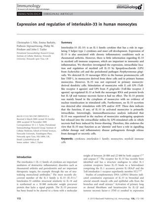
  • 4. IL-33 in monocytes mounted on microscope slides with Dako fluorescent (a) 200 Unstimulated E. coli LPS P. gingivalis LPS mounting medium (DAKO, Ely, UK). The cells were exam- 175 * IL-33 mRNA expression ined using a Leica TCS SP UV confocal laser scanning 150 * microscope (Leica, Wetzlar, Germany). A series of Z-stack 125 images were taken and each image was analysed individu- 100 * * ally using Leica LCS LITE software (Leica) and findings were 75 verified by an independent expert observer. Specificity of 50 the IL-33 monoclonal antibody was confirmed by complete 25 abrogation of fluorescent labelling after pre-incubation 0 with a fivefold concentration of blocking peptide 0·5 3 6 9 24 48 Time (hr) (IL-33112–270) (Axxora) (data not shown). (b) 50 Unstimulated E. coli LPS P. gingivalis LPS 45 IL-33 mRNA expression Statistical analysis 40 Statistical analysis was performed using Mann–Whitney 35 * 30 tests (cytokine expression analysis) and Student’s t-test 25 * (real-time RT-PCR).21 A P-value of < 0Á05 was consid- 20 ered to be statistically significant. 15 10 5 Results 0 3 9 24 Time (hr) IL-33 expression by myeloid cells (c) 50 Data from analysis of a cDNA library indicated that mod- 45 Unstimulated TNF-a IL-1b IL-33 mRNA expression est levels of IL-33 mRNA are expressed by human mono- 40 35 cytes and dendritic cells.2 However, to date, the expression 30 of IL-33 by myeloid cells has not been fully characterized. 25 We therefore investigated the expression of IL-33 by 20 monocytes in response to bacterial LPS and the cytokines 15 IL-1b and TNF-a using quantitative real-time PCR 10 5 (Fig. 1). We found that IL-33 mRNA was significantly up- 0 regulated in LPS-stimulated THP-1 monocytes (Fig. 1a) 0·5 1 3 6 24 and primary monocytes isolated from three separate Time (hr) donors (Fig. 1b), in comparison to unstimulated controls, Figure 1. Interleukin-33 (IL-33) messenger RNA (mRNA) expression with maximal transcript levels observed after 6–9 hr of is up-regulated by monocytes in response to lipopolysaccharide incubation with LPS. Conversely, real-time PCR per- (LPS) but not tumour necrosis factor-a (TNF-a) and IL-1b. Quanti- formed on cDNA isolated from MDDCs derived from pri- tative real-time polymerase chain reaction analysis was used to deter- mary macrophages of three separate donors showed a lack mine IL-33 expression by THP-1 monocytes (a) and primary human of IL-33 expression (data not shown). In contrast to IL-33, monocytes (b) in response to stimulation with 100 ng/ml Escherichia the ST2 and HMGB1 genes exhibited no up-regulation in coli or Porphyromonas gingivalis LPS. IL-33 mRNA expression by expression levels in myeloid cells upon stimulation with THP-1 monocytes stimulated with 100 ng/ml TNF-a and 100 pg/ml bacterial LPS (data not shown). The up-regulation of IL-1b was also measured (c). Levels of mRNA were normalized IL-33 in monocytes by LPS may be the result of transcrip- against RNA polymerase II mRNA levels and fold changes (‘IL-33 tional up-regulation via Toll-like receptor (TLR) signalling expression’) were calculated relative to unstimulated cells at each individual time-point using the DDCt method.20 The data represent or may be an indirect, secondary effect of LPS-stimulated the mean values (± SD) from three independent experiments. Signif- TNF-a or IL-1b secretion. To address this issue, THP-1 icant differences we determined using Student’s t-test on the DCt monocytes were stimulated with TNF-a and IL-1b and values.20 *P < 0Á05 relative to unstimulated cells. IL-33 mRNA was quantified by real-time RT-PCR; the results do not provide any evidence to suggest that these cytokines up-regulate IL-33 (Fig. 1c). failed to detect any IL-33 secretion from viable cells either at 9 hr (Fig. 2) or during the full time–course of our experiments (0–48 hr) (data not shown). There is a IL-33 is not secreted from viable cells suggestion that pro-IL-33 is processed by the action of The LPS-stimulated IL-33 mRNA levels were maximal caspase-13,4,6 and ATP has been shown to enhance after 6–9 hr of monocyte culture but we consistently the activity of caspase-1, enhancing the processing and Ó 2010 Blackwell Publishing Ltd, Immunology, 130, 172–180 175
  • 5. C. J. Nile et al. 70 Unstimulated E. coli LPS P. gingivalis LPS natants of LPS-stimulated cells incubated with ATP for 60 9–24 hr, when IL-33 mRNA expression was maximal (not 50 shown). Previous reports have indicated that IL-33 is pri- marily an intracellular cytokine7 and HMGB1 has been IL-33 (pg/ml) 40 shown to be released by cells undergoing necrosis22 so we 30 therefore induced necrosis in LPS-stimulated cells and 20 investigated IL-33 release (Fig. 2). There was no IL-33 10 released by necrotic monocytes that had not been stimu- 0 ND ND ND ND ND ND ND ND ND ND lated by LPS, suggesting that this protein was not consti- Live control 3 6 9 24 48 tutively synthesized by these cells or at least was below Time (hr) the level of detection of the ELISA (Fig. 2). However, Figure 2. Interleukin-33 (IL-33) is not secreted by viable THP-1 IL-33 was released into the culture medium by necrotic monocytes but is released from necrotic cells. THP-1 monocytes cells stimulated by LPS and the levels of IL-33 protein were either stimulated with 100 ng/ml Escherichia coli or Porphyro- detected reflected the kinetics of IL-33 mRNA stimulation monas gingivalis lipopolysaccharide (LPS) for 9 hr (live control), or by LPS (Fig. 2a) with maximal levels of IL-33 protein stimulated with 100 ng/ml E. coli or P. gingivalis LPS for 3–48 hr fol- being released by monocytes stimulated with LPS for 9 hr lowed by induction of necrosis by five rounds of freezing () 70°) (Fig. 2). Stimulation of the cells in these experiments was and thawing (38°). Stimulation of the cells in these experiments was confirmed by detection of TNF-a in the isolated super- confirmed by detection of tumour necrosis factor-a in the isolated natants using ELISA (data not shown). Immunocyto- supernatants using enzyme-linked immunosorbent assay (not chemistry confirmed that IL-33 protein was barely shown). ND = not detected. The data represent the mean values expressed in unstimulated THP-1 monocytes and was not (± SD) from three independent experiments. expressed at all in unstimulated primary monocytes but there was a prominent expression of intracellular IL-33 Table 1. Effect of adenosine triphosphate (ATP) on interleukin-33 protein by LPS-stimulated monocytes (Fig. 3a,b). Count- (IL-33) and IL-18 secretion by THP-1 monocytes erstaining of cells with the nuclear stain DAPI indicated that IL-33 was predominantly located in the cytoplasmic IL-33 IL-18 compartment in LPS-stimulated monocytes as confirmed Treatment (pg/ml) (pg/ml) by the absence of co-localization of green and blue fluorescence (Fig. 3a,b). Unstimulated 0 27Á0 ± 10Á7 ATP (6 mm) 0 110Á1 ± 52Á0 E. coli LPS (1 ng/ml) 0 228Á2 ± 96Á5 IL-33 is sequestered in the nucleus of apoptotic E. coli LPS (1 ng/ml) + ATP (6 mm) 0 1635Á5 ± 524Á5* monocytes E. coli LPS (1 ng/ml) + freeze–thawing 66Á37 ± 8Á1 – In cells induced to undergo programmed cell death IL-33 secretion from THP-1 monocytes is not activated by ATP. HMGB1 was found to remain closely associated with THP-1 monocytes were stimulated with 1 ng/ml Escherichia coli lipo- nuclear chromatin.24 We therefore investigated the effect polysaccharide (LPS) for 3 hr in the presence of 6 mm ATP for the of apoptosis on IL-33 in monocytes. The LPS-stimulated final 30 min of this incubation. Cells without additives (unstimu- monocytes induced to undergo apoptosis by UVB irradia- lated) and with ATP or LPS alone served as controls. The IL-33 and tion did not release any IL-33 into the culture medium IL-18 were measured by specific enzyme-linked immunosorbent assay. The data represent the median values (± interquartile range) over a 0–48-hr time–course (data not shown). Immuno- from three independent experiments. IL-18 secretion was signifi- cytochemistry was then used to determine the intracellu- cantly increased by stimulation of the monocytes with E. coli LPS in lar location of IL-33 in stimulated cells (Fig. 4). Nuclear combination with ATP as compared with LPS alone; Mann–Whitney morphology in control cell cultures confirms the presence test, *P < 0Á05. of apoptotic cells (Fig. 4a) and staining for IL-33 using FITC-conjugated antibodies clearly shows that the intra- cellular IL-33 protein is tightly associated with the secretion of IL-1b and IL-18.23 We therefore investigated nucleus of cells undergoing apoptosis (Fig. 4b). whether ATP stimulated IL-33 secretion, but we were again unable to detect any extracellular IL-33 in mono- Discussion cytes exposed to ATP either alone or in combination with LPS (Table 1). Similar experiments confirmed ATP The IL-1 family of cytokines have an established role enhancement of IL-18 secretion in LPS-stimulated cells in immune regulation and inflammatory processes.1,17 over the same time–course, confirming that ATP-stimu- Inappropriate activity of IL-1 cytokines is a feature of a lated caspase-1 was functional in this system (Table 1). number of immune-mediated pathologies and therefore Furthermore, IL-33 was not detected in the culture super- pathways activated by these cytokines are rational therapeutic 176 Ó 2010 Blackwell Publishing Ltd, Immunology, 130, 172–180
  • 6. IL-33 in monocytes (a) Control Ec LPS Pg LPS IL-33 47·62 µm 47·62 µm 47·62 µm Figure 3. Interleukin-33 (IL-33) protein is expressed in the cytoplasm of lipopolysaccha- ride (LPS) -stimulated THP-1 monocytes (a) IL-33 + and primary monocytes (b). Immunocyto- DAPI chemistry was performed on monocytes (con- trol) and cells stimulated with 100 ng/ml 47·62 µm 47·62 µm 47·62 µm Escherichia coli (Ec) or Porphyromonas gingiva- lis (Pg) LPS for 9 hr. The IL-33 was located (b) Control Ec LPS Pg LPS using an IL-33 antibody and secondary anti- body conjugated to fluorescein isothiocyanate (FITC; green) or tetramethyl rhodamine iso- thiocyanate (TRITC; red) and the nuclei were stained with 40 -60 ,diamidino-2-phenylindole IL-33 (DAPI; blue). A series of Z-stack images were taken to confirm the cellular location of IL-33 and the image depicted is representative of 47·62 µm 47·62 µm 47·62 µm findings throughout the Z stack. The upper panels (a) show IL-33 expression alone (green FITC) and the lower panels (b) show IL-33 (red TRITC) and nuclei (blue DAPI) in com- IL-33 + bination. The results are representative of three DAPI independent stimulation experiments and have been verified by an independent observer. Scale 47·62 µm 47·62 µm 47·62 µm as depicted on images. (a) Control Ec LPS Pg LPS IL-33 12·30 µm 8·7 µm 11·50 µm (b) IL-33 + DAPI 12·30 µm 8·7 µm 11·50 µm Figure 4. Interleukin-33 (IL-33) protein is closely associated with the nucleus in THP-1 monocytes undergoing apoptosis. Immunocytochemistry was performed on unstimulated THP-1 monocytes (control) and on cells stimulated with 100 ng/ml Escherichia coli (Ec) or Porphyromonas gingivalis (Pg) lipopolysaccharide (LPS) for 9 hr followed by exposure to UVB-irradiation to induce apoptosis. The IL-33 was located using an IL-33 antibody and secondary antibody conjugated to fluorescein isothiocyanate (FITC; green). The nuclei were stained with 40 -60 ,diamidino- 2-phenylindole (DAPI; blue). The upper panels (a) show IL-33 expression alone (green FITC) and the lower panels (b) show IL-33 (green FITC) and nuclei (blue DAPI) in combination. Arrows indicate examples of apoptotic cells. The results are representative of three independent stimu- lation experiments and have been verified by an independent observer. Scale as depicted on the images. Ó 2010 Blackwell Publishing Ltd, Immunology, 130, 172–180 177
  • 7. C. J. Nile et al. targets.1 Interleukin-33 is a recently described member of onstrate any effect of these cytokines on IL-33 expression. the IL-1 family and has attracted attention because, Previously, expression of IL-33 in human primary lung uniquely among IL-1 cytokines, it stimulates the produc- fibroblasts as well as in dermal fibroblasts and keratino- tion of Th2 cytokines such as IL-5 and IL-13, activates cytes was shown to be enhanced by a combination of Th2 cells, and has a role in mast cell development and TNF-a and IL-1b.2 Also, TNF-a either alone or in combi- function.2,11–16 As a result, IL-33 has been implicated in nation with IL-1b enhances IL-33 expression in human the pathogenesis of immune-mediated disorders which primary synovial fibroblasts.9,32 However, TNF-a and feature Th2 responses and mast cell dysregulation, and in IL-1b had no effect on IL-33 expression in rat cardiac particular those which involve pulmonary and mucosal fibroblasts or rat cardiomyocytes.18 The reasons for these inflammation.2,16,25 Studies of the action of recombinant inconsistencies are not clear, although the data may IL-33 and the expression of ST2 (a subunit of the IL-33 reflect variable responsiveness of different cell types. receptor) suggest that IL-33 has roles in immune The function, if any, of IL-33 in the myeloid lineage responses and immunopathogenesis in addition to those remains obscure but a key question is whether or not mediated by mast cells and Th2 cells. Hence, eosinophils IL-33 is secreted or remains in the intracellular compart- and basophils express ST2 and are stimulated by exoge- ment. In terms of myeloid cell IL-33, we consistently nous IL-33.26–29 Interleukin-33 also enhanced cytokine failed to detect IL-33 in culture supernatants even after production by invariant natural killer (NK) T cells and LPS stimulation. Although there is much indirect human NK cells.29 Also, IL-33 and ST2 are widely evidence for an immunological role of IL-33 in vitro and expressed in vascular cells and tissues, in fibroblasts and in vivo, only a limited number of reports have described in the central nervous system.6,7,9,18 the detection of soluble, endogenous IL-33.6,18 Hence, We have investigated the expression, intracellular loca- although rat cardiac fibroblasts synthesized IL-33 protein, tion and regulation of IL-33 in monocytes and MDDCs. unstimulated cells did not secrete this protein but IL-33 In experiments using both conventional RT-PCR (not secretion (as detected by Western blot of culture super- shown) and quantitative RT-PCR, IL-33 was not natants) was induced by stimulation with phorbol expressed in MDDCs and stimulation with LPS had no 12-myristate 13-acetate.18 Similarly, in a study of mixed effect on IL-33 expression in MDDCs. This is in contrast glial cell cultures and astrocyte-enriched cultures, IL-33 to Schmitz et al.2 who reported no expression in ‘resting’ was only found to be secreted in cells stimulated with MDDC but IL-33 expression (albeit at very modest levels PAMPs and ATP together.6 Recently, the release of IL-33 as compared with smooth muscle cells and fibroblasts) from primary human endothelial cells in conditions of was detected when these cells were activated by LPS. Sim- mechanical stress or injury was demonstrated.3 The physio- ilarly, whereas Schmitz et al.2 only detected IL-33 mRNA logical relevance of these data remains unclear, but this in LPS-activated monocytes, we detected IL-33 mRNA in does suggest that under certain circumstances cells may the human promonocytic cell line THP-1, in monocytes secrete IL-33 in response to the appropriate signals. derived from these cells and in primary human mono- It is possible that IL-33 may be secreted in a form that cytes by both conventional RT-PCR (not shown) and is not detectable by ELISA. For example, the soluble quantitative RT-PCR. However, analysis of protein form of the IL-33 receptor subunit ST2, sST2 interacts expression using immunocytochemistry demonstrated with IL-33 and blocks IL-33 signalling18,25 and sST2 is only very low levels of constitutive IL-33 expression in induced by LPS and cytokines in monocytes.33 Although THP-1 monocytes and none in primary cells. The differ- we have not analysed sST2 in our cultures, soluble IL-33 ences between our findings and those of Schmitz et al.2 was readily detectable by ELISA after induction of necro- with respect to mRNA expression may reflect technical sis in monocyte cultures previously stimulated by LPS. differences or differences in the sources of cDNA. Interleukin-33 may require other signals, in addition to We investigated regulation of IL-33 expression in LPS, to induce efficient extracellular release. For example, monocytes: LPS from E. coli as well as LPS from the peri- ATP is known to enhance both IL-1b and IL-18 release odontal pathogen P. gingivalis both enhanced IL-33 via activation of a multiprotein complex (the inflamma- expression. The LPS from E. coli and P. gingivalis repre- some) containing caspase-1, the enzyme responsible for sent different pathogen-associated molecular patterns intracellular processing of the pro- form of these cyto- (PAMPs): LPS from these species stimulate host cells via kines.23 It is not yet clear whether IL-33 is processed TLR4 and TLR2, respectively, which results in overlapping in vivo via a caspase-1 pathway: although there is evidence but distinct cytokine responses.30,31 Our data suggest that that in-vitro-translated IL-33 is cleaved by incubation with engagement of both TLR2 and TLR4 pathways stimulates caspase-1,2 this was not confirmed by experiments analy- IL-33 indicating a potential role for this cytokine in sing intracellular IL-33.7 Furthermore, the predicted cleav- immune responses to diverse pathogens and PAMPs. age site for caspase-1-induced IL-33 is not conserved in We also investigated the regulation of IL-33 in mono- orthologues from other species.7 Recently, it was demon- cytes by both TNF-a and IL-1b but were unable to dem- strated that IL-33 is probably cleaved by both caspase-1 178 Ó 2010 Blackwell Publishing Ltd, Immunology, 130, 172–180
  • 8. IL-33 in monocytes and caspase-3 to yield a 20 000–22 000 molecular weight cytoplasmic compartment in viable monocytes and we form of IL-33 and that this cleavage took place at an did not observe prominent nuclear translocation of IL-33. amino acid residue (Asp178) distinct from that previously Palmer et al.32 reported that IL-33 was expressed in both proposed (Ser111).3 Significantly, unlike the full-length the nucleus and cytoplasm of synovial fibroblasts and IL-331–270, the 20 000–22 000 form was not biologically ‘mononuclear inflammatory cells’. The sub-cellular locali- active (in terms of binding and activation of the ST2 zation of IL-33 may reflect intrinsic differences between receptor), suggesting that IL-33 was inactivated by caspase cell types. Also, there are differences in IL-33 expression cleavage.3 The antibodies used for the ELISA and in smooth muscle cells cultured in vitro and IL-33 expres- immunocytochemistry in the present report do not distin- sion in these cells in vivo.2,10 Significantly, the expression guish between the different forms of IL-33 described by pattern of IL-33 changes in different cell culture condi- Cayrol et al.;3 it will therefore be interesting to investigate tions.8 Therefore, data from in vitro culture models in detail the molecular structures of IL-33 expressed in should be interpreted with care and it will be important monocytes, the effect of LPS on IL-33 maturation, and the to extend studies of IL-33 expression in immune cells specific forms of IL-33 released from necrotic monocytes. in vitro to investigate the expression of this cytokine in Addition of ATP to mixed glial cell cultures and astro- tissue sections from patients with inflammatory disease. cyte-enriched cultures stimulated the release of IL-33 We also generated preliminary evidence to suggest that from these cells whereas these reagents had no effect in during apoptosis, IL-33 is associated with the nucleus in isolation.6 Significantly, we failed to demonstrate any monocytes. In this regard it is interesting to note that, effect of ATP on IL-33 secretion from monocytes, either apoptosis in endothelial cells stimulated IL-33 cleavage alone or in combination with LPS, although enhancement (into a form which does not bind the extracellular ST2 of IL-18 secretion was measured in the same experiments receptor) suggesting that this reaction existed to inactivate (Table 1). Intriguingly, it has been speculated that IL-33 IL-33 after apoptosis.3 In addition, we demonstrated may be processed by an inflammasome distinct from that release of IL-33 as the result of necrosis in the same cells. which processes IL-1b and IL-18.34 There is evidence that In similar experiments it was recently demonstrated that IL-33 may function as a so-called dual function cytokine necrosis in primary endothelial cells was associated with having a role as an (extracellular) pro-inflammatory cyto- the release of biologically active IL-33.3 The behaviour of kine and an (intracellular) nuclear factor with transcrip- IL-33 during apoptosis and necrosis is similar to the so- tional regulatory action.7 called ‘alarmins’, which are a group of biologically active These authors draw analogy with other ‘dual function’ mediators that include the dual function cytokines cytokines such as IL-1a and HMGB1.7 In support of this HMGB1 and IL-1a.36 Alarmins are endogenous signalling hypothesis they found that in high endothelial venules, molecules that, like exogenous PAMPs, trigger similar IL-33 was found to be primarily an intracellular protein pathways and have a common purpose as damage-associ- that closely associates with eukaryotic chromatin7 The ated molecule patterns (DAMPS).36 These findings nuclear localization was found to be mediated by an evo- endorse the view that IL-33 has functions beyond that of lutionarily conserved homeodomain-like helix-turn-helix a conventional extracellular, immunoregulatory cytokine (HTH) motif which is located within the N-terminal por- and may be classified as a DAMP.34 The recent finding of tion of the IL-33 precursor protein.7 More recent data widespread expression of IL-33 in the vasculature, in the provide a molecular model for the chromatin interactions skin and mucosal epithelial cells supports this hypo- of IL-33 and suggest that IL-33 binds to the histone thesis.10 Interestingly, in addition to IL-1a and IL-33, an dimer H2A-H2B and hence regulates chromatin compac- isoform of IL-7 (IL-7b), also an IL-1 family cytokine, has tion by influencing nucleosome–nucleosome interac- recently been shown to translocate to the nucleus in acti- tions.35 Interleukin-33 is therefore shown to confer potent vated monocytes and to down-regulate the synthesis of transcriptional repressor activity although the target genes pro-inflammatory cytokines.37 remain to be characterized.7 In summary, the function of IL-33 in activated mono- Significantly, it was demonstrated that endothelial cells cytes is hypothesized to be primarily intracellular; and as in inflamed tissues from patients with Crohn’s disease such IL-33 may act as an ‘alarmin’ and have a role in sig- and rheumatoid arthritis express high levels of IL-33 and nalling cellular damage and inflammatory disease patho- constitute the major source of IL-33 in these tissues.7 The genesis through release from damaged or necrotic cells. high expression of IL-33 in the endothelial cells of normal and inflamed synovial tissue has recently been con- Acknowledgements firmed.32 Xu et al.9 also confirmed the high expression of IL-33 (and ST2) in the rheumatoid synovium and in This work was supported by a grant from the Newcastle fibrobasts isolated from this tissue. Although we have Healthcare charity and a UK Department of Health Clini- demonstrated that IL-33 is not secreted from stimulated cian Scientist Fellowship DHCS/03/G121/46 awarded to monocytes, IL-33 is predominantly associated with the Philip Preshaw. Ó 2010 Blackwell Publishing Ltd, Immunology, 130, 172–180 179
  • 9. C. J. Nile et al. Disclosures 17 Arend WP, Palmer G, Gabay C. IL-1, IL-18, and IL-33 families of cytokines. Immunol Rev 2008; 223:20–38. 18 Sanada S, Hakuno D, Higgins LJ, Schreiter ER, McKenzie AN, Lee RT. IL-33 and ST2 The authors declare no conflict of interest. comprise a critical biomechanically induced and cardioprotective signaling system. J Clin Invest 2007; 117:1538–49. 19 Baekkevold ES, Roussigne M, Yamanaka T et al. Molecular characterization of NF-HEV, References a nuclear factor preferentially expressed in human high endothelial venules. Am J Pathol 1 Barksby HE, Lea SR, Preshaw PM, Taylor JJ. The expanding family of interleukin-1 2003; 163:69–79. cytokines and their role in destructive inflammatory disorders. Clin Exp Immunol 2007; 20 Foster N, Cheetham J, Taylor JJ, Preshaw PM. VIP inhibits Porphyromonas gingivalis 149:217–25. LPS-induced immune responses in human monocytes. J Dent Res 2005; 84:999–1004. 2 Schmitz J, Owyang A, Oldham E et al. IL-33, an interleukin-1-like cytokine that signals 21 Yuan JS, Reed A, Chen F, Stewart CN Jr. Statistical analysis of real-time PCR data. via the IL-1 receptor-related protein ST2 and induces T helper type 2-associated cyto- BMC Bioinformatics 2006; 7:85. kines. Immunity 2005; 23:479–90. 22 Rovere-Querini P, Capobianco A, Scaffidi P et al. HMGB1 is an endogenous immune 3 Cayrol C, Girard JP. The IL-1-like cytokine IL-33 is inactivated after maturation by adjuvant released by necrotic cells. EMBO Rep 2004; 5:825–30. caspase-1. Proc Natl Acad Sci USA 2009; 106:9021–6. 23 Sutterwala FS, Ogura Y, Flavell RA. The inflammasome in pathogen recognition and 4 Chackerian AA, Oldham ER, Murphy EE, Schmitz J, Pflanz S, Kastelein RA. IL-1 recep- inflammation. J Leukoc Biol 2007; 82:259–64. tor accessory protein and ST2 comprise the IL-33 receptor complex. J Immunol 2007; 24 Scaffidi P, Misteli T, Bianchi ME. Release of chromatin protein HMGB1 by necrotic 179:2551–5. cells triggers inflammation. Nature 2002; 418:191–5. 5 Ali S, Huber M, Kollewe C, Bischoff SC, Falk W, Martin MU. IL-1 receptor accessory 25 Hayakawa H, Hayakawa M, Kume A, Tominaga S. Soluble ST2 blocks interleukin-33 protein is essential for IL-33-induced activation of T lymphocytes and mast cells. Proc signaling in allergic airway inflammation. J Biol Chem 2007; 282:26369–80. Natl Acad Sci USA 2007; 104:18660–5. 26 Cherry WB, Yoon J, Bartemes KR, Iijima K, Kita H. A novel IL-1 family cytokine, IL- 6 Hudson CA, Christophi GP, Gruber RC, Wilmore JR, Lawrence DA, Massa PT. Induc- 33, potently activates human eosinophils. J Allergy Clin Immunol 2008; 121:1484–90. tion of IL-33 expression and activity in central nervous system glia. J Leukoc Biol 2008; 27 Suzukawa M, Iikura M, Koketsu R et al. An IL-1 cytokine member, IL-33, induces 84:631–43. human basophil activation via its ST2 receptor. J Immunol 2008; 181:5981–9. 7 Carriere V, Roussel L, Ortega N et al. IL-33, the IL-1-like cytokine ligand for ST2 28 Suzukawa M, Koketsu R, Iikura M et al. Interleukin-33 enhances adhesion, CD11b receptor, is a chromatin-associated nuclear factor in vivo. Proc Natl Acad Sci USA 2007; expression and survival in human eosinophils. Lab Invest 2008; 88:1245–53. 104:282–7. 29 Smithgall MD, Comeau MR, Yoon BR, Kaufman D, Armitage R, Smith DE. IL-33 8 Kuchler AM, Pollheimer J, Balogh J et al. Nuclear interleukin-33 is generally expressed amplifies both Th1- and Th2-type responses through its activity on human basophils, in resting endothelium but rapidly lost upon angiogenic or proinflammatory activation. allergen-reactive Th2 cells, iNKT and NK cells. Int Immunol 2008; 20:1019–30. Am J Pathol 2008; 173:1229–42. 30 Chen C, Coats SR, Bumgarner RE, Darveau RP. Hierarchical gene expression profiles of 9 Xu D, Jiang HR, Kewin P et al. IL-33 exacerbates antigen-induced arthritis by activat- HUVEC stimulated by different lipid A structures obtained from Porphyromonas gingi- ing mast cells. Proc Natl Acad Sci USA 2008; 105:10913–8. valis and Escherichia coli. Cell Microbiol 2007; 9:1028–38. 10 Moussion C, Ortega N, Girard JP. The IL-1-like cytokine IL-33 is constitutively expres- 31 Barksby HE, Nile CJ, Jaedicke KM, Taylor JJ, Preshaw PM. Differential expression of sed in the nucleus of endothelial cells and epithelial cells in vivo: a novel ‘alarmin’? immunoregulatory genes in monocytes in response to Porphyromonas gingivalis and PLoS ONE 2008; 3:e3331. Escherichia coli lipopolysaccharide. Clin Exp Immunol 2009; 156:479–87. 11 Allakhverdi Z, Smith DE, Comeau MR, Delespesse G. Cutting Edge: the ST2 ligand 32 Palmer G, Talabot-Ayer D, Lamacchia C et al. Inhibition of interleukin-33 signaling IL-33 potently activates and drives maturation of human mast cells. J Immunol 2007; attenuates the severity of experimental arthritis. Arthritis Rheum 2009; 60:738–49. 179:2051–4. 33 Trajkovic V, Sweet MJ, Xu D. T1/ST2 – an IL-1 receptor-like modulator of immune 12 Ho LH, Ohno T, Oboki K et al. IL-33 induces IL-13 production by mouse mast cells responses. Cytokine Growth Factor Rev 2004; 15:87–95. independently of IgE-FceRI signals. J Leukoc Biol 2007; 82:1481–90. 34 Gadina M, Jefferies CA IL-33: a sheep in wolf’s clothing? Sci STKE 2007; 2007:pe31. 13 Iikura M, Suto H, Kajiwara N et al. IL-33 can promote survival, adhesion and cytokine 35 Roussel L, Erard M, Cayrol C, Girard JP. Molecular mimicry between IL-33 and KSHV production in human mast cells. Lab Invest 2007; 87:971–8. for attachment to chromatin through the H2A-H2B acidic pocket. EMBO Rep 2008; 14 Komai-Koma M, Xu D, Li Y, McKenzie AN, McInnes IB, Liew FY. IL-33 is a chemo- 9:1006–12. attractant for human Th2 cells. Eur J Immunol 2007; 37:2779–86. 36 Bianchi ME. DAMPs, PAMPs and alarmins: all we need to know about danger. J Leu- 15 Moulin D, Donze O, Talabot-Ayer D, Mezin F, Palmer G, Gabay C. Interleukin (IL)-33 koc Biol 2007; 81:1–5. induces the release of pro-inflammatory mediators by mast cells. Cytokine 2007; 37 Sharma S, Kulk N, Nold MF et al. The IL-1 family member 7b translocates to the 40:216–25. nucleus and down-regulates proinflammatory cytokines. J Immunol 2008; 180:5477–82. 16 Humphreys NE, Xu D, Hepworth MR, Liew FY, Grencis RK. IL-33, a potent inducer of adaptive immunity to intestinal nematodes. J Immunol 2008; 180:2443–9. 180 Ó 2010 Blackwell Publishing Ltd, Immunology, 130, 172–180