X rays discovered on nov
•Descargar como PPTX, PDF•
1 recomendación•838 vistas

Collected from Internet, text books, etc

Recomendados
Más contenido relacionado
La actualidad más candente
La actualidad más candente (20)
Destacado
Destacado (20)
Similar a X rays discovered on nov
Similar a X rays discovered on nov (20)
Más de Rekha Pathak
Más de Rekha Pathak (20)
Último
Último (20)
X rays discovered on nov
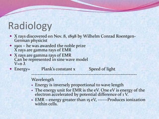
- 1. Radiology X rays discovered on Nov. 8, 1898 by Wilhelm Conrad Roentgen- German physicist 1901 – he was awarded the noble prize X rays are gamma rays of EMR X rays are gamma rays of EMR Can be represented in sine wave model V=n ƛ Energy= Plank’s constant x Speed of light ------------------------------------------------------------------ Wavelength Energy is inversely proportional to wave length The energy unit for EMR is the eV. One eV is energy of the electron accelerated by potential difference of 1 V. EMR – energy greater than 15 eV, ------Produces ionization within cells.
- 2. Ionisation causes ion pairs to form A photon is a discrete bundle of EMR Ionisation in DNA may result in biologic amplification—ie affecting the progeny of future generations Increased rate of mutation Rate of abortion or fetal abnormalities Susceptibility to diseases Shortened life span Cancer Cataracts
- 3. Radiation protection:Roentgen, rad and rem---units to quantify radiation exposure, absorption and equivalent dose respectively. SI unit is coulomb per kg and joule per kg. EMR have speed of light Properties: No charge, no mass, speed of light, invisible, cannot be felt, travel in a st. Line , cannot be deflected by magnetic field, penetrate all matter to some degree, cause certain substance to fluoresce, can ionize atoms. The amount of radiation exposure is quantified by measuring the no. Of ionization / electric charge produced by x-rays in air.
- 4. C/kg is based on no. Of ion pairs produced in air by the incoming radiation. 1 Roentgen=production of 2.58x10⁻⁴ C/kg in air. The high absorber is bone and low absorber is air: absorption is difference between the incident photon and output photon after passing through the object. Absorption of the same dose in Gy from different types of radiations may not produce the same biologic effect. For eg damage from particulate radiation such as alpha particles and neutrons is greater on a Gy for Gy basis than damage from the same dose of x rays. This is related to differences in ionization density for different types of radiation.
- 5. A high mass , heavily charged particle such as an alpha particle creates many ionizations that are very close to one another in the tissue compared with low mass lightly charged particle such as electron. So deposition of 1 Gy from alpha particle absorption does more ;biologic damage than deposition of 1 Gy from x ray absorption.
- 6. Absorbed dose: varies with object to object. SI unit for the absorbed dose is gray. Amount of radiation such that the absorbed energy is 1 joule j/kg of tissue. Before the unit of absorbed dose was the rad which is = 100 ergs/g. 1Gy=100 rad. The differential between the exposure dose and absorbed dose is inversely proportional to photon energy.
- 7. Radiation Damage may be compared by weighting factor which is a numeric factor describing the relative effectiveness of a particular type of radiation to photons. The weighting factor for photons is 1 and it is greater than 1 for the charged or particulate types of radiation such as e, n or alpha particles in the SI system, the unit of dose equivalency is the Sievert (Sv). 1 sv= absorbed dose in Gy x weighting factor REM= ABSORBED dose in rads x weighting factor 1 Gy=100 rads, 1 Sv = 100 rem. 1 Gy=1 sv
- 8. NRC nuclear regulatory commission is the official source for establishing the guidelines for radiation protection. Recommended that the annual occupational radiation dose to individual adults should be limited to a maximum of 0.05 Sv (5 rem) No upper ;limit for the cumulative exposure
- 9. NCRP national council on radiation protection : to prevent clinically significant radiation induced effects by adhering to dose limits that are below the apparent or practical threshold To limit the risk of cancer and heritable effects to a reasonable level in relation to societal needs and values and benefits gained.
- 10. Max. Permissible dose is the max amt. Of absorbed radiation that can be delivered to an individual as a whole body dose or as a dose to a specific organ and still be considered safe. Safe means no evidence of harmful immediate or long term effects to the body as a whole or to any individual structure or organ ALARA principle: as low as reasonably achievable. But as no threshold can be established for smokers as to how much frequency of smoking there will not be any damage similarly no threshold for radiation has been established. NCRP recommendations :
- 11. Lifetime effective dose should not exceed age in years x 10 m Sv (age in yrs x 1 REM) and no exposure is permitted upto 18 yrs. So in REMS calculation the lifetime effective dose equivalent in Rems should not exceed the value of his her age in yrs The effective dose in any 1 yr should not exceed 50 mSv (5 rem) For general public should not exceed 1mSv (0.1 rem) Pregnancy once declared should not exceed 0.5mSv (0.05 rem) For occupationally exposed woman it is yet to be declared
- 12. cosmic 8% terrestrial 8% Internal 11% Man made 18% Medical x rays 11% Nuclear medicine 4% others 3% Ionizing radiation damage to DNA: Important target of ionization Because body contains lot of water, lot of free radical formation is there leads to DNA damage
- 13. Muscle is not affected easily but growing fetus, BM, gonadal cells are very sensitive to ionization damage. DNA strand breakage, DNA base damage (nucleotide), DNA cross linkage This causes biologic amplification ie passes from one generation to other Protective lead aprons and gloves have 0.5mm thickness of lead Used against secondary radiations
- 14. •Radiation Badges may contain film (exposure is read by observing the blackness and it may contain thermoluminiscence dosimeter TLD) The incoming radiations cause electron cloud to form and depending on the no. Of electrons formed radiation dose is assessed. •Radiation badges must be assessed at least quarterly
- 15. •When protective apron is worn the badge should be worn on outside of the apron for monitoring the radiation environment but inside when the estimate of body exposure is desired. •Protective glasses provide 0.25 mm of Pb equivalent •The thyroid shield , scrotal shield, gloves, aprons etc protect from the radiations •Primary beam should be properly collimated and should not exceed the size of the cassette •Smaller the focal spot better the detail on the RG
- 16. •Braking radiations or Bremstrauling radiation: two mechanisms •1. collision: high energy oncoming electron strikes the inner shells and eject the electrons having high BE outside, the next higher shell electrons / outer most orbit drops into the void of inner shell: this produces radiating x ray which has the energy proportional to difference in the BE of the two shells. So high energy incident photon produce high energy x rays. (KV increases the energy of the x rays). This produces a characteristic x-ray
- 18. •2. Radiative/ braking : Just like applying of the brakes. When oncoming electron brakes and bends around the nucleus because of the charge difference it bends and changes its direction this releases energy in the form of x rays. When the deflected electron decelerates it releases EMR in the form of an x ray. Because deflected electrons may pass within various distances of the nucleus braking radiation has a spectrum of energies.
- 20. Interaction of radiation with matter 1. Coherent scattering: Photon interacts with an object and changes its direction but the subject does not absorb the photon and a change in photon energy does not occur. This is very small in the patient. It may strike the x ray film and degrade the quality of the image or increase the personnel exposure 2. Photoelectric effect: very effectively results in an image formation . Characteristic radiation is given off. The energy of the characteristic radiation is a function of the atomic number of the atom from which it arises. Thus with a large atomic no. Atom such as tungsten the target of the x ray tube the characteristic x ray is actually part of the useful x ray beam .
- 21. The probability of photoelectric interaction is directly proportional to the cube of the atomic number and inversely proportional to the cube of the photon energy . • There is lack of scattered radiations with photoelectric effect because most of it is absorbed. The decrease in probability of photoelectric absorption as the energy of the photon beam increases result sin a loss of contrast between tissue of various types when very high energy photon are use for radiography. 3. Comptons scattering : mainly contributes to the fogging. The outer shell electron with low energy is ejected and radiation of low energy is is radiated when this void is filled. It fogs and spoils the quality of film. 4. Photodisintegration 5. Pair production Pair production and photodisintegration has no relevance with diagnostic radiology