SlideShare una empresa de Scribd logo
1 de 23
Descargar para leer sin conexión
41
Animal Nutrition
The Need to Feed
Dinnertime has arrived for the sea otter in Figure 41.1 (and for the crab,
though in quite a different sense). The muscles and other organs of the crab
will be chewed into pieces, broken down by acid and enzymes in the otter’s diges-
tive system, and finally absorbed as small molecules into the body of the otter.
Such a process is what is meant by animal nutrition: food being taken in, taken
apart, and taken up.
Although dining on fish, crabs, urchins, and abalone is the sea otter’s specialty,
all animals eat other organisms—dead or alive, piecemeal or whole. Unlike plants,
animals must consume food for both energy and the organic molecules used to as-
semble new molecules, cells, and tissues. Despite this shared need, animals have
diverse diets. Herbivores, such as cattle, sea slugs, and caterpillars, dine mainly
on plants or algae. Carnivores, such as sea otters, hawks, and spiders, mostly eat
other animals. Rats and other omnivores (from the Latin omnis, all) don’t in fact
eat everything, but they do regularly consume animals as well as plants or algae. We
humans are typically omnivores, as are cockroaches and crows.
The terms herbivore, carnivore, and omnivore represent the kinds of food an ani-
mal usually eats. Keep in mind, however, that most animals are opportunistic feed-
ers, eating foods outside their standard diet when their usual foods aren’t available.
▲ Figure 41.1 How does a crab help an otter
make fur?
K E Y C O N C E P T S
41.1 An animal’s diet must supply
chemical energy, organic
molecules, and essential
nutrients
41.2 The main stages of food
processing are ingestion,
digestion, absorption, and
elimination
41.3 Organs specialized for
sequential stages of
food processing form the
mammalian digestive system
41.4 Evolutionary adaptations of
vertebrate digestive systems
correlate with diet
41.5 Feedback circuits regulate
digestion, energy storage, and
appetite
892
CHAPT ER 41 Animal Nutrition 893
For example, deer are herbivores, but in addition to feed-
ing on grass and other plants, they occasionally eat insects,
worms, or bird eggs. Note as well that microorganisms are an
unavoidable “supplement” in every animal’s diet.
Animals must eat. But to survive and reproduce, they must
also balance their consumption, storage, and use of food.
Sea otters, for example, support a high rate of metabolism
by eating up to 25% of their body mass each day. Eating too
little food, too much food, or the wrong mixture of foods can
endanger an animal’s health. In this chapter, we’ll survey the
nutritional requirements of animals, explore diverse evolu-
tionary adaptations for obtaining and processing food, and
investigate the regulation of energy intake and expenditure.
proteins, and lipids, for use in cellular respiration and en-
ergy storage.
In addition to providing fuel for ATP production, an
animal’s diet must supply the raw materials needed for bio-
synthesis. To build the complex molecules it needs to grow,
maintain itself, and reproduce, an animal must obtain two
types of organic precursors from its food. Animals need a
source of organic carbon (such as sugar) and a source of or-
ganic nitrogen (such as protein). Starting with these materials,
animals can construct a great variety of organic molecules.
Essential Nutrients
Some cellular processes require materials that an animal
cannot assemble from simpler organic precursors. These
materials—preassembled organic molecules and miner-
als—are called essential nutrients. Obtained from an ani-
mal’s diet, essential nutrients include essential amino acids
and fatty acids, vitamins, and minerals. Essential nutrients
have key functions in cells, including serving as substrates
of enzymes, as coenzymes, and as cofactors in biosynthetic
reactions (Figure 41.2). Needs for particular nutrients vary
among species. For instance, ascorbic acid (vitamin C) is an
essential nutrient for humans and other primates, as well as
guinea pigs, but not for many other animals.
Essential Amino Acids
Animals require 20 amino acids to make proteins (see
Figure 5.14). Most animal species have the enzymes to
synthesize about half of these amino acids, as long as their
diet includes sulfur and organic nitrogen. The remaining
amino acids must be obtained from food in prefab-
ricated form and are therefore called essential
amino acids. Many animals, including adult
humans, require eight amino acids in their
diet: isoleucine, leucine, lysine, methio-
nine, phenylalanine, threonine, trypto-
phan, and valine. (Human infants also
need a ninth, histidine.)
The proteins in animal products
such as meat, eggs, and cheese are
“complete,” which means that they
provide all the essential amino acids in
their proper proportions. In contrast,
most plant proteins are “incomplete,”
being deficient in one or more es-
sential amino acids. Corn (maize), for
example, is deficient in tryptophan
and lysine, whereas beans are lacking
in methionine. However, vegetarians
can easily obtain all of the essential
amino acids by eating a varied diet of
plant proteins.
C O N C E P T 41.1
An animal’s diet must supply chemical
energy, organic molecules, and
essential nutrients
Overall, an adequate diet must satisfy three nutritional
needs: chemical energy for cellular processes, organic build-
ing blocks for macromolecules, and essential nutrients.
The activities of cells, tissues, organs, and whole animals
depend on sources of chemical energy in the diet. This en-
ergy is used to produce ATP, which powers processes rang-
ing from DNA replication and cell division to vision and
flight. To meet the continuous requirement for ATP, ani-
mals ingest and digest nutrients, including carbohydrates,
Linoleic acid γ-Linoleic acid
Fatty acid desaturase
Phospholipids
(cell membrane components)
Prostaglandins
(used in cell signaling)
NADH
ESSENTIAL
AMINO ACIDS
(monomers of
polypeptide)
VITAMIN
(coenzyme)
ESSENTIAL
FATTY ACID
(substrate of
enzyme)
Vitamin B3
MINERAL
(cofactor)
Iron
Tyr
Glu
Gly
Phe
Phe
Leu
Ile
▲ Figure 41.2 Roles of essential nutrients.Linoleic acid is converted by the enzyme fatty acid
desaturase to γ-linoleic acid, a precursor for phospholipids and prostaglandins. This biosynthetic reac-
tion illustrates common functions of the four classes of essential nutrients, labeled in blue. Note that
nearly every enzyme or other protein in animal bodies contains some essential amino acids, as indi-
cated in the partial sequence shown for fatty acid desaturase.
894 UNIT SEVEN Animal Form and Function
Vitamins are classified as water-soluble or fat-soluble
(Table 41.1). B vitamins, which generally act as coenzymes,
are water-soluble. So is vitamin C, which is required for the
production of connective tissue. Fat-soluble vitamins in-
clude vitamin A, which is incorporated into visual pigments
of the eye, and vitamin D, which aids in calcium absorption
and bone formation. The dietary requirement for vitamin
D is variable in humans because we can synthesize it from
other molecules when our skin is exposed to sunlight.
For people with imbalanced diets, taking vitamin supple-
ments that provide recommended daily levels is certainly
reasonable. It is far less clear that massive doses of vita-
mins confer any health benefits or are even safe. Moderate
overdoses of water-soluble vitamins are probably harmless
because excesses are excreted in urine. However, excesses
of fat-soluble vitamins are deposited in body fat, so overcon-
sumption may cause them to accumulate to toxic levels.
Minerals
Dietary minerals are inorganic nutrients, such as iron and
sulfur, that are usually required in small amounts—from less
than 1 mg to about 2,500 mg per day. As shown in Table 41.2,
Essential Fatty Acids
Animals require fatty acids to synthesize a variety of cellular
components, including membrane phospholipids, signaling
molecules, and storage fats. Although animals can synthe-
size many fatty acids, they lack the enzymes to form the
double bonds found in certain required fatty acids. Instead,
these molecules must be obtained from the diet and are
considered essential fatty acids. In mammals, they include
linoleic acid (see Figure 41.2). Because seeds, grains, and
vegetables generally furnish ample quantities of essential
fatty acids, deficiencies in this class of nutrients are rare.
Vitamins
As Albert Szent-Györgyi, the discoverer of vitamin C, once
quipped, “A vitamin is a substance that makes you ill if you
don’t eat it.” Vitamins are organic molecules that are re-
quired in the diet in very small amounts. They have diverse
functions. Vitamin B2, for example, is converted in the body
to FAD, a coenzyme used in many metabolic processes, in-
cluding cellular respiration (see Figure 9.12). For humans,
13 vitamins have been identified. Depending on the vitamin,
the required amount ranges from 0.01 to 100 mg per day.
Vitamin Major Dietary Sources Major Functions in the Body Symptoms of Deficiency
Water-Soluble Vitamins
B1 (thiamine) Pork, legumes, peanuts, whole
grains
Coenzyme used in removing CO2
from organic compounds
Beriberi (tingling, poor coordina-
tion, reduced heart function)
B2 (riboflavin) Dairy products, meats, enriched
grains, vegetables
Component of coenzymes FAD
and FMN
Skin lesions, such as cracks at cor-
ners of mouth
B3 (niacin) Nuts, meats, grains Component of coenzymes NAD+
and NADP+
Skin and gastrointestinal lesions,
delusions, confusion
B5 (pantothenic acid) Meats, dairy products, whole
grains, fruits, vegetables
Component of coenzyme A Fatigue, numbness, tingling of
hands and feet
B6 (pyridoxine) Meats, vegetables, whole grains Coenzyme used in amino acid
metabolism
Irritability, convulsions, muscular
twitching, anemia
B7 (biotin) Legumes, other vegetables, meats Coenzyme in synthesis of fat, gly-
cogen, and amino acids
Scaly skin inflammation, neuro-
muscular disorders
B9 (folic acid) Green vegetables, oranges, nuts,
legumes, whole grains
Coenzyme in nucleic acid and
amino acid metabolism
Anemia, birth defects
B12 (cobalamin) Meats, eggs, dairy products Production of nucleic acids and
red blood cells
Anemia, numbness, loss of balance
C (ascorbic acid) Citrus fruits, broccoli, tomatoes Used in collagen synthesis;
antioxidant
Scurvy (degeneration of skin and
teeth), delayed wound healing
Fat-Soluble Vitamins
A (retinol) Dark green and orange vegetables
and fruits, dairy products
Component of visual pigments;
maintenance of epithelial tissues
Blindness, skin disorders, impaired
immunity
D Dairy products, egg yolk Aids in absorption and use of cal-
cium and phosphorus
Rickets (bone deformities) in chil-
dren, bone softening in adults
E (tocopherol) Vegetable oils, nuts, seeds Antioxidant; helps prevent dam-
age to cell membranes
Nervous system degeneration
K (phylloquinone) Green vegetables, tea; also made
by colon bacteria
Important in blood clotting Defective blood clotting
Table 41.1 Vitamin Requirements of Humans
CHAPT ER 41 Animal Nutrition 895
Mineral Major Dietary Sources Major Functions in the Body Symptoms of Deficiency
Morethan200mgperdayrequired      
Calcium (Ca) Dairy products, dark green vegeta-
bles, legumes
Bone and tooth formation, blood
clotting, nerve and muscle function
Impaired growth, loss of bone mass
Phosphorus (P) Dairy products, meats, grains Bone and tooth formation, acid-
base balance, nucleotide synthesis
Weakness, loss of minerals from
bone, calcium loss
Sulfur (S) Proteins from many sources Component of certain amino acids Impaired growth, fatigue, swelling
Potassium (K) Meats, dairy products, many fruits
and vegetables, grains
Acid-base balance, water balance,
nerve function
Muscular weakness, paralysis, nau-
sea, heart failure
Chlorine (Cl) Table salt Acid-base balance, formation of
gastric juice, nerve function, osmotic
balance
Muscle cramps, reduced appetite
Sodium (Na) Table salt Acid-base balance, water balance,
nerve function
Muscle cramps, reduced appetite
Magnesium (Mg) Whole grains, green leafy
vegetables
Enzyme cofactor; ATP bioenergetics Nervous system disturbances
Iron (Fe) Meats, eggs, legumes, whole grains,
green leafy vegetables
Component of hemoglobin and of
electron carriers; enzyme cofactor
Iron-deficiency anemia, weakness,
impaired immunity
Fluorine (F) Drinking water, tea, seafood Maintenance of tooth structure Higher frequency of tooth decay
Iodine (I) Seafood, iodized salt Component of thyroid hormones Goiter (enlarged thyroid gland)
*Additional minerals required in trace amounts include cobalt (Co), copper (Cu), manganese (Mn), molybdenum (Mo),
selenium (Se), and zinc (Zn). All of these minerals, as well as those in the table, can be harmful in excess.
Table 41.2 Mineral Requirements of Humans*
i
minerals have diverse functions in animal physiology. Some
are assembled into the structure of proteins; iron, for ex-
ample, is incorporated into the oxygen carrier hemoglobin as
well as some enzymes (see Figure 41.2). In contrast, sodium,
potassium, and chloride are important in the functioning of
nerves and muscles and in maintaining osmotic balance be-
tween cells and the surrounding body fluid. In vertebrates, the
mineral iodine is incorporated into thyroid hormone, which
regulates metabolic rate. Vertebrates also require relatively
large quantities of calcium and phosphorus for building and
maintaining bone.
Ingesting large amounts of some minerals can upset ho-
meostatic balance and impair health. For example, excess
salt (sodium chloride) can contribute to high blood pressure.
This is a particular problem in the United States, where the
typical person consumes enough salt to provide about 20
times the required amount of sodium. Packaged (prepared)
foods often contain large amounts of sodium chloride, even
if they do not taste very salty.
Dietary Deficiencies
A diet that lacks one or more essential nutrients or consis-
tently supplies less chemical energy than the body requires
results in malnutrition, a failure to obtain adequate nutri-
tion. Malnutrition resulting from either type of dietary defi-
ciency can have negative impacts on health and survival and
affects one out of four children worldwide.
▲ Figure 41.3 Obtaining essential nutrients from an unusual
source.A juvenile chamois (Rupicapra rupicapra), an herbivore, licks
salts from exposed rocks in its alpine habitat. This behavior is common
among herbivores that live where soils and plants provide insufficient
amounts of minerals, such as sodium, calcium, phosphorus, and iron.
Deficiencies in Essential Nutrients
Insufficient intake of essential nutrients can cause defor-
mities, disease, and even death. For example, cattle, deer,
and other herbivores may develop dangerously fragile
bones if they graze on plants growing in soil that lacks
phosphorus. In such environments, some grazing animals
obtain missing nutrients by consuming concentrated
sources of salt or other minerals (Figure 41.3).
Similarly, some birds supplement
their diet with snail shells, and
certain tortoises ingest
stones.
896 UNIT SEVEN Animal Form and Function
spinal cord (see Concept 47.2). The English scientist Richard
Smithells thought that malnutrition among these women
might be responsible. As described in Figure 41.4, he found
that vitamin supplementation greatly reduced the risk of
neural tube defects. In other studies, he obtained evidence
that folic acid (vitamin B9) was the specific vitamin responsi-
ble, a finding confirmed by other researchers. Based on this
evidence, the United States in 1998 began to require that
folic acid be added to enriched grain products used to make
bread, cereals, and other foods. Follow-up studies have doc-
umented the effectiveness of this program in reducing the
frequency of neural tube defects. Thus, at a time when mi-
crosurgery and sophisticated diagnostic imaging dominate
Like other animals, humans sometimes suffer from diets
lacking in essential nutrients. A diet that provides insufficient
amounts of one or more essential amino acids causes protein
deficiency, the most common type of malnutrition among
humans. In children, protein deficiency may arise if their diet
shifts from breast milk to foods that contain relatively little
protein, such as rice. Such children, if they survive infancy,
often have impaired physical and mental development.
In populations subsisting on simple rice diets, individuals
are often deficient in vitamin A, which can result in blind-
ness or death. To overcome this problem, scientists have
engineered “Golden Rice,” a strain of rice that synthesizes
the orange-colored pigment beta-carotene, which the body
converts to vitamin A (see Concept 38.3).
Undernutrition
A diet that fails to provide adequate sources of chemical
energy results in undernutrition. When an animal is un-
dernourished, a series of events unfold: The body uses up
stored carbohydrates and fat and then begins breaking down
its own proteins for fuel; muscles begin to decrease in size;
and the brain may become protein-deficient. If energy in-
take remains less than energy expenditures, the animal will
eventually die. Even if a seriously undernourished animal
survives, some of the damage may be irreversible.
Human undernutrition is most common when drought,
war, or another crisis severely disrupts the food supply. In
sub-Saharan Africa, where the AIDS epidemic has crippled
both rural and urban communities, approximately 200 mil-
lion children and adults cannot obtain enough food.
Sometimes undernutrition occurs within well-fed human
populations as a result of eating disorders. For example,
anorexia nervosa leads individuals, usually female, to starve
themselves compulsively.
Assessing Nutritional Needs
Determining the ideal diet for the human population is an
important but difficult problem for scientists. As objects of
study, people present many challenges. Unlike laboratory
animals, humans are genetically diverse. They also live in
settings far more varied than the stable and uniform envi-
ronment that scientists use to facilitate comparisons in labo-
ratory experiments. Ethical concerns present an additional
barrier. For example, it is not acceptable to investigate the
nutritional needs of children in a way that might harm a
child’s growth or development.
Many insights into human nutrition have come from
epidemiology, the study of human health and disease at the
population level. In the 1970s, for instance, researchers dis-
covered that children born to women of low socioeconomic
status were more likely to have neural tube defects, which
occur when tissue fails to enclose the developing brain and
Inquiry
Can diet influence the frequency of birth
defects?
▼ Figure 41.4
Experiment Richard Smithells, of the University of Leeds, in England,
examined the effect of vitamin supplementation on the risk of neural
tube defects. Women who had had one or more babies with such
a defect were put into two study groups. The experimental group
consisted of those who were planning a pregnancy and began taking
a multivitamin at least four weeks before attempting conception. The
control group, who were not given vitamins, included women who
declined them and women who were already pregnant. The numbers
of neural tube defects resulting from the pregnancies were recorded
for each group.
Results
Group
Number of
Infants/Fetuses
Studied
Infants/Fetuses
with a Neural
Tube Defect
Vitamin supplements
(experimental group)
141 1
No vitamin supple-
ments (control group)
204 12
Conclusion This controlled study provided evidence that vitamin
supplementation protects against neural tube defects, at least after
the first pregnancy. Follow-up trials demonstrated that folic acid alone
provided an equivalent protective effect.
Source: R. W. Smithells et al., Possible prevention of neural-tube defects by pericon-
ceptional vitamin supplementation, Lancet 315:339–340 (1980).
Inquiry in Action Read and analyze the original paper in Inquiry in
Action: Interpreting Scientific Papers.
I N T E R P R E T T H E DATA After folic acid supplementation became
standard in the U.S., the frequency of neural tube defects dropped to an
average of just 1 in 5,000 live births. Propose two explanations why the
observed frequency was much higher in the experimental group of the
Smithells study.
W H AT I F ? Subsequent studies were designed to learn if folic acid
supplements prevent neural tube defects during first-time pregnancies. To
determine the required number of subjects, what type of additional infor-
mation did the researchers need?
CHAPT ER 41 Animal Nutrition 897
the headlines, a simple dietary change such as folic acid
supplementation may be among the greatest contributors to
human health.
that have evolved in animals. We will focus in this chapter
on the shared aspects of food processing, pausing periodi-
cally to consider some adaptations to particular diets or
environments.
During digestion, the second stage of food processing,
food is broken down into molecules small enough for the
body to absorb. Mechanical digestion, such as chewing,
typically precedes chemical digestion. Mechanical diges-
tion breaks food into smaller pieces, increasing the surface
area available for chemical processes. Chemical digestion is
necessary because animals cannot directly use the proteins,
carbohydrates, nucleic acids, fats, and phospholipids in
food. One problem is that these molecules are too large to
pass through membranes and enter the cells of the animal.
In addition, the large molecules in food are not all identi-
cal to those the animal needs for its particular tissues and
functions. When large molecules in food are broken down
into their components, however, the animal can use these
smaller molecules to assemble the large molecules it needs.
For example, although fruit flies and humans have very dif-
ferent diets, both convert proteins in their food to the same
20 amino acids from which they assemble all of the specific
proteins in their bodies.
A cell makes a macromolecule or fat by linking together
smaller components; it does so by removing a molecule of
water for each new covalent bond formed. Chemical diges-
tion by enzymes reverses this process by breaking bonds
through the addition of water (see Figure 5.2). This splitting
process is called enzymatic hydrolysis. A variety of enzymes
catalyze the digestion of large molecules in food. Polysac-
charides and disaccharides are split into simple sugars; pro-
teins are broken down into amino acids; and nucleic acids
are cleaved into nucleotides and their components. Enzy-
matic hydrolysis also releases fatty acids and other compo-
nents from fats and phospholipids. In many animals, such
as the gorilla in Figure 41.5, digestion of some materials is
accomplished by bacteria living in the digestive system.
The last two stages of food processing occur after the
food is digested. In the third stage, absorption, the animal’s
cells take up (absorb) small molecules such as amino acids
and simple sugars. Elimination completes the process as
undigested material passes out of the digestive system.
Digestive Compartments
In our overview of food processing, we have seen that diges-
tive enzymes hydrolyze the same biological materials (such
as proteins, fats, and carbohydrates) that make up the bod-
ies of the animals themselves. How, then, are animals able
to digest food without digesting their own cells and tissues?
The evolutionary adaptation that allows animals to avoid
self-digestion is the processing of food within specialized
intracellular or extracellular compartments.
C O N C E P T C H E C K 4 1 . 1
1. All 20 amino acids are needed to make animal proteins.
Why aren’t they all essential to animal diets?
2. M A K E C O N N E C T I O N S Considering the role of en-
zymes in metabolic reactions (see Concept 8.4), explain
why vitamins are required in very small amounts in the diet.
3. W H AT I F ? If a zoo animal eating ample food shows
signs of malnutrition, how might a researcher determine
which nutrient is lacking in its diet?
For suggested answers, see Appendix A.
C O N C E P T 41.2
The main stages of food processing are
ingestion, digestion, absorption, and
elimination
In this section, we turn from nutritional requirements to the
mechanisms by which animals process food. Food process-
ing can be divided into four distinct stages: ingestion, diges-
tion, absorption, and elimination (Figure 41.5).
The first stage, ingestion, is the act of eating or feeding.
Given the variation in food sources among animal species,
it is not surprising that strategies for extracting resources
from food also differ widely. Figure 41.6, on the next page,
surveys and classifies the principal feeding mechanisms
Mechanical
digestion
Chemical
digestion
(enzymatic
hydrolysis)
Nutrient
molecules
enter
body cells
Undigested
material
INGESTION1
DIGESTION2
ABSORPTION3
ELIMINATION4
▲ Figure 41.5 The stages of food processing.
Filter Feeding
Many aquatic animals are filter feeders, which strain small organ-
isms or food particles from the surrounding medium. The humpback
whale, shown above, is one example. Attached to the whale’s upper
jaw are comblike plates called baleen, which remove small inverte-
brates and fish from enormous volumes of water and sometimes mud.
Filter feeding in water is a type of suspension feeding, which also
includes removing suspended food particles from the surrounding
medium by capture or trapping mechanisms.
Substrate Feeding
Substrate feeders are animals
that live in or on their food
source. This leaf miner cater-
pillar, the larva of a moth, is
eating through the soft tissue of
an oak leaf, leaving a dark trail
of feces in its wake. Other sub-
strate feeders include maggots
(fly larvae), which burrow into
animal carcasses.
Caterpillar Feces
Fluid Feeding
Fluid feeders suck nutrient-
rich fluid from a living host.
This mosquito has pierced the
skin of its human host with
hollow, needlelike mouthparts
and is consuming a blood meal
(colorized SEM). Similarly,
aphids are fluid feeders that tap
the phloem sap of plants. In
contrast to such parasites, some
fluid feeders actually benefit
their hosts. For example, hummingbirds and bees move pollen
between flowers as they fluid-feed on nectar.
Bulk Feeding
it whole—even if the prey is much bigger than the diameter of the
snake. They can do so because the lower jaw is loosely hinged to
the skull by an elastic ligament that permits the mouth and throat
to open very wide. After swallowing its prey, which may take more
than an hour, the python will spend two weeks or longer digesting
its meal.
Baleen
Most animals, including humans, are bulk feeders, which eat
relatively large pieces of food. Their adaptations include tentacles,
pincers, claws, venomous fangs, jaws, and teeth that kill their prey
or tear off pieces of meat or vegetation. In this amazing scene, a
rock python is beginning to ingest a gazelle it has captured and
killed. Snakes cannot chew their food into pieces and must swallow
▼ Figure 41.6
Exploring Four Main Feeding Mechanisms of Animals
898 UNIT SEVEN Animal Form and Function
CHAPT ER 41 Animal Nutrition 899
Intracellular Digestion
Food vacuoles—cellular organelles in which hydrolytic en-
zymes break down food—are the simplest digestive com-
partments. The hydrolysis of food inside vacuoles, called
intracellular digestion, begins after a cell engulfs solid food by
phagocytosis or liquid food by pinocytosis (see Figure 7.19).
Newly formed food vacuoles fuse with lysosomes, organelles
containing hydrolytic enzymes. This fusion of organelles
brings food in contact with the enzymes, allowing digestion to
occur safely within a compartment enclosed by a protective
membrane. A few animals, such as sponges, digest their food
entirely by this intracellular mechanism (see Figure 33.4).
Extracellular Digestion
In most animal species, hydrolysis occurs largely by extra-
cellular digestion, the breakdown of food in compartments
that are continuous with the outside of the animal’s body.
Having one or more extracellular compartments for diges-
tion enables an animal to devour much larger pieces of food
than can be ingested by phagocytosis.
Many animals with relatively simple body plans have a
digestive compartment with a single opening (Figure 41.7).
This pouch, called a gastrovascular cavity, functions in
digestion as well as in the distribution of nutrients through-
out the body (hence the vascular part of the term). The
cnidarians called hydras provide a good example of how a
gastrovascular cavity works. A carnivore, the hydra uses its
tentacles to stuff captured prey through its mouth into its
gastrovascular cavity. Specialized gland cells of the hydra’s
gastrodermis, the tissue layer that lines the cavity, then se-
crete digestive enzymes that break the soft tissues of the prey
into tiny pieces. Other cells of the gastrodermis engulf these
food particles, and most of the hydrolysis of macromolecules
occurs intracellularly, as in sponges. After the hydra has
digested its meal, undigested materials that remain in its gas-
trovascular cavity, such as exoskeletons of small crustaceans,
are eliminated through its mouth. Many flatworms also have
a gastrovascular cavity (see Figure 33.10).
In contrast with cnidarians and flatworms, most animals
have a digestive tube extending between two openings, a
mouth and an anus (Figure 41.8). Such a tube is called a
complete digestive tract or, more commonly, an alimentary
canal. Because food moves along the alimentary canal in a
Mouth
Esophagus
Pharynx
Crop
Gizzard
Intestine
Anus
Mouth
Esophagus
Crop
Gizzard
Intestine
Anus
Stomach
(a)
Mouth
Esophagus
Foregut
Crop
Anus
Rectum
Gastric cecae
Midgut Hindgut
(b) Grasshopper. A grasshopper has several digestive chambers
grouped into three main regions: a foregut, with an esophagus and
crop; a midgut; and a hindgut. Food is moistened and stored in the
crop, but most digestion occurs in the midgut. Pouches called
gastric cecae (singular, ceca) extend from the beginning of the
midgut and function in digestion and absorption.
Earthworm. The alimentary canal of an earthworm includes a
muscular pharynx that sucks food in through the mouth. Food
passes through the esophagus and is stored and moistened in the
crop. Mechanical digestion occurs in the muscular gizzard, which
pulverizes food with the aid of small bits of sand and gravel.
Further digestion and absorption occur in the intestine.
(c) Bird. Many birds have a crop for storing food and a stomach and
gizzard for mechanically digesting it. Chemical digestion and
absorption of nutrients occur in the intestine.
▲ Figure 41.8 Alimentary canals.These examples illustrate varia-
tion in the organization and structure of compartments that carry out
stepwise digestion, storage, and absorption in different animals.
3 Food
particles are
engulfed and
digested in food
vacuoles.
2 Enzymes
break food
down into
small particles.
Mouth
GastrodermisEpidermis
Food (Daphnia,
a water flea)
Tentacles
1 Digestive
enzymes are
released from a
gland cell.
3
2
▲ Figure 41.7 Digestion in a hydra.Digestion begins in the gas-
trovascular cavity and is completed intracellularly after small food par-
ticles are engulfed by specialized cells of the gastrodermis.
900 UNIT SEVEN Animal Form and Function
mammals, the digestive system consists of the alimentary
canal and various accessory glands that secrete digestive
juices through ducts into the canal (Figure 41.9). The ac-
cessory glands of the mammalian digestive system are
three pairs of salivary glands, the pancreas, the liver, and
the gallbladder.
Food is pushed along the alimentary canal by peristalsis,
alternating waves of contraction and relaxation in the
smooth muscles lining the canal. At some of the junctions
between specialized compartments, the muscular layer
forms ringlike valves called sphincters. Acting like draw-
strings to close off the alimentary canal, sphincters regulate
the passage of material between compartments.
Using the human digestive system as a model, let’s now
follow a meal through the alimentary canal. As we do so,
we’ll examine in more detail what happens to the food in
each digestive compartment along the way.
The Oral Cavity, Pharynx, and Esophagus
Ingestion and the initial steps of digestion occur in the
mouth, or oral cavity. Mechanical digestion begins as teeth
of various shapes cut, mash, and grind food, making the
food easier to swallow and increasing its surface area. Mean-
while, the salivary glands deliver saliva through ducts to the
oral cavity. The release of saliva when food enters the mouth
is a reflex, an automatic reaction mediated by the nervous
system. Saliva may also be released before food enters the
mouth, triggered by a learned association between eating
and the time of day, a cooking odor, or another stimulus.
Tongue Oral cavity
Large
intestine
Rectum
Anus
Salivary
glands Pharynx
Gall-
bladder
Pancreas
Small
intestine
Salivary
glands
Mouth
Stomach
Small
intestine
Pancreas
Liver
Large
intestine
Rectum
Anus
Esophagus
A schematic diagram of the
human digestive system
(accessory glands in purple)
Gall-
bladder
Esophagus
Sphincter
Stomach
Duodenum of
small intestine
Sphincter
Liver
© Pearson Education, Inc.
© 1996 Cengage Learning, Inc.
▶ Figure 41.9 The human
digestive system.After food
is chewed and swallowed, it
takes 5–10 seconds for it to
pass down the esophagus and
into the stomach, where it
spends 2–6 hours being partially
digested. Final digestion and
nutrient absorption occur in the
small intestine over a period of
5–6 hours. Within 12–24 hours,
any undigested material passes
through the large intestine, and
feces are expelled through the
anus.
C O N C E P T C H E C K 4 1 . 2
1. Distinguish the overall structure of a gastrovascular cavity
from that of an alimentary canal.
2. In what sense are nutrients from a recently ingested meal
not really “inside” your body prior to the absorption stage
of food processing?
3. W H AT I F ? Thinking in broad terms, what similarities
can you identify between digestion in an animal body
and the breakdown of gasoline in an automobile? (You
don’t have to know about auto mechanics.)
For suggested answers, see Appendix A.
C O N C E P T 41.3
Organs specialized for sequential stages
of food processing form the mammalian
digestive system
Because most animals, including mammals, have an ali-
mentary canal, the mammalian digestive system can serve
to illustrate the general principles of food processing. In
single direction, the tube can be organized into specialized
compartments that carry out digestion and nutrient absorp-
tion in a stepwise fashion. An animal with an alimentary
canal can ingest food while earlier meals are still being di-
gested, a feat that is likely to be difficult or inefficient for an
animal with a gastrovascular cavity. In the next section, we’ll
explore the organization of a mammalian alimentary canal.
CHAPT ER 41 Animal Nutrition 901
vigorous coughing, a series of back slaps, or a forced upward
thrust of the diaphragm (the Heimlich maneuver).
Digestion in the Stomach
The stomach, which is located just below the diaphragm,
stores food and begins digestion of proteins. With accordion-
like folds and a very elastic wall, this organ can stretch to
accommodate about 2 L of food and fluid. The stomach se-
cretes a digestive fluid called gastric juice and mixes it with
the food through a churning action. This mixture of ingested
food and gastric juice is called chyme.
Chemical Digestion in the Stomach
Two components of gastric juice carry out chemical diges-
tion. One is hydrochloric acid (HCl), which disrupts the
extracellular matrix that binds cells together in meat and
plant material. The concentration of HCl is so high that the
pH of gastric juice is about 2, acidic enough to dissolve iron
nails (and to kill most bacteria). This low pH denatures (un-
folds) proteins in food, increasing exposure of their peptide
bonds. The exposed bonds are attacked by the second com-
ponent of gastric juice—a protease, or protein-digesting
enzyme, called pepsin. Unlike most enzymes, pepsin works
best in a very acidic environment. By breaking peptide
bonds, it cleaves proteins into smaller polypeptides. Fur-
ther digestion to individual amino acids occurs in the small
intestine.
Why doesn’t gastric juice destroy the stomach cells that
make it? The answer is that the ingredients of gastric juice
are kept inactive until they are released into the lumen (cav-
ity) of the stomach.
The components of gastric juice are produced by two
types of cells in the gastric glands of the stomach. Parietal
cells use an ATP-driven pump to expel hydrogen ions into
the lumen. At the same time, chloride ions diffuse into the
Saliva initiates chemical digestion while also protecting
the oral cavity. The enzyme amylase, found in saliva, hydro-
lyzes starch (a glucose polymer from plants) and glycogen (a
glucose polymer from animals) into smaller polysaccharides
and the disaccharide maltose. Much of the protective effect
of saliva is provided by mucus, a viscous mixture of water,
salts, cells, and slippery glycoproteins (carbohydrate-protein
complexes) called mucins. Mucus in saliva protects the
lining of the mouth from abrasion and lubricates food for
easier swallowing. Additional components of saliva include
buffers, which help prevent tooth decay by neutralizing acid,
and antimicrobial agents (such as lysozyme; see Figure 5.16),
which protect against bacteria that enter the mouth with
food.
Much as a doorman screens and assists people enter-
ing a fancy hotel, the tongue aids digestive processes by
evaluating ingested material and then enabling its further
passage. When food arrives at the oral cavity, the tongue
plays a critical role in distinguishing which foods should be
processed further. (See Chapter 50 for a discussion of the
sense of taste.) After food is deemed acceptable and chew-
ing commences, tongue movements manipulate the mix-
ture of saliva and food, helping shape it into a ball called a
bolus. During swallowing, the tongue provides further help,
pushing the bolus to the back of the oral cavity and into the
pharynx.
The pharynx, or throat region, opens to two passageways:
the trachea (windpipe) and the esophagus (Figure 41.10).
The trachea leads to the lungs (see Figure 42.23), whereas
the esophagus connects to the stomach. Once food enters
the esophagus, peristaltic contractions of smooth muscle
move each bolus to the stomach.
Swallowing must be carefully choreographed to keep food
and liquids from entering the trachea and causing chok-
ing, a blockage of the trachea. The resulting lack of airflow
into the lungs can be fatal if the material is not dislodged by
Larynx
Trachea
To lungs To stomach
Bolus of
food
Epiglottis
up
Glottis up
and closed
Esophageal
sphincter
contracted
Esophageal
sphincter
relaxed
Esophagus
Tongue
Pharynx
Glottis
Epiglottis
down
(a) Trachea open (b) Esophagus open
▶ Figure 41.10 Intersection of the
human airway and digestive tract.In hu-
mans, the pharynx connects to the trachea and
the esophagus. (a) At most times, a contracted
sphincter seals off the esophagus while the
trachea remains open. (b) When a food bolus
arrives at the pharynx, the swallowing reflex is
triggered. Movement of the larynx, the upper
part of the airway, tips a flap of tissue called
the epiglottis down, preventing food from
entering the trachea. At the same time, the
esophageal sphincter relaxes, allowing the
bolus to pass into the esophagus. The trachea
then reopens, and peristaltic contractions of
the esophagus move the bolus to the stomach.
902 UNIT SEVEN Animal Form and Function
After hydrochloric acid converts a small amount of pep-
sinogen to pepsin, pepsin itself helps activate the remain-
ing pepsinogen. Pepsin, like HCl, can clip pepsinogen to
expose the enzyme’s active site. This generates more pep-
sin, which activates more pepsinogen. This series of events
is an example of positive feedback (see Concept 40.2).
Why don’t HCl and pepsin eat through the lining of the
stomach? For one thing, mucus secreted by cells in gastric
glands protects against self-digestion (see Figure 41.11). In
addition, cell division adds a new epithelial layer every three
days, replacing cells before they are fully eroded by diges-
tive juices. Under certain circumstances, however, damaged
areas of the stomach lining called gastric ulcers can appear.
It had been thought that they were caused by psychologi-
cal stress and resulting excess acid secretion. However,
Australian researchers Barry Marshall and Robin Warren
discovered that infection by the acid-tolerant bacterium
Helicobacter pylori causes ulcers. They also demonstrated
that an antibiotic could cure most gastric ulcers. For these
findings, they were awarded the Nobel Prize in 2005.
Stomach Dynamics
Chemical digestion by gastric juice is facilitated by the
churning action of the stomach. This coordinated series
of muscle contractions and relaxations mixes the stomach
contents about every 20 seconds. As a result of mixing and
enzyme action, what begins as a recently swallowed meal
becomes the acidic, nutrient-rich broth known as chyme.
Most of the time, sphincters close off the stomach at both
ends (see Figure 41.9). The sphincter between the esophagus
and the stomach normally opens only when a bolus arrives.
Occasionally, however, a person experiences acid reflux, a
backflow of chyme from the stomach into the lower end of
the esophagus. The resulting irritation of the esophagus is
commonly called “heartburn.”
Peristaltic contractions typically empty the contents of
the stomach into the small intestine within 2–6 hours after a
meal. The sphincter located where the stomach opens to the
small intestine helps regulate passage into the small intes-
tine, allowing only one squirt of chyme at a time.
Digestion in the Small Intestine
Although chemical digestion of some nutrients begins in
the oral cavity or stomach, most enzymatic hydrolysis of
the macromolecules from food occurs in the small intestine
(Figure 41.12). The small intestine is the alimentary canal’s
longest compartment—over 6 m (20 feet) long in humans!
Its name refers to its small diameter, compared with that of
the large intestine. The first 25 cm (10 inches) or so of the
small intestine forms the duodenum. It is here that chyme
from the stomach mixes with digestive juices from the pan-
creas, liver, and gallbladder, as well as from gland cells of the
Epithelium
Stomach
Pepsinogen and HCl
are introduced into the
lumen of the stomach.
The production of
gastric juice
Pepsin then activates
more pepsinogen,
starting a chain
reaction. Pepsin
begins the chemical
digestion of proteins.
HCl converts
pepsinogen to pepsin.
1
3
21
2
3
Pepsin
(active enzyme)
Chief
cell
Parietal
cell
Mucous cells secrete mucus,
which lubricates and protects
the cells lining the stomach.
Chief cells secrete pepsinogen,
an inactive form of the
digestive enzyme pepsin.
Parietal cells produce
the components of
hydrochloric acid (HCl).
Interior surface of stomach.
The interior surface of the
stomach wall is highly folded
and dotted with pits leading
into tubular gastric glands.
Pepsinogen
HCl
H+
Cl–
Gastric gland. The gastric
glands have three types of
cells that secrete different
components of the gastric
juice: mucous cells, chief cells,
and parietal cells.
▲ Figure 41.11 The stomach and its secretions.
lumen through specific membrane channels of the parietal
cells. It is therefore only within the lumen that hydrogen
and chloride ions combine to form HCl (Figure 41.11).
Meanwhile, chief cells release pepsin into the lumen
in an inactive form called pepsinogen. HCl
converts pepsinogen to active pepsin by
clipping off a small portion of the
molecule and exposing its active site.
Through these processes, both HCl
and pepsin form in the lumen of the
stomach, not within the cells of
the gastric glands.
CHAPT ER 41 Animal Nutrition 903
that is made in the liver. Bile contains bile salts, which act as
emulsifiers (detergents) that aid in digestion and absorption
of lipids. Bile is stored and concentrated in the gallbladder.
Bile production is integral to one of the other vital func-
tions of the liver: the destruction of red blood cells that are
no longer fully functional. In producing bile, the liver incor-
porates some pigments that are by-products of red blood
cell disassembly. These bile pigments are then eliminated
from the body with the feces. In some liver or blood disor-
ders, bile pigments accumulate in the skin, resulting in a
characteristic yellowing called jaundice.
Secretions of the Small Intestine
The epithelial lining of the duodenum is the source of sev-
eral digestive enzymes (see Figure 41.12). Some are secreted
into the lumen of the duodenum, whereas others are bound
to the surface of epithelial cells.
intestinal wall itself. As you will see in Concept 41.5, hor-
mones released by the stomach and duodenum control the
digestive secretions into the alimentary canal.
Pancreatic Secretions
The pancreas aids chemical digestion by producing an alka-
line solution rich in bicarbonate as well as several enzymes
(see Figure 41.12). The bicarbonate neutralizes the acidity of
chyme and acts as a buffer. Among the pancreatic enzymes
are trypsin and chymotrypsin, proteases secreted into the
duodenum in inactive forms. In a chain reaction similar to
the activation of pepsin, they are activated when safely lo-
cated in the lumen of the duodenum.
Bile Production by the Liver
Digestion of fats and other lipids begins in the small intestine
and relies on the production of bile, a mixture of substances
ORAL
CAVITY,
PHARYNX,
ESOPHAGUS
Polysaccharides
(starch, glycogen)
Salivary amylase
Pepsin
Pancreatic carboxypeptidase
Nucleotidases
Nucleosidases
and
phosphatases
Pancreatic lipase
Pancreatic trypsin and
chymotrypsin (These protein-
digesting enzymes, or proteases,
cleave bonds adjacent to certain
amino acids.)
Pancreatic
nucleases
Pancreatic amylases
Disaccharidases
Proteins
Small polypeptides
DNA, RNA
Glycerol, fatty acids,
monoglycerides
Nucleotides
Nucleosides
Nitrogenous bases,
sugars, phosphates
Smaller
polypeptides
Smaller
polysaccharides
Maltose
Disaccharides
Monosaccharides Amino acids
Small peptides
Disaccharides
(sucrose, lactose)
STOMACH
SMALL
INTESTINE
(enzymes
from
pancreas)
SMALL
INTESTINE
(enzymes
from
intestinal
epithelium)
Dipeptidases, carboxy-
peptidase, and
aminopeptidase (These
proteases each split off one amino
acid at a time from a dipeptide or
polypeptide.)
Fat (triglycerides)
(in droplets coated with
bile salts)
CARBOHYDRATE DIGESTION
PROTEIN DIGESTION
NUCLEIC ACID DIGESTION FAT DIGESTION
▼ Figure 41.12 Chemical digestion in the human digestive
system.The timing and location of chemical breakdown are specific to
each class of nutrients.
? Pepsin is resistant to the denaturing effect of the
low pH environment of the stomach. Thinking about
the different digestive processes that occur in the small
intestine, describe an adaptation shared by the diges-
tive enzymes in that compartment.
904 UNIT SEVEN Animal Form and Function
intestine into the epithelial cells. From there, fructose exits
the basal surface and is absorbed into microscopic blood
vessels, or capillaries, at the core of each villus. Other nutri-
ents, including amino acids, small peptides, vitamins, and
most glucose molecules, are pumped against concentration
gradients into the epithelial cells of the villus. This active
transport allows much more absorption of those nutrients
than would be possible with passive diffusion alone.
The capillaries and veins that carry nutrient-rich blood
away from the villi converge into the hepatic portal vein, a
blood vessel that leads directly to the liver. From the liver,
blood travels to the heart and then to other tissues and or-
gans. This arrangement serves two major functions. First, it
allows the liver to regulate the distribution of nutrients to
the rest of the body. Because the liver can interconvert many
organic molecules, blood that leaves the liver may have a
very different nutrient balance than the blood that entered.
Second, the arrangement allows the liver to remove toxic
substances before the blood circulates broadly. The liver is
the primary site for the detoxification of many organic mol-
ecules, including drugs, that are foreign to the body.
Although many nutrients leave the small intestine
through the bloodstream, some products of fat (triglyceride,
also known as triacylglycerol) digestion take a different path
(Figure 41.14). Hydrolysis of fats by lipase in the small intes-
tine generates fatty acids and monoglycerides. (A monoglyc-
eride is a single fatty acid joined to glycerol.) These products
are absorbed by epithelial cells and recombined into triglyc-
erides. They are then coated with phospholipids, cholesterol,
While enzymatic hydrolysis proceeds, peristalsis moves
the mixture of chyme and digestive juices along the small in-
testine. Most digestion is completed in the duodenum. The
remaining regions of the small intestine, called the jejunum
and ileum, are the major sites for absorption of nutrients, as
discussed next.
Absorption in the Small Intestine
To reach body tissues, nutrients in the lumen must first be
absorbed across the lining of the alimentary canal. Most of
this absorption occurs at the highly folded surface of the
small intestine, as illustrated in Figure 41.13. Large folds in
the lining encircle the intestine and are studded with finger-
like projections called villi. In turn, each epithelial cell of a
villus has on its apical surface many microscopic projections,
or microvilli, that are exposed to the intestinal lumen. The
many side-by-side microvilli give cells of the intestinal epi-
thelium a brush-like appearance that is reflected in the name
brush border. Together, the folds, villi, and microvilli of the
small intestine have a surface area of 200–300 m2
, roughly
the size of a tennis court. This enormous surface area is an
evolutionary adaptation that greatly increases the rate of
nutrient absorption (see Figure 33.9 for more discussion and
examples of maximizing surface area in diverse organisms).
Depending on the nutrient, transport across the epithe-
lial cells can be passive or active (see Chapter 7). The sugar
fructose, for example, moves by facilitated diffusion down
its concentration gradient from the lumen of the small
Muscle layers
Intestinal
wall
Villi
Epithelial
cells
Villi Large
circular
folds
Vein carrying
blood to liver
Blood
capillaries
(toward
capillary)
Epithelial
cells
Lymph
vessel
Basal
surface
Microvilli (brush
border) at apical
(lumenal) surface Lumen
Lacteal
Nutrient
absorption
Key
▲ Figure 41.13 Nutrient absorption in the small intestine.
? Tapeworms sometimes infect the human alimentary canal, anchoring themselves to the wall of the
small intestine. Based on how digestion is compartmentalized along the mammalian alimentary canal,
what digestive functions would you expect these parasites to have?
Visit the Study Area
in MasteringBiology for the
BioFlix®
3-D Animation on Mem-
brane Transport.
A N I M AT I O N
CHAPT ER 41 Animal Nutrition 905
recovery occurring in the small intestine. There is no mech-
anism for active transport of water. Instead, water is reab-
sorbed by osmosis when sodium and other ions are pumped
out of the lumen of the intestine.
Processing in the Large Intestine
The alimentary canal ends
with the large intestine,
which includes the colon,
cecum, and rectum. The
small intestine connects to
the large intestine at a
T-shaped junction
(Figure 41.15). One arm
of the T is the 1.5-m-long
colon, which leads to the
rectum and anus. The
other arm is a pouch called
the cecum. The cecum is
important for fermenting
ingested material, especially
in animals that eat large
amounts of plant material.
Compared with many other mammals, humans have a small
cecum. The appendix, a finger-like extension of the human
cecum, has a minor and dispensable role in immunity.
The colon completes the reabsorption of water that
began in the small intestine. What remain are the feces, the
wastes of the digestive system, which become increasingly
solid as they are moved along the colon by peristalsis. It
takes approximately 12–24 hours for material to travel the
length of the colon. If the lining of the colon is irritated—by
a viral or bacterial infection, for instance—less water than
normal may be reabsorbed, resulting in diarrhea. The op-
posite problem, constipation, occurs when the feces move
along the colon too slowly. Too much water is reabsorbed,
and the feces become compacted.
The undigested material in feces includes cellulose fiber.
Although it provides no caloric value (energy) to humans,
fiber helps move food along the alimentary canal.
A rich community of mostly harmless bacteria lives on
the unabsorbed organic material in the human colon, con-
tributing approximately one-third of the dry weight of feces.
As by-products of their metabolism, many colon bacteria
generate gases, including methane and hydrogen sulfide, the
latter of which has an offensive odor. These gases and in-
gested air are expelled through the anus.
The terminal portion of the large intestine is the rectum,
where the feces are stored until they can be eliminated. Be-
tween the rectum and the anus are two sphincters, the inner
one being involuntary and the outer one being voluntary.
Periodically, strong contractions of the colon create an urge
and proteins, forming globules called chylomicrons. Being
water soluble, chylomicrons can dissolve in the blood and
travel via the circulatory system.
Before reaching the bloodstream, chylomicrons are first
transported from an epithelial cell in the intestine into a
lacteal, a vessel at the core of each villus (see Figures 41.13
and 41.14). Lacteals are part of the vertebrate lymphatic
system, which is a network of vessels filled with a clear fluid
called lymph. Starting at the lacteals, lymph containing the
chylomicrons passes into the larger vessels of the lymphatic
system and eventually into large veins that return the blood
to the heart.
In addition to absorbing nutrients, the small intestine
has an important function in the recovery of water and
ions. Each day we consume about 2 L of water and secrete
another 7 L in digestive juices. Typically all but 0.1 L of
the water is reabsorbed in the intestines, with most of the
LUMEN
OF SMALL
INTESTINE
Epithelial
cell
Lacteal
Phospholipids,
cholesterol,
and proteins
Chylomicron
Fatty acids
Triglycerides
Monoglycerides
Triglycerides 1 In the lumen,
triglycerides (fat
molecules) exposed on
the surface of fat
droplets (not shown)
are subject to
enzymatic hydrolysis.
The enzyme lipase
breaks the triglycerides
down to fatty acids
and monoglycerides.
2 After diffusing into
epithelial cells,
monoglycerides and
fatty acids are
re-formed into triglyc-
erides. (Some glycerol
and fatty acids pass
directly into capillaries.)
3 Triglycerides are
incorporated into
water-soluble globules
called chylomicrons.
4 Chylomicrons leave
epithelial cells by
exocytosis and enter
lacteals, where they
are carried away by
the lymph and later
pass into large veins.
▲ Figure 41.14 Absorption of fats.Because fats are insoluble in
water, adaptations are needed to digest and absorb them. Bile salts (not
shown) break up large fat droplets and maintain a small droplet size in
the intestinal lumen, exposing more of the fat at the surface to enzymatic
hydrolysis. The fatty acids and monoglycerides released by hydrolysis can
diffuse into epithelial cells, where fats are reassembled and incorporated
into water-soluble chylomicrons that enter the lymphatic system.
Ascending
portion
of colon
Appendix
Cecum
Small
intestine
▲ Figure 41.15 Junction of
the small and large intestines.
906 UNIT SEVEN Animal Form and Function
to defecate. Because filling of the stomach triggers a reflex
that increases the rate of contractions in the colon, the urge
to defecate often follows a meal.
We have followed a meal from one opening (the mouth)
of the alimentary canal to the other (the anus). Next we’ll
look at some adaptations of this general digestive plan in dif-
ferent animals.
C O N C E P T C H E C K 4 1 . 3
1. Explain why a proton pump inhibitor, such as the drug
Prilosec, relieves the symptoms of acid reflux.
2. Thinking about our nutritional needs and feeding behav-
ior, propose an evolutionary explanation for why amylase,
unlike other digestive enzymes, is secreted into the mouth.
3. W H AT I F ? If you mixed gastric juice with crushed food
in a test tube, what would happen?
For suggested answers, see Appendix A.
C O N C E P T 41.4
Evolutionary adaptations of vertebrate
digestive systems correlate with diet
E VO L U T I O N The digestive systems of mammals and other
vertebrates are variations on a common plan, but there are
many intriguing adaptations, often associated with the ani-
mal’s diet. To highlight how form fits function, we’ll exam-
ine a few of them.
Dental Adaptations
Dentition, an animal’s assortment of teeth, is one example
of structural variation reflecting diet (Figure 41.16). The
Carnivore
Carnivores, such as members of the dog
and cat families, generally have large,
pointed incisors and canines that can be
used to kill prey and rip or cut away pieces
of flesh. The jagged premolars and molars
crush and shred food.
Incisors MolarsCanines PremolarsKey
Herbivore
Herbivores, such as horses and deer, usually
have premolars and molars with broad,
ridged surfaces that grind tough plant
material. The incisors and canines are
generally modified for biting off pieces of
vegetation. In some herbivores, canines
are absent.
As omnivores, humans are adapted to
eating both plants and meat. Adults have
32 teeth. From front to back along either
side of the mouth are four bladelike incisors
for biting, a pair of pointed canines for
tearing, four premolars for grinding, and six
molars for crushing (see inset, top view).
Omnivore
▼ Figure 41.16 Dentition and diet.
evolutionary adaptation of teeth for processing different
kinds of food is one of the major reasons mammals have
been so successful. For example, the sea otter in Figure
41.1 uses its sharp canine teeth to tear apart prey such as
crabs and its slightly rounded molars to crush their shells.
Nonmammalian vertebrates generally have less specialized
dentition, but there are interesting exceptions. Venomous
snakes, such as rattlesnakes, have fangs, modified teeth that
inject venom into prey. Some fangs are hollow, like syringes,
whereas others drip the toxin along grooves on the surfaces
of the teeth.
Stomach and Intestinal Adaptations
Evolutionary adaptations to differences in diet are some-
times apparent as variations in the dimensions of digestive
organs. For example, large, expandable stomachs are com-
mon in carnivorous vertebrates, which may wait a long time
between meals and must eat as much as they can when they
do catch prey. An expandable stomach enables a rock py-
thon to ingest a whole gazelle (see Figure 41.6) and a 200-kg
African lion to consume 40 kg of meat in one meal!
Adaptation is also apparent in the length of the digestive
system in different vertebrates. In general, herbivores and
omnivores have longer alimentary canals relative to their
body size than do carnivores. Plant matter is more difficult to
digest than meat because it contains cell walls. A longer diges-
tive tract furnishes more time for digestion and more surface
area for the absorption of nutrients. As an example, consider
the coyote and koala in Figure 41.17. Although these two
mammals are about the same size, the koala’s intestines are
much longer, enhancing the processing of fibrous, protein-
poor eucalyptus leaves from which the koala obtains nearly all
of its nutrients and water.
CHAPT ER 41 Animal Nutrition 907
One recent microbiome study provided an important
clue as to why the bacterium H. pylori disrupts stomach
health, leading to ulcers. After collecting stomach tissue
from uninfected and H. pylori-infected adults, research-
ers identified all the bacterial species in each sample. What
they found was remarkable: H. pylori infection led to a near
complete elimination from the stomach of all other bacte-
rial species (Figure 41.18). Such studies on differences in the
microbiome as a result of particular diseases holds promise
for the development of new and more effective therapies.
Mutualistic Adaptations in Herbivores
Mutualistic symbiosis is particularly important in herbi-
vores. Much of the chemical energy in herbivore diets comes
from the cellulose of plant cell walls, but animals do not
produce enzymes that hydrolyze cellulose. Instead, many
vertebrates (as well as termites, whose wooden diets consist
largely of cellulose) host large populations of mutualistic
bacteria and protists in fermentation chambers in their ali-
mentary canals. These microorganisms have enzymes that
can digest cellulose to simple sugars and other compounds
that the animal can absorb. In many cases, the microorgan-
isms also use the sugars from digested cellulose in the pro-
duction of a variety of nutrients essential to the animal, such
as vitamins and amino acids.
In horses, koalas, and elephants, mutualistic microorgan-
isms are housed in a large cecum. In contrast, the hoatzin,
an herbivorous bird found in South American rain forests,
Percentofsampledstomachbacteria
0
100
80
60
40
20
Phylum
Actinobacteria
Firm
icutesBacteroidetes
ProteobacteriaFusobacteria
Uninfected
individuals
Individuals with
H. pylori infection
H. pylori
▲ Figure 41.18 The stomach microbiome.By copying and se-
quencing bacterial DNA in samples obtained from human stomachs,
researchers characterized the bacterial community that makes up
the stomach microbiome. In samples from individuals infected with
Helicobacter pylori, more than 95% of the sequences were from that
species, which belongs to the phylum Proteobacteria. The stomach mi-
crobiome in uninfected individuals was much more diverse.
Mutualistic Adaptations
An estimated 10–100 trillion bacteria live in the human
digestive system. One bacterial inhabitant, Escherichia coli,
is so common in the digestive system that its presence in
lakes and streams is a useful indicator of contamination by
untreated sewage.
The coexistence of humans and many of these bacteria
involves mutualistic symbiosis, a mutually beneficial interac-
tion between two species (see Concept 54.1). For example,
some intestinal bacteria produce vitamins, such as vitamin
K, biotin, and folic acid, that supplement our dietary intake
when absorbed into the blood. Intestinal bacteria also regu-
late the development of the intestinal epithelium and the
function of the innate immune system.
Recently, we have greatly expanded our knowledge of
the collection of bacteria, called the microbiome, in the
human digestive system. To identify these bacteria, both
beneficial and harmful, scientists are using a DNA se-
quencing approach based on the polymerase chain reaction
(see Figure 20.8). They have found more than 400 bacterial
species in the human digestive tract, a far greater number
than had been identified through approaches relying on
laboratory culture and characterization.
Stomach
Small intestine
Small
intestine
Cecum
Colon
(large
intestine)Carnivore
Herbivore
▲ Figure 41.17 The alimentary canals of a carnivore (coyote)
and herbivore (koala).The relatively short digestive tract of the
coyote is sufficient for digesting meat and absorbing its nutrients. In
contrast, the koala’s long alimentary canal is specialized for digesting
eucalyptus leaves. Extensive chewing chops the leaves into tiny pieces,
increasing exposure to digestive juices. In the long cecum and the
upper portion of the colon, symbiotic bacteria further digest the shred-
ded leaves, releasing nutrients that the koala can absorb.
908 UNIT SEVEN Animal Form and Function
hosts microorganisms in a large, muscular crop (an esopha-
geal pouch; see Figure 41.8). Hard ridges in the wall of the
crop grind plant leaves into small fragments, and the micro-
organisms break down cellulose.
In rabbits and some rodents, mutualistic bacteria live
in the large intestine as well as in the cecum. Since most
nutrients are absorbed in the small intestine, nourishing by-
products of fermentation by bacteria in the large intestine
are initially lost with the feces. Rabbits and rodents recover
these nutrients by coprophagy (from the Greek, meaning
“dung eating”), feeding on some of their feces and then pass-
ing the food through the alimentary canal a second time.
The familiar rabbit “pellets,” which are not reingested, are
the feces eliminated after food has passed through the diges-
tive tract twice.
The most elaborate adaptations for an herbivorous diet
have evolved in the animals called ruminants, the cud-
chewing animals that include deer, sheep, and cattle
(Figure 41.19).
Although we have focused our discussion on vertebrates,
adaptations related to digestion
are also widespread among other
animals. Some of the most re-
markable examples are the giant
tubeworms (over 3 m long) that
live at pressures as high as 260
atmospheres around deep-sea
hydrothermal vents (see
Figure 52.14). These worms have
no mouth or digestive system.
Instead, they obtain all of their
energy and nutrients from mu-
tualistic bacteria that live within
their bodies. The bacteria carry
out chemoautotrophy (see
Intestine
Esophagus
RumenReticulum
Abomasum
Omasum
1
2
3
4
◀ Figure 41.19 Ruminant digestion.The
stomach of a cow, a ruminant, has four chambers.
1 Chewed food first enters the rumen and reticu-
lum, where mutualistic microorganisms digest cel-
lulose in the plant material. 2 Periodically, the cow
regurgitates and rechews “cud” from the reticulum,
further breaking down fibers and thereby enhancing
microbial action. 3 The reswallowed cud passes
to the omasum, where some water is removed.
4 It then passes to the abomasum, for digestion
by the cow’s enzymes. In this way, the cow obtains
significant nutrients from both the grass and the
mutualistic microorganisms, which maintain a stable
population in the rumen.
C O N C E P T C H E C K 4 1 . 4
1. What are two advantages of a longer alimentary canal
for processing plant material that is difficult to digest?
2. What features of a mammal’s digestive system make it an
attractive habitat for mutualistic microorganisms?
3. W H AT I F ? “Lactose-intolerant” people have a shortage
of lactase, the enzyme that breaks down lactose in milk.
As a result, they sometimes develop cramps, bloating, or
diarrhea after consuming dairy products. Suppose such
a person ate yogurt containing bacteria that produce
lactase. Why would eating yogurt likely provide at best
only temporary relief of the symptoms?
For suggested answers, see Appendix A.
C O N C E P T 41.5
Feedback circuits regulate digestion,
energy storage, and appetite
The processes that enable an animal to obtain nutrients
are matched to the organism’s circumstances and need
for energy—an example of evolutionary adaptation.
Regulation of Digestion
Many animals have long intervals between meals and do
not need their digestive systems to be active continuously.▲ Giant tubeworm
Concept 27.3) using the carbon dioxide, oxygen, hydrogen
sulfide, and nitrate available at the vents. Thus, for inver-
tebrates and vertebrates alike, mutualistic symbiosis has
evolved as an adaptation that expands the sources of nutri-
tion available to animals. Having examined how animals
optimize their extraction of nutrients from food, we’ll next
turn to the challenge of balancing the use of these nutrients.  
CHAPT ER 41 Animal Nutrition 909
Secretin
and CCK
CCK
CCK
Secretin
+
+
+
–
1 As food arrives at the stomach, it stretches the stomach walls,
triggering release of the hormone gastrin. Gastrin circulates via
the bloodstream back to the stomach, where it stimulates
production of gastric juices.
3 If the chyme is rich in fats, the high levels of secretin and CCK
released act on the stomach to inhibit peristalsis and secretion of
gastric juices, thereby slowing digestion.
Gallbladder
Chyme
Liver Food
Stomach
Pancreas
Gastrin
Duodenum of
small intestine
Gastric
juices
Bile
HCO3
–, enzymes
Gastric
juices
+
Key Stimulation Inhibition+ –
2 Chyme—an acidic mixture of partially digested food—eventually
passes from the stomach to the duodenum. The duodenum
responds to amino acids or fatty acids in the chyme by releasing
the digestive hormones cholecystokinin and secretin.
Cholecystokinin (CCK) stimulates the release of digestive enzymes
from the pancreas and of bile from the gallbladder. Secretin
stimulates the pancreas to release bicarbonate (HCO3
–), which
neutralizes chyme.
▲ Figure 41.20 Hormonal control of digestion.
Instead, each step in processing is activated as food reaches
a new compartment in the alimentary canal. The arrival of
food triggers the secretion of substances that promote the
next stage of chemical digestion, as well as muscular con-
tractions that propel food farther along the canal. For exam-
ple, you learned earlier that nervous reflexes stimulate the
release of saliva when food enters the oral cavity and orches-
trate swallowing when a bolus of food reaches the pharynx.
Similarly, the arrival of food in the stomach triggers churn-
ing and the release of gastric juices. A branch of the nervous
system called the enteric division, which is dedicated to the
digestive organs, regulates these events as well as peristalsis
in the small and large intestines.
The endocrine system also plays a critical role in control-
ling digestion. As described in Figure 41.20, a series of hor-
mones released by the stomach and duodenum help ensure
that digestive secretions are present only when needed. Like
all hormones, they are transported through the bloodstream.
This is true even for the hormone gastrin, whose target (the
stomach) is the same organ that secretes it.
Regulation of Energy Storage
When an animal takes in more energy-rich molecules than
it needs for metabolism and activity, it stores the excess en-
ergy (see Concept 40.4). In concluding our overview of nu-
trition, we’ll examine some ways in which animals manage
their energy allocation.
In humans, the first sites used for energy storage are liver
and muscle cells. In these cells, excess energy from the diet is
stored in glycogen, a polymer made up of many glucose units
(see Figure 5.6b). Once glycogen depots are full, any addi-
tional excess energy is usually stored in fat in adipose cells.
When fewer calories are taken in than are expended—
perhaps because of sustained heavy exercise or lack of
food—the human body generally expends liver glycogen first
and then draws on muscle glycogen and fat. Fats are espe-
cially rich in energy; oxidizing a gram of fat liberates about
twice the energy liberated from a gram of carbohydrate or
protein. For this reason, adipose tissue provides the most
space-efficient way for the body to store large amounts of
energy. Most healthy people have enough stored fat to sus-
tain them through several weeks without food.
Glucose Homeostasis
The synthesis and breakdown of glycogen are central not
only to energy storage, but also to maintaining metabolic
balance through glucose homeostasis. In humans, the nor-
mal range for the concentration of glucose in the blood is
70–110 mg/100 mL. Because glucose is a major fuel for cel-
lular respiration and a key source of carbon skeletons for
biosynthesis, maintaining blood glucose concentrations near
this normal range is critical.
910 UNIT SEVEN Animal Form and Function
pancreatic islets. Each pancreatic islet has alpha cells, which
make glucagon, and beta cells, which make insulin. Like all
hormones, insulin and glucagon are secreted into the inter-
stitial fluid and enter the circulatory system.
Overall, hormone-secreting cells make up only 1–2%
of the mass of the pancreas. Other cells in the pancreas
produce and secrete bicarbonate ions and the digestive en-
zymes active in the small intestine (see Figure 41.12). These
secretions are released into small ducts that empty into the
pancreatic duct, which leads to the small intestine. Thus, the
pancreas has functions in both the endocrine and digestive
systems.
Diabetes Mellitus
In discussing the role of insulin and glucagon in glucose
homeostasis, we have focused exclusively on a healthy meta-
bolic state. However, a number of disorders can disrupt
glucose homeostasis with potentially serious consequences,
especially for the heart, blood vessels, eyes, and kidneys. The
best known and most prevalent of these disorders is diabetes
mellitus.
The disease diabetes mellitus is caused by a deficiency
of insulin or a decreased response to insulin in target tis-
sues. Blood glucose levels rise, but cells are unable to take
up enough glucose to meet metabolic needs. Instead, fat be-
comes the main substrate for cellular respiration. In severe
Glucose homeostasis relies predominantly on the an-
tagonistic (opposing) effects of two hormones, insulin and
glucagon (Figure 41.21). When the blood glucose level rises
above the normal range, the secretion of insulin triggers the
uptake of glucose from the blood into body cells, decreasing
the blood glucose concentration. When the blood glucose
level drops below the normal range, the secretion of
glucagon promotes the release of glucose into the blood
from energy stores, such as liver glycogen, increasing the
blood glucose concentration.
The liver is a key site for insulin and glucagon action.
After a carbohydrate-rich meal, for example, rising levels
of insulin promote biosynthesis of glycogen from glucose
entering the liver in the hepatic portal vein. Between meals,
when blood in the hepatic portal vein has a much lower glu-
cose concentration, glucagon stimulates the liver to break
down glycogen, convert amino acids and glycerol to glucose,
and release glucose into the blood.
Insulin also acts on nearly all body cells to stimulate glu-
cose uptake from blood. A major exception is brain cells,
which can take up glucose whether or not insulin is pres-
ent. This evolutionary adaptation ensures that the brain al-
most always has access to circulating fuel, even if supplies
are low.
Glucagon and insulin are both produced in the pancreas.
Scattered throughout this organ are cell clusters called
Blood glucose
level rises
(such as after eating).
Blood glucose
level falls
(such as after fasting).
Beta cells of the
pancreas secrete
the hormone
insulin into the
blood.
Glucagon promotes
the breakdown of
glycogen in the liver
and the release of
glucose into the blood.
Alpha cells of the
pancreas secrete
the hormone
glucagon
into the blood.
Insulin enhances
the transport of
glucose into
body cells and
stimulates the liver
to store glucose
as glycogen.
Insulin
Glucagon
Blood glucose
level falls.
Blood glucose
level rises.
NORMAL BLOOD GLUCOSE
(70–110 mg glucose/
100 mL)
▶ Figure 41.21 Homeostatic regulation
of cellular fuel.After a meal is digested, glu-
cose and other monomers are absorbed into
the blood from the digestive tract. The human
body regulates the use and storage of glucose,
a major cellular fuel.
M A K E C O N N E C T I O N S What form of
feedback control does each of these regulatory
circuits reflect (see Concept 40.2)?
Visit the Study
Area in MasteringBiology for
the BioFlix®
3-D Animation on
Homeostasis: Regulating Blood
Sugar.
A N I M AT I O N
CHAPT ER 41 Animal Nutrition 911
Regulation of Appetite and Consumption
Consuming more calories than the body needs for normal
metabolism, or overnourishment, can lead to obesity, the
excessive accumulation of fat. Obesity, in turn, contributes
to a number of health problems, including type 2 diabetes,
cancer of the colon and breast, and cardiovascular disease
that can result in heart attacks and strokes. It is estimated
that obesity is a factor in about 300,000 deaths per year in
the United States alone.
Researchers have discovered several homeostatic mecha-
nisms that operate as feedback circuits controlling the stor-
age and metabolism of fat. A network of neurons relays and
integrates information from the digestive system to regulate
secretion of hormones that regulate long-term and short-
term appetite. The target for these hormones is a “satiety
center” in the brain (Figure 41.22). For example, ghrelin,
a hormone secreted by the stomach wall, triggers feel-
ings of hunger before meals. In contrast, both insulin and
PYY, a hormone secreted by the small intestine after meals,
Type 2 Diabetes Non-insulin-dependent diabetes, or
type 2 diabetes, is characterized by a failure of tar-
get cells to respond normally to insulin. Insulin is
produced, but target cells fail to take up glucose
from the blood, and blood glucose levels remain
elevated. Although heredity can play a role in type
2 diabetes, excess body weight and lack of exercise
significantly increase the risk. This form of diabetes
generally appears after age 40, but even children can
develop the disease, particularly if they are overweight
Ghrelin
Satiety
center
+
Insulin
–
Leptin
–
PYY
–
Secreted by the stomach wall,
ghrelin is one of the signals
that triggers feelings of
hunger as mealtimes
approach. In dieters who lose
weight, ghrelin levels
increase, which may be one
reason it’s so hard to stay on
a diet.
A rise in blood sugar level
after a meal stimulates the
pancreas to secrete insulin. In
addition to its other functions,
insulin suppresses appetite by
acting on the brain.
Produced by adipose (fat)
tissue, leptin suppresses
appetite. When the amount of
body fat decreases, leptin
levels fall, and appetite
increases.
The hormone PYY, secreted
by the small intestine after
meals, acts as an appetite
suppressant that counters
the appetite stimulant
ghrelin.
© 2003 AAAS
▲ Figure 41.22 A few of the appetite-regulating hormones.
Secreted by various organs and tissues, the hormones reach the brain
via the bloodstream. These signals act on a region of the brain that
in turn controls the “satiety center,” which generates the nervous
impulses that make us feel either hungry or satiated (“full”). The
hormone ghrelin is an appetite stimulant; the other three hormones
shown here are appetite suppressants.
and sedentary. More than 90% of people with diabetes have
type 2. Many can control their blood glucose levels with reg-
ular exercise and a healthy diet; some require medications.
Nevertheless, type 2 diabetes is the seventh most common
cause of death in the United States and a growing public
health problem worldwide.
The resistance to insulin signaling in type 2 diabetes is
sometimes due to a genetic defect in the insulin receptor
or the insulin response pathway. In many cases, however,
events in target cells suppress activity of an otherwise func-
tional response pathway. One source of this suppression
appears to be inflammatory signals generated by the innate
immune system (see Chapter 43). How obesity and inactivity
relate to this suppression is being studied in both humans
and laboratory animals.
cases, acidic metabolites formed during fat breakdown ac-
cumulate in the blood, threatening life by lowering blood pH
and depleting sodium and potassium ions from the body.
In people with diabetes mellitus, the level of glucose in
the blood may exceed the capacity of the kidneys to re-
absorb this nutrient. Glucose that remains in the kidney
filtrate is excreted. For this reason, the presence of sugar in
urine is one test for this disorder. As glucose is concentrated
in the urine, more water is excreted along with it, result-
ing in excessive volumes of urine. Diabetes (from the Greek
diabainein, to pass through) refers to this copious urination,
and mellitus (from the Greek meli, honey) refers to the pres-
ence of sugar in urine.
Type 1 Diabetes There are two main types of diabetes mel-
litus. Each is marked by high blood glucose levels, but with
very different causes. Type 1 diabetes, or insulin-dependent
diabetes, is an autoimmune disorder in which the immune
system destroys the beta cells of the pancreas. Type 1 dia-
betes, which usually appears during childhood, destroys the
person’s ability to produce insulin. Treatment consists of
insulin injections, typically given several times daily. In the
past, insulin was extracted from animal pancreases, but now
human insulin can be obtained from genetically engineered
bacteria, a relatively inexpensive source (see Figure 20.2).
Stem cell research may someday provide a cure for type 1
diabetes by generating replacement beta cells that restore
insulin production by the pancreas.
912 UNIT SEVEN Animal Form and Function
C O N C E P T C H E C K 4 1 . 5
1. Explain how people can become obese even if their in-
take of dietary fat is relatively low compared with carbo-
hydrate intake.
2. W H AT I F ? Suppose you were studying two groups of
obese people with genetic abnormalities in the leptin
pathway. In one group, the leptin levels are abnormally
high; in the other group, they are abnormally low. How
would each group’s leptin levels change if they ate a
low-calorie diet for an extended period? Explain.
3. W H AT I F ? An insulinoma is a cancerous mass of pan-
creatic beta cells that secrete insulin but do not respond
to feedback mechanisms. How you would expect an in-
sulinoma to affect blood glucose levels and liver activity?
For suggested answers, see Appendix A.
S C I E N T I F I C S K I L L S E X E R C I S E
What Are the Roles of the ob and db Genes in Appetite Regu-
lation? A mutation that disrupts a physiological process is often used to
study the normal function of the mutated gene. Ideally, researchers use
a standard set of conditions and compare animals that differ genetically
only in whether a particular gene is mutant (nonfunctional) or wild-type
(normal). In this way, a difference in phenotype, the physiological prop-
erty being measured, can be attributed to a difference in genotype, the
presence or absence of the mutation. To study the role of specific genes
in regulating appetite, researchers used laboratory animals with known
mutations in those genes.
Mice in which recessive mutations inactivate both copies of either the
ob gene or the db gene eat voraciously and grow much more massive
than wild-type mice. In the photograph below, the mouse on the right is
wild-type, whereas the obese mouse on the left has an inactivating mu-
tation in both copies of the ob gene.
One hypothesis for the normal role of the ob and db genes is that
they participate in a hormone pathway that suppresses appetite when
caloric intake is sufficient. Before setting out to isolate the potential hor-
mone, researchers explored this hypothesis genetically.
How the Experiment Was Done The researchers measured the mass
of young subject mice of various genotypes and surgically linked the
circulatory system of each one to that of another mouse. This procedure
ensured that any factor circulating in the bloodstream of either mouse
would be transferred to the other in the pair. After eight weeks, they
again measured the mass of each subject mouse.
Interpreting Data from Experiments with Genetic Mutants
Data from the Experiment
Genotype Pairing (red type
indicates mutant genes) Average Change
in Body Mass
of Subject (g)
 
  Subject Paired with
(a) ob+
/ob+
, db+
/db+
ob+
/ob+
, db+
/db+
8.3
(b) ob/ob, db+
/db+
ob/ob, db+
/db+
38.7
(c) ob/ob, db+
/db+
ob+
/ob+
, db+
/db+
8.2
(d) ob/ob, db+
/db+
ob+
/ob+
, db/db -14.9*
* Due to pronounced weight loss and weakening, subjects in this pairing were
remeasured after less than eight weeks.
Interpret the Data
1.First, practice reading the genotype information given in the data
table. For example, pairing (a) joined two mice that each had the
wild-type version of both genes. Describe the two mice in pairing (b),
pairing (c), and pairing (d). Explain how each pairing contributed to
the experimental design.
2.Compare the results observed for pairing (a) and pairing (b) in terms
of phenotype. If the results had been identical for these two pair-
ings, what would that outcome have implied about the experimental
design?
3.Compare the results observed for pairing (c) to those observed for
pairing (b). Based on these results, does the ob+
gene product appear
to promote or suppress appetite? Explain your answer.
4.Describe the results observed for pairing (d). Note how these results
differ from those for pairing (b). Suggest a hypothesis to explain this
difference. How could you test your hypothesis using the kinds of
mice in this study?
A version of this Scientific Skills Exercise can be assigned in
MasteringBiology.
Data from D. L. Coleman, Effects of parabiosis of obese mice with diabetes and normal
mice, Diabetologia 9:294–298 (1973).
suppress appetite. Leptin, a hormone produced by adipose
(fat) tissue, also suppresses appetite and appears to play a
major role in regulating body fat levels. In the Scientific
Skills Exercise, you’ll interpret data from an experiment
studying genes that affect leptin production and function in
mice.
Obtaining food, digesting it, and absorbing nutrients are
part of the larger story of how animals fuel their activities.
Provisioning the body also involves distributing nutrients
(circulation), and using nutrients for metabolism requires
exchanging respiratory gases with the environment. These
processes and the adaptations that facilitate them are the
focus of Chapter 42.
CHAPT ER 41 Animal Nutrition 913
C O N C E P T 41.3
Organs specialized for sequential stages of food processing
form the mammalian digestive system (pp. 900–906)
Mouth Stomach
Small intestine
Lipids
Absorbed food
(except lipids)
Absorbed
water
Lymphatic system
Veins to heart
Anus
Liver
Hepatic portal vein
RectumLarge
intestine
Secretions from
gastric glands
Secretions
from salivary
glands Secretions from pancreas
Secretions from liver
Esophagus
? What structural feature of the small intestine makes it better suited
for absorption of nutrients than the stomach?
C O N C E P T 41.4
Evolutionary adaptations of vertebrate digestive systems
correlate with diet (pp. 906–908)
Vertebrate digestive systems display many evolutionary adapta-
tions associated with diet. For example, dentition, which is the
assortment of teeth, generally correlates with diet. In a form of
mutualism, many herbivores, including cows, have fermentation
chambers where microorganisms digest cellulose. Herbivores
also usually have longer alimentary canals than carnivores, re-
flecting the longer time needed to digest vegetation.
? How does human anatomy indicate that our primate ancestors
were not strict vegetarians?
C O N C E P T 41.5
Feedback circuits regulate digestion, energy storage, and
appetite (pp. 908–912)
Nutrition is regulated at multiple levels. Food in the alimentary
canal triggers nervous and hormonal responses that control the
secretion of digestive juices and that promote the movement of
ingested material through the canal. The availability of glucose
for energy production is regulated by the hormones insulin
and glucagon, which control the synthesis and breakdown of
glycogen.
Vertebrates store excess calories in glycogen (in liver and muscle
cells) and in fat (in adipose cells). These energy stores can be
tapped when an animal expends more calories than it consumes.
If, however, an animal consumes more calories than it needs for
normal metabolism, the resulting overnourishment can lead to
the serious health problem of obesity.
Several hormones, including leptin and insulin, regulate appetite
by affecting the brain’s satiety center.
? Explain why your stomach might make growling noises when you
skip a meal.
SUMMARY OF KEY CONCEPTS
Animals have diverse diets. Herbivores mainly eat plants;
carnivores mainly eat other animals; and omnivores eat both.
In meeting their nutritional needs, animals must balance con-
sumption, storage, and use of food.
C O N C E P T 41.1
An animal’s diet must supply chemical energy, organic
molecules, and essential nutrients (pp. 893–897)
Food provides animals with energy for ATP production, carbon
skeletons for biosynthesis, and essential nutrients—nutrients
that must be supplied in preassembled form. Essential nutrients
include certain amino acids and fatty acids that animals cannot
synthesize; vitamins, which are organic molecules; and minerals,
which are inorganic substances.
Animals can suffer from two types of malnutrition: an inad-
equate intake of essential nutrients and a deficiency in sources of
chemical energy. Studies of disease at the population level help
researchers determine human dietary requirements.
? How can an enzyme cofactor needed for a process that is vital to
all animals be an essential nutrient (vitamin) for only some?
C O N C E P T 41.2
The main stages of food processing are ingestion, digestion,
absorption, and elimination (pp. 897–900)
Chapter Review41
Undigested
material
INGESTION
(eating)
Stages of food
processing
1
DIGESTION
(enzymatic breakdown
of large molecules)
2
ABSORPTION
(uptake of nutrients
by cells)
3
ELIMINATION
(passage of undigested
materials out of the
body in feces)
4
Animals differ in the ways they obtain and ingest food. Many an-
imals are bulk feeders, eating large pieces of food. Other strate-
gies include filter feeding, suspension feeding, and fluid feeding.
Compartmentalization is necessary to avoid self-digestion. In
intracellular digestion, food particles are engulfed by endocyto-
sis and digested within food vacuoles that have fused with lyso-
somes. In extracellular digestion, which is used by most animals,
enzymatic hydrolysis occurs outside cells in a gastrovascular
cavity or alimentary canal.
? Propose an artificial diet that would eliminate the need for one of
the first three steps in food processing.
914 UNIT SEVEN Animal Form and Function
TEST YOUR UNDERSTANDING
LEVEL 1: KNOWLEDGE/COMPREHENSION
1. Fat digestion yields fatty acids and glycerol, whereas protein
digestion yields amino acids. Both digestive processes
a. occur inside cells in most animals.
b. add a water molecule to break bonds.
c. require a low pH resulting from HCl production.
d. consume ATP.
2. The mammalian trachea and esophagus both connect to the
a. pharynx.
b. stomach.
c. large intestine.
d. rectum.
3. Which of the following organs is incorrectly paired with its
function?
a. stomach—protein digestion
b. large intestine—bile production
c. small intestine—nutrient absorption
d. pancreas—enzyme production
4. Which of the following is not a major activity of the stomach?
a. mechanical digestion
b. HCl production
c. nutrient absorption
d. enzyme secretion
LEVEL 2: APPLICATION/ANALYSIS
5. After surgical removal of an infected gallbladder, a person
must be especially careful to restrict dietary intake of
a. starch.
b. protein.
c. sugar.
d. fat.
6. If you were to jog 1 km a few hours after lunch, which stored
fuel would you probably tap?
a. muscle proteins
b. muscle and liver glycogen
c. fat in the liver
d. fat in adipose tissue
LEVEL 3: SYNTHESIS/EVALUATION
7. D R AW I T Make a flowchart of the events that occur after
partially digested food leaves the stomach. Use the following
terms: bicarbonate secretion, circulation, decrease in acidity,
increase in acidity, secretin secretion, signal detection. Next to
each term, indicate the compartment(s) involved. You may use
terms more than once.
8. EVOLUTION CONNECTION
The human esophagus and trachea share a passage leading
from the mouth and nasal passages, which can cause problems.
After reviewing vertebrate evolution (see Chapter 34), explain
how the evolutionary concept of descent with modification ex-
plains this “imperfect” anatomy.
Students Go to MasteringBiology for assignments, the eText, and the
Study Area with practice tests, animations, and activities.
Instructors Go to MasteringBiology for automatically graded tutorials and
questions that you can assign to your students, plus Instructor Resources.
Hummingbirds are well adapted to obtain sugary nectar from
flowers, but they use some of the energy obtained from nectar
when they forage for insects and spiders. Explain why this for-
aging is necessary.
For selected answers, see Appendix A.
9. SCIENTIFIC INQUIRY
In human populations of northern European origin, the dis-
order called hemochromatosis causes excess iron uptake from
food and affects one in 200 adults. Among adults, men are ten
times as likely as women to suffer from iron overload. Taking
into account the existence of a menstrual cycle in humans, de-
vise a hypothesis that explains this difference.
10. WRITE ABOUT A THEME: ORGANIZATION
Hair is largely made up of the protein keratin. In a short essay
(100–150 words), explain why a shampoo containing protein is
not effective in replacing the protein in damaged hair.
11. SYNTHESIZE YOUR KNOWLEDGE

Más contenido relacionado

La actualidad más candente

La actualidad más candente (15)

Ch41
Ch41Ch41
Ch41
 
Fisheries Presentation
Fisheries PresentationFisheries Presentation
Fisheries Presentation
 
Fish culture system
Fish culture systemFish culture system
Fish culture system
 
Phytoplankton in aquaculture ponds, friends or foes?
Phytoplankton in aquaculture ponds, friends or foes?Phytoplankton in aquaculture ponds, friends or foes?
Phytoplankton in aquaculture ponds, friends or foes?
 
Flight Muscles of Birds
Flight Muscles of BirdsFlight Muscles of Birds
Flight Muscles of Birds
 
WING TYPES AND VENATION SSNAIK
WING TYPES AND VENATION SSNAIKWING TYPES AND VENATION SSNAIK
WING TYPES AND VENATION SSNAIK
 
Small-Scale Cricket Farming in Ban Hathviangkham, Laos
Small-Scale Cricket Farming in Ban Hathviangkham, LaosSmall-Scale Cricket Farming in Ban Hathviangkham, Laos
Small-Scale Cricket Farming in Ban Hathviangkham, Laos
 
Ch44
Ch44Ch44
Ch44
 
activity 2 - amphioxus
activity 2 - amphioxusactivity 2 - amphioxus
activity 2 - amphioxus
 
Chapter 27 Nutrition and Digestion
Chapter 27 Nutrition and DigestionChapter 27 Nutrition and Digestion
Chapter 27 Nutrition and Digestion
 
Amphibians Introduction Notes
Amphibians  Introduction NotesAmphibians  Introduction Notes
Amphibians Introduction Notes
 
Chapter 27 reproduction [compatibility mode]
Chapter 27   reproduction [compatibility mode]Chapter 27   reproduction [compatibility mode]
Chapter 27 reproduction [compatibility mode]
 
Excretion in other organisms
Excretion in other organismsExcretion in other organisms
Excretion in other organisms
 
Acid Base Balance In Fishes
Acid Base Balance In FishesAcid Base Balance In Fishes
Acid Base Balance In Fishes
 
Hatchery
HatcheryHatchery
Hatchery
 

Similar a 41 animal nutrition

Unit i nutrition and health
Unit i nutrition and healthUnit i nutrition and health
Unit i nutrition and healthmaysoonslika
 
Mechanism of digestion of various nutrients in poultry
Mechanism of digestion of various nutrients in poultryMechanism of digestion of various nutrients in poultry
Mechanism of digestion of various nutrients in poultryDr. Muhammad Awais
 
2 the only diet you will ever need tp_2013
2 the only diet you will ever need tp_20132 the only diet you will ever need tp_2013
2 the only diet you will ever need tp_2013Dili
 
2 the only diet you will ever need tp_2013
2 the only diet you will ever need tp_20132 the only diet you will ever need tp_2013
2 the only diet you will ever need tp_2013Dili
 
IGCSE CIE Nutrition Biology PowerPoint Presentation
IGCSE CIE Nutrition Biology PowerPoint PresentationIGCSE CIE Nutrition Biology PowerPoint Presentation
IGCSE CIE Nutrition Biology PowerPoint Presentationcmotlhagodi2009
 
role of carbohydrade, lipids and water in insect digetion
role of carbohydrade, lipids and water in insect digetionrole of carbohydrade, lipids and water in insect digetion
role of carbohydrade, lipids and water in insect digetionGaurang Rudani
 
Scope Of Biochemistry In Human Nutrition [Autosaved].pptx
Scope Of Biochemistry In Human Nutrition [Autosaved].pptxScope Of Biochemistry In Human Nutrition [Autosaved].pptx
Scope Of Biochemistry In Human Nutrition [Autosaved].pptxMDRAKIBULISLAM326836
 
Concept of Diet Nutrition According To Modern Age
Concept of Diet Nutrition According To Modern AgeConcept of Diet Nutrition According To Modern Age
Concept of Diet Nutrition According To Modern AgeKaruna Yoga Vidya Peetham
 
Chapter 30.2
Chapter 30.2Chapter 30.2
Chapter 30.2fj560
 
Proteins classification, source, function & RDA
Proteins classification, source, function & RDA Proteins classification, source, function & RDA
Proteins classification, source, function & RDA Dhaka Gaurav
 
ANIMAL-NUTRITION POWERPOINT GRADE 12 GEN BIO 2
ANIMAL-NUTRITION POWERPOINT GRADE 12 GEN BIO 2ANIMAL-NUTRITION POWERPOINT GRADE 12 GEN BIO 2
ANIMAL-NUTRITION POWERPOINT GRADE 12 GEN BIO 2DimpleKateTamaris
 
Nutrition Health grade 7 2nd Quarter
Nutrition Health grade 7 2nd QuarterNutrition Health grade 7 2nd Quarter
Nutrition Health grade 7 2nd QuarterElmer Llames
 
Fish nutrition and feed technology 4th sem (full sylabus)
Fish nutrition and feed technology 4th sem (full sylabus)Fish nutrition and feed technology 4th sem (full sylabus)
Fish nutrition and feed technology 4th sem (full sylabus)SHUBHAM PATIDAR FISHERIES ADDAA
 
Healthy diet most potent medicine
Healthy diet most potent medicineHealthy diet most potent medicine
Healthy diet most potent medicineNilesh Jadhav
 

Similar a 41 animal nutrition (20)

Unit i nutrition and health
Unit i nutrition and healthUnit i nutrition and health
Unit i nutrition and health
 
Zoology CHEMISTRY OF LIFE
Zoology CHEMISTRY OF LIFEZoology CHEMISTRY OF LIFE
Zoology CHEMISTRY OF LIFE
 
Mechanism of digestion of various nutrients in poultry
Mechanism of digestion of various nutrients in poultryMechanism of digestion of various nutrients in poultry
Mechanism of digestion of various nutrients in poultry
 
2 the only diet you will ever need tp_2013
2 the only diet you will ever need tp_20132 the only diet you will ever need tp_2013
2 the only diet you will ever need tp_2013
 
2 the only diet you will ever need tp_2013
2 the only diet you will ever need tp_20132 the only diet you will ever need tp_2013
2 the only diet you will ever need tp_2013
 
IGCSE CIE Nutrition Biology PowerPoint Presentation
IGCSE CIE Nutrition Biology PowerPoint PresentationIGCSE CIE Nutrition Biology PowerPoint Presentation
IGCSE CIE Nutrition Biology PowerPoint Presentation
 
role of carbohydrade, lipids and water in insect digetion
role of carbohydrade, lipids and water in insect digetionrole of carbohydrade, lipids and water in insect digetion
role of carbohydrade, lipids and water in insect digetion
 
Scope Of Biochemistry In Human Nutrition [Autosaved].pptx
Scope Of Biochemistry In Human Nutrition [Autosaved].pptxScope Of Biochemistry In Human Nutrition [Autosaved].pptx
Scope Of Biochemistry In Human Nutrition [Autosaved].pptx
 
Minerals,water and vitamins( umar)
Minerals,water and vitamins( umar)Minerals,water and vitamins( umar)
Minerals,water and vitamins( umar)
 
Concept of Diet Nutrition According To Modern Age
Concept of Diet Nutrition According To Modern AgeConcept of Diet Nutrition According To Modern Age
Concept of Diet Nutrition According To Modern Age
 
Chapter 30.2
Chapter 30.2Chapter 30.2
Chapter 30.2
 
Proteins classification, source, function & RDA
Proteins classification, source, function & RDA Proteins classification, source, function & RDA
Proteins classification, source, function & RDA
 
ANIMAL-NUTRITION POWERPOINT GRADE 12 GEN BIO 2
ANIMAL-NUTRITION POWERPOINT GRADE 12 GEN BIO 2ANIMAL-NUTRITION POWERPOINT GRADE 12 GEN BIO 2
ANIMAL-NUTRITION POWERPOINT GRADE 12 GEN BIO 2
 
PPT on Nutrition.pptx
PPT on Nutrition.pptxPPT on Nutrition.pptx
PPT on Nutrition.pptx
 
Nutrients classification
Nutrients classificationNutrients classification
Nutrients classification
 
You & your health
You & your healthYou & your health
You & your health
 
Nutrition Health grade 7 2nd Quarter
Nutrition Health grade 7 2nd QuarterNutrition Health grade 7 2nd Quarter
Nutrition Health grade 7 2nd Quarter
 
Fish nutrition and feed technology 4th sem (full sylabus)
Fish nutrition and feed technology 4th sem (full sylabus)Fish nutrition and feed technology 4th sem (full sylabus)
Fish nutrition and feed technology 4th sem (full sylabus)
 
Nutrients.pdf
Nutrients.pdfNutrients.pdf
Nutrients.pdf
 
Healthy diet most potent medicine
Healthy diet most potent medicineHealthy diet most potent medicine
Healthy diet most potent medicine
 

Más de Renee Ariesen

48 neurons, synapses, and signaling
48   neurons, synapses, and signaling48   neurons, synapses, and signaling
48 neurons, synapses, and signalingRenee Ariesen
 
46 animal reproduction
46   animal reproduction46   animal reproduction
46 animal reproductionRenee Ariesen
 
45 hormones and the endocrine system
45   hormones and the endocrine system45   hormones and the endocrine system
45 hormones and the endocrine systemRenee Ariesen
 
32 an overview of animal diversity
32   an overview of animal diversity32   an overview of animal diversity
32 an overview of animal diversityRenee Ariesen
 
30 plant diversity ii-the evolution of seed plants
30   plant diversity ii-the evolution of seed plants30   plant diversity ii-the evolution of seed plants
30 plant diversity ii-the evolution of seed plantsRenee Ariesen
 
29 plant diversity I, how plants colonized land
29   plant diversity I, how plants colonized land29   plant diversity I, how plants colonized land
29 plant diversity I, how plants colonized landRenee Ariesen
 
26 phylogeny and the tree of life
26   phylogeny and the tree of life26   phylogeny and the tree of life
26 phylogeny and the tree of lifeRenee Ariesen
 
21 genomes and their evolution
21   genomes and their evolution21   genomes and their evolution
21 genomes and their evolutionRenee Ariesen
 
13 meiosis and sexual life cycles
13   meiosis and sexual life cycles13   meiosis and sexual life cycles
13 meiosis and sexual life cyclesRenee Ariesen
 

Más de Renee Ariesen (11)

49 nervous systems
49   nervous systems49   nervous systems
49 nervous systems
 
48 neurons, synapses, and signaling
48   neurons, synapses, and signaling48   neurons, synapses, and signaling
48 neurons, synapses, and signaling
 
46 animal reproduction
46   animal reproduction46   animal reproduction
46 animal reproduction
 
45 hormones and the endocrine system
45   hormones and the endocrine system45   hormones and the endocrine system
45 hormones and the endocrine system
 
32 an overview of animal diversity
32   an overview of animal diversity32   an overview of animal diversity
32 an overview of animal diversity
 
30 plant diversity ii-the evolution of seed plants
30   plant diversity ii-the evolution of seed plants30   plant diversity ii-the evolution of seed plants
30 plant diversity ii-the evolution of seed plants
 
29 plant diversity I, how plants colonized land
29   plant diversity I, how plants colonized land29   plant diversity I, how plants colonized land
29 plant diversity I, how plants colonized land
 
26 phylogeny and the tree of life
26   phylogeny and the tree of life26   phylogeny and the tree of life
26 phylogeny and the tree of life
 
21 genomes and their evolution
21   genomes and their evolution21   genomes and their evolution
21 genomes and their evolution
 
13 meiosis and sexual life cycles
13   meiosis and sexual life cycles13   meiosis and sexual life cycles
13 meiosis and sexual life cycles
 
12 the cell cycle
12   the cell cycle12   the cell cycle
12 the cell cycle
 

Último

Sociology 101 Demonstration of Learning Exhibit
Sociology 101 Demonstration of Learning ExhibitSociology 101 Demonstration of Learning Exhibit
Sociology 101 Demonstration of Learning Exhibitjbellavia9
 
SOC 101 Demonstration of Learning Presentation
SOC 101 Demonstration of Learning PresentationSOC 101 Demonstration of Learning Presentation
SOC 101 Demonstration of Learning Presentationcamerronhm
 
Food safety_Challenges food safety laboratories_.pdf
Food safety_Challenges food safety laboratories_.pdfFood safety_Challenges food safety laboratories_.pdf
Food safety_Challenges food safety laboratories_.pdfSherif Taha
 
Wellbeing inclusion and digital dystopias.pptx
Wellbeing inclusion and digital dystopias.pptxWellbeing inclusion and digital dystopias.pptx
Wellbeing inclusion and digital dystopias.pptxJisc
 
How to Give a Domain for a Field in Odoo 17
How to Give a Domain for a Field in Odoo 17How to Give a Domain for a Field in Odoo 17
How to Give a Domain for a Field in Odoo 17Celine George
 
Python Notes for mca i year students osmania university.docx
Python Notes for mca i year students osmania university.docxPython Notes for mca i year students osmania university.docx
Python Notes for mca i year students osmania university.docxRamakrishna Reddy Bijjam
 
Jamworks pilot and AI at Jisc (20/03/2024)
Jamworks pilot and AI at Jisc (20/03/2024)Jamworks pilot and AI at Jisc (20/03/2024)
Jamworks pilot and AI at Jisc (20/03/2024)Jisc
 
Exploring_the_Narrative_Style_of_Amitav_Ghoshs_Gun_Island.pptx
Exploring_the_Narrative_Style_of_Amitav_Ghoshs_Gun_Island.pptxExploring_the_Narrative_Style_of_Amitav_Ghoshs_Gun_Island.pptx
Exploring_the_Narrative_Style_of_Amitav_Ghoshs_Gun_Island.pptxPooja Bhuva
 
Unit 3 Emotional Intelligence and Spiritual Intelligence.pdf
Unit 3 Emotional Intelligence and Spiritual Intelligence.pdfUnit 3 Emotional Intelligence and Spiritual Intelligence.pdf
Unit 3 Emotional Intelligence and Spiritual Intelligence.pdfDr Vijay Vishwakarma
 
2024-NATIONAL-LEARNING-CAMP-AND-OTHER.pptx
2024-NATIONAL-LEARNING-CAMP-AND-OTHER.pptx2024-NATIONAL-LEARNING-CAMP-AND-OTHER.pptx
2024-NATIONAL-LEARNING-CAMP-AND-OTHER.pptxMaritesTamaniVerdade
 
Salient Features of India constitution especially power and functions
Salient Features of India constitution especially power and functionsSalient Features of India constitution especially power and functions
Salient Features of India constitution especially power and functionsKarakKing
 
How to Add New Custom Addons Path in Odoo 17
How to Add New Custom Addons Path in Odoo 17How to Add New Custom Addons Path in Odoo 17
How to Add New Custom Addons Path in Odoo 17Celine George
 
REMIFENTANIL: An Ultra short acting opioid.pptx
REMIFENTANIL: An Ultra short acting opioid.pptxREMIFENTANIL: An Ultra short acting opioid.pptx
REMIFENTANIL: An Ultra short acting opioid.pptxDr. Ravikiran H M Gowda
 
HMCS Vancouver Pre-Deployment Brief - May 2024 (Web Version).pptx
HMCS Vancouver Pre-Deployment Brief - May 2024 (Web Version).pptxHMCS Vancouver Pre-Deployment Brief - May 2024 (Web Version).pptx
HMCS Vancouver Pre-Deployment Brief - May 2024 (Web Version).pptxmarlenawright1
 
Interdisciplinary_Insights_Data_Collection_Methods.pptx
Interdisciplinary_Insights_Data_Collection_Methods.pptxInterdisciplinary_Insights_Data_Collection_Methods.pptx
Interdisciplinary_Insights_Data_Collection_Methods.pptxPooja Bhuva
 
TỔNG ÔN TẬP THI VÀO LỚP 10 MÔN TIẾNG ANH NĂM HỌC 2023 - 2024 CÓ ĐÁP ÁN (NGỮ Â...
TỔNG ÔN TẬP THI VÀO LỚP 10 MÔN TIẾNG ANH NĂM HỌC 2023 - 2024 CÓ ĐÁP ÁN (NGỮ Â...TỔNG ÔN TẬP THI VÀO LỚP 10 MÔN TIẾNG ANH NĂM HỌC 2023 - 2024 CÓ ĐÁP ÁN (NGỮ Â...
TỔNG ÔN TẬP THI VÀO LỚP 10 MÔN TIẾNG ANH NĂM HỌC 2023 - 2024 CÓ ĐÁP ÁN (NGỮ Â...Nguyen Thanh Tu Collection
 
General Principles of Intellectual Property: Concepts of Intellectual Proper...
General Principles of Intellectual Property: Concepts of Intellectual  Proper...General Principles of Intellectual Property: Concepts of Intellectual  Proper...
General Principles of Intellectual Property: Concepts of Intellectual Proper...Poonam Aher Patil
 
How to Create and Manage Wizard in Odoo 17
How to Create and Manage Wizard in Odoo 17How to Create and Manage Wizard in Odoo 17
How to Create and Manage Wizard in Odoo 17Celine George
 
Beyond_Borders_Understanding_Anime_and_Manga_Fandom_A_Comprehensive_Audience_...
Beyond_Borders_Understanding_Anime_and_Manga_Fandom_A_Comprehensive_Audience_...Beyond_Borders_Understanding_Anime_and_Manga_Fandom_A_Comprehensive_Audience_...
Beyond_Borders_Understanding_Anime_and_Manga_Fandom_A_Comprehensive_Audience_...Pooja Bhuva
 

Último (20)

Sociology 101 Demonstration of Learning Exhibit
Sociology 101 Demonstration of Learning ExhibitSociology 101 Demonstration of Learning Exhibit
Sociology 101 Demonstration of Learning Exhibit
 
SOC 101 Demonstration of Learning Presentation
SOC 101 Demonstration of Learning PresentationSOC 101 Demonstration of Learning Presentation
SOC 101 Demonstration of Learning Presentation
 
Food safety_Challenges food safety laboratories_.pdf
Food safety_Challenges food safety laboratories_.pdfFood safety_Challenges food safety laboratories_.pdf
Food safety_Challenges food safety laboratories_.pdf
 
Wellbeing inclusion and digital dystopias.pptx
Wellbeing inclusion and digital dystopias.pptxWellbeing inclusion and digital dystopias.pptx
Wellbeing inclusion and digital dystopias.pptx
 
How to Give a Domain for a Field in Odoo 17
How to Give a Domain for a Field in Odoo 17How to Give a Domain for a Field in Odoo 17
How to Give a Domain for a Field in Odoo 17
 
Mehran University Newsletter Vol-X, Issue-I, 2024
Mehran University Newsletter Vol-X, Issue-I, 2024Mehran University Newsletter Vol-X, Issue-I, 2024
Mehran University Newsletter Vol-X, Issue-I, 2024
 
Python Notes for mca i year students osmania university.docx
Python Notes for mca i year students osmania university.docxPython Notes for mca i year students osmania university.docx
Python Notes for mca i year students osmania university.docx
 
Jamworks pilot and AI at Jisc (20/03/2024)
Jamworks pilot and AI at Jisc (20/03/2024)Jamworks pilot and AI at Jisc (20/03/2024)
Jamworks pilot and AI at Jisc (20/03/2024)
 
Exploring_the_Narrative_Style_of_Amitav_Ghoshs_Gun_Island.pptx
Exploring_the_Narrative_Style_of_Amitav_Ghoshs_Gun_Island.pptxExploring_the_Narrative_Style_of_Amitav_Ghoshs_Gun_Island.pptx
Exploring_the_Narrative_Style_of_Amitav_Ghoshs_Gun_Island.pptx
 
Unit 3 Emotional Intelligence and Spiritual Intelligence.pdf
Unit 3 Emotional Intelligence and Spiritual Intelligence.pdfUnit 3 Emotional Intelligence and Spiritual Intelligence.pdf
Unit 3 Emotional Intelligence and Spiritual Intelligence.pdf
 
2024-NATIONAL-LEARNING-CAMP-AND-OTHER.pptx
2024-NATIONAL-LEARNING-CAMP-AND-OTHER.pptx2024-NATIONAL-LEARNING-CAMP-AND-OTHER.pptx
2024-NATIONAL-LEARNING-CAMP-AND-OTHER.pptx
 
Salient Features of India constitution especially power and functions
Salient Features of India constitution especially power and functionsSalient Features of India constitution especially power and functions
Salient Features of India constitution especially power and functions
 
How to Add New Custom Addons Path in Odoo 17
How to Add New Custom Addons Path in Odoo 17How to Add New Custom Addons Path in Odoo 17
How to Add New Custom Addons Path in Odoo 17
 
REMIFENTANIL: An Ultra short acting opioid.pptx
REMIFENTANIL: An Ultra short acting opioid.pptxREMIFENTANIL: An Ultra short acting opioid.pptx
REMIFENTANIL: An Ultra short acting opioid.pptx
 
HMCS Vancouver Pre-Deployment Brief - May 2024 (Web Version).pptx
HMCS Vancouver Pre-Deployment Brief - May 2024 (Web Version).pptxHMCS Vancouver Pre-Deployment Brief - May 2024 (Web Version).pptx
HMCS Vancouver Pre-Deployment Brief - May 2024 (Web Version).pptx
 
Interdisciplinary_Insights_Data_Collection_Methods.pptx
Interdisciplinary_Insights_Data_Collection_Methods.pptxInterdisciplinary_Insights_Data_Collection_Methods.pptx
Interdisciplinary_Insights_Data_Collection_Methods.pptx
 
TỔNG ÔN TẬP THI VÀO LỚP 10 MÔN TIẾNG ANH NĂM HỌC 2023 - 2024 CÓ ĐÁP ÁN (NGỮ Â...
TỔNG ÔN TẬP THI VÀO LỚP 10 MÔN TIẾNG ANH NĂM HỌC 2023 - 2024 CÓ ĐÁP ÁN (NGỮ Â...TỔNG ÔN TẬP THI VÀO LỚP 10 MÔN TIẾNG ANH NĂM HỌC 2023 - 2024 CÓ ĐÁP ÁN (NGỮ Â...
TỔNG ÔN TẬP THI VÀO LỚP 10 MÔN TIẾNG ANH NĂM HỌC 2023 - 2024 CÓ ĐÁP ÁN (NGỮ Â...
 
General Principles of Intellectual Property: Concepts of Intellectual Proper...
General Principles of Intellectual Property: Concepts of Intellectual  Proper...General Principles of Intellectual Property: Concepts of Intellectual  Proper...
General Principles of Intellectual Property: Concepts of Intellectual Proper...
 
How to Create and Manage Wizard in Odoo 17
How to Create and Manage Wizard in Odoo 17How to Create and Manage Wizard in Odoo 17
How to Create and Manage Wizard in Odoo 17
 
Beyond_Borders_Understanding_Anime_and_Manga_Fandom_A_Comprehensive_Audience_...
Beyond_Borders_Understanding_Anime_and_Manga_Fandom_A_Comprehensive_Audience_...Beyond_Borders_Understanding_Anime_and_Manga_Fandom_A_Comprehensive_Audience_...
Beyond_Borders_Understanding_Anime_and_Manga_Fandom_A_Comprehensive_Audience_...
 

41 animal nutrition

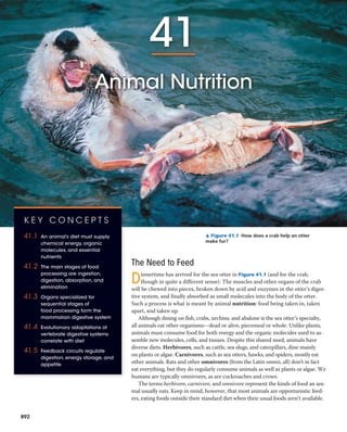
  • 1. 41 Animal Nutrition The Need to Feed Dinnertime has arrived for the sea otter in Figure 41.1 (and for the crab, though in quite a different sense). The muscles and other organs of the crab will be chewed into pieces, broken down by acid and enzymes in the otter’s diges- tive system, and finally absorbed as small molecules into the body of the otter. Such a process is what is meant by animal nutrition: food being taken in, taken apart, and taken up. Although dining on fish, crabs, urchins, and abalone is the sea otter’s specialty, all animals eat other organisms—dead or alive, piecemeal or whole. Unlike plants, animals must consume food for both energy and the organic molecules used to as- semble new molecules, cells, and tissues. Despite this shared need, animals have diverse diets. Herbivores, such as cattle, sea slugs, and caterpillars, dine mainly on plants or algae. Carnivores, such as sea otters, hawks, and spiders, mostly eat other animals. Rats and other omnivores (from the Latin omnis, all) don’t in fact eat everything, but they do regularly consume animals as well as plants or algae. We humans are typically omnivores, as are cockroaches and crows. The terms herbivore, carnivore, and omnivore represent the kinds of food an ani- mal usually eats. Keep in mind, however, that most animals are opportunistic feed- ers, eating foods outside their standard diet when their usual foods aren’t available. ▲ Figure 41.1 How does a crab help an otter make fur? K E Y C O N C E P T S 41.1 An animal’s diet must supply chemical energy, organic molecules, and essential nutrients 41.2 The main stages of food processing are ingestion, digestion, absorption, and elimination 41.3 Organs specialized for sequential stages of food processing form the mammalian digestive system 41.4 Evolutionary adaptations of vertebrate digestive systems correlate with diet 41.5 Feedback circuits regulate digestion, energy storage, and appetite 892
  • 2. CHAPT ER 41 Animal Nutrition 893 For example, deer are herbivores, but in addition to feed- ing on grass and other plants, they occasionally eat insects, worms, or bird eggs. Note as well that microorganisms are an unavoidable “supplement” in every animal’s diet. Animals must eat. But to survive and reproduce, they must also balance their consumption, storage, and use of food. Sea otters, for example, support a high rate of metabolism by eating up to 25% of their body mass each day. Eating too little food, too much food, or the wrong mixture of foods can endanger an animal’s health. In this chapter, we’ll survey the nutritional requirements of animals, explore diverse evolu- tionary adaptations for obtaining and processing food, and investigate the regulation of energy intake and expenditure. proteins, and lipids, for use in cellular respiration and en- ergy storage. In addition to providing fuel for ATP production, an animal’s diet must supply the raw materials needed for bio- synthesis. To build the complex molecules it needs to grow, maintain itself, and reproduce, an animal must obtain two types of organic precursors from its food. Animals need a source of organic carbon (such as sugar) and a source of or- ganic nitrogen (such as protein). Starting with these materials, animals can construct a great variety of organic molecules. Essential Nutrients Some cellular processes require materials that an animal cannot assemble from simpler organic precursors. These materials—preassembled organic molecules and miner- als—are called essential nutrients. Obtained from an ani- mal’s diet, essential nutrients include essential amino acids and fatty acids, vitamins, and minerals. Essential nutrients have key functions in cells, including serving as substrates of enzymes, as coenzymes, and as cofactors in biosynthetic reactions (Figure 41.2). Needs for particular nutrients vary among species. For instance, ascorbic acid (vitamin C) is an essential nutrient for humans and other primates, as well as guinea pigs, but not for many other animals. Essential Amino Acids Animals require 20 amino acids to make proteins (see Figure 5.14). Most animal species have the enzymes to synthesize about half of these amino acids, as long as their diet includes sulfur and organic nitrogen. The remaining amino acids must be obtained from food in prefab- ricated form and are therefore called essential amino acids. Many animals, including adult humans, require eight amino acids in their diet: isoleucine, leucine, lysine, methio- nine, phenylalanine, threonine, trypto- phan, and valine. (Human infants also need a ninth, histidine.) The proteins in animal products such as meat, eggs, and cheese are “complete,” which means that they provide all the essential amino acids in their proper proportions. In contrast, most plant proteins are “incomplete,” being deficient in one or more es- sential amino acids. Corn (maize), for example, is deficient in tryptophan and lysine, whereas beans are lacking in methionine. However, vegetarians can easily obtain all of the essential amino acids by eating a varied diet of plant proteins. C O N C E P T 41.1 An animal’s diet must supply chemical energy, organic molecules, and essential nutrients Overall, an adequate diet must satisfy three nutritional needs: chemical energy for cellular processes, organic build- ing blocks for macromolecules, and essential nutrients. The activities of cells, tissues, organs, and whole animals depend on sources of chemical energy in the diet. This en- ergy is used to produce ATP, which powers processes rang- ing from DNA replication and cell division to vision and flight. To meet the continuous requirement for ATP, ani- mals ingest and digest nutrients, including carbohydrates, Linoleic acid γ-Linoleic acid Fatty acid desaturase Phospholipids (cell membrane components) Prostaglandins (used in cell signaling) NADH ESSENTIAL AMINO ACIDS (monomers of polypeptide) VITAMIN (coenzyme) ESSENTIAL FATTY ACID (substrate of enzyme) Vitamin B3 MINERAL (cofactor) Iron Tyr Glu Gly Phe Phe Leu Ile ▲ Figure 41.2 Roles of essential nutrients.Linoleic acid is converted by the enzyme fatty acid desaturase to γ-linoleic acid, a precursor for phospholipids and prostaglandins. This biosynthetic reac- tion illustrates common functions of the four classes of essential nutrients, labeled in blue. Note that nearly every enzyme or other protein in animal bodies contains some essential amino acids, as indi- cated in the partial sequence shown for fatty acid desaturase.
  • 3. 894 UNIT SEVEN Animal Form and Function Vitamins are classified as water-soluble or fat-soluble (Table 41.1). B vitamins, which generally act as coenzymes, are water-soluble. So is vitamin C, which is required for the production of connective tissue. Fat-soluble vitamins in- clude vitamin A, which is incorporated into visual pigments of the eye, and vitamin D, which aids in calcium absorption and bone formation. The dietary requirement for vitamin D is variable in humans because we can synthesize it from other molecules when our skin is exposed to sunlight. For people with imbalanced diets, taking vitamin supple- ments that provide recommended daily levels is certainly reasonable. It is far less clear that massive doses of vita- mins confer any health benefits or are even safe. Moderate overdoses of water-soluble vitamins are probably harmless because excesses are excreted in urine. However, excesses of fat-soluble vitamins are deposited in body fat, so overcon- sumption may cause them to accumulate to toxic levels. Minerals Dietary minerals are inorganic nutrients, such as iron and sulfur, that are usually required in small amounts—from less than 1 mg to about 2,500 mg per day. As shown in Table 41.2, Essential Fatty Acids Animals require fatty acids to synthesize a variety of cellular components, including membrane phospholipids, signaling molecules, and storage fats. Although animals can synthe- size many fatty acids, they lack the enzymes to form the double bonds found in certain required fatty acids. Instead, these molecules must be obtained from the diet and are considered essential fatty acids. In mammals, they include linoleic acid (see Figure 41.2). Because seeds, grains, and vegetables generally furnish ample quantities of essential fatty acids, deficiencies in this class of nutrients are rare. Vitamins As Albert Szent-Györgyi, the discoverer of vitamin C, once quipped, “A vitamin is a substance that makes you ill if you don’t eat it.” Vitamins are organic molecules that are re- quired in the diet in very small amounts. They have diverse functions. Vitamin B2, for example, is converted in the body to FAD, a coenzyme used in many metabolic processes, in- cluding cellular respiration (see Figure 9.12). For humans, 13 vitamins have been identified. Depending on the vitamin, the required amount ranges from 0.01 to 100 mg per day. Vitamin Major Dietary Sources Major Functions in the Body Symptoms of Deficiency Water-Soluble Vitamins B1 (thiamine) Pork, legumes, peanuts, whole grains Coenzyme used in removing CO2 from organic compounds Beriberi (tingling, poor coordina- tion, reduced heart function) B2 (riboflavin) Dairy products, meats, enriched grains, vegetables Component of coenzymes FAD and FMN Skin lesions, such as cracks at cor- ners of mouth B3 (niacin) Nuts, meats, grains Component of coenzymes NAD+ and NADP+ Skin and gastrointestinal lesions, delusions, confusion B5 (pantothenic acid) Meats, dairy products, whole grains, fruits, vegetables Component of coenzyme A Fatigue, numbness, tingling of hands and feet B6 (pyridoxine) Meats, vegetables, whole grains Coenzyme used in amino acid metabolism Irritability, convulsions, muscular twitching, anemia B7 (biotin) Legumes, other vegetables, meats Coenzyme in synthesis of fat, gly- cogen, and amino acids Scaly skin inflammation, neuro- muscular disorders B9 (folic acid) Green vegetables, oranges, nuts, legumes, whole grains Coenzyme in nucleic acid and amino acid metabolism Anemia, birth defects B12 (cobalamin) Meats, eggs, dairy products Production of nucleic acids and red blood cells Anemia, numbness, loss of balance C (ascorbic acid) Citrus fruits, broccoli, tomatoes Used in collagen synthesis; antioxidant Scurvy (degeneration of skin and teeth), delayed wound healing Fat-Soluble Vitamins A (retinol) Dark green and orange vegetables and fruits, dairy products Component of visual pigments; maintenance of epithelial tissues Blindness, skin disorders, impaired immunity D Dairy products, egg yolk Aids in absorption and use of cal- cium and phosphorus Rickets (bone deformities) in chil- dren, bone softening in adults E (tocopherol) Vegetable oils, nuts, seeds Antioxidant; helps prevent dam- age to cell membranes Nervous system degeneration K (phylloquinone) Green vegetables, tea; also made by colon bacteria Important in blood clotting Defective blood clotting Table 41.1 Vitamin Requirements of Humans
  • 4. CHAPT ER 41 Animal Nutrition 895 Mineral Major Dietary Sources Major Functions in the Body Symptoms of Deficiency Morethan200mgperdayrequired       Calcium (Ca) Dairy products, dark green vegeta- bles, legumes Bone and tooth formation, blood clotting, nerve and muscle function Impaired growth, loss of bone mass Phosphorus (P) Dairy products, meats, grains Bone and tooth formation, acid- base balance, nucleotide synthesis Weakness, loss of minerals from bone, calcium loss Sulfur (S) Proteins from many sources Component of certain amino acids Impaired growth, fatigue, swelling Potassium (K) Meats, dairy products, many fruits and vegetables, grains Acid-base balance, water balance, nerve function Muscular weakness, paralysis, nau- sea, heart failure Chlorine (Cl) Table salt Acid-base balance, formation of gastric juice, nerve function, osmotic balance Muscle cramps, reduced appetite Sodium (Na) Table salt Acid-base balance, water balance, nerve function Muscle cramps, reduced appetite Magnesium (Mg) Whole grains, green leafy vegetables Enzyme cofactor; ATP bioenergetics Nervous system disturbances Iron (Fe) Meats, eggs, legumes, whole grains, green leafy vegetables Component of hemoglobin and of electron carriers; enzyme cofactor Iron-deficiency anemia, weakness, impaired immunity Fluorine (F) Drinking water, tea, seafood Maintenance of tooth structure Higher frequency of tooth decay Iodine (I) Seafood, iodized salt Component of thyroid hormones Goiter (enlarged thyroid gland) *Additional minerals required in trace amounts include cobalt (Co), copper (Cu), manganese (Mn), molybdenum (Mo), selenium (Se), and zinc (Zn). All of these minerals, as well as those in the table, can be harmful in excess. Table 41.2 Mineral Requirements of Humans* i minerals have diverse functions in animal physiology. Some are assembled into the structure of proteins; iron, for ex- ample, is incorporated into the oxygen carrier hemoglobin as well as some enzymes (see Figure 41.2). In contrast, sodium, potassium, and chloride are important in the functioning of nerves and muscles and in maintaining osmotic balance be- tween cells and the surrounding body fluid. In vertebrates, the mineral iodine is incorporated into thyroid hormone, which regulates metabolic rate. Vertebrates also require relatively large quantities of calcium and phosphorus for building and maintaining bone. Ingesting large amounts of some minerals can upset ho- meostatic balance and impair health. For example, excess salt (sodium chloride) can contribute to high blood pressure. This is a particular problem in the United States, where the typical person consumes enough salt to provide about 20 times the required amount of sodium. Packaged (prepared) foods often contain large amounts of sodium chloride, even if they do not taste very salty. Dietary Deficiencies A diet that lacks one or more essential nutrients or consis- tently supplies less chemical energy than the body requires results in malnutrition, a failure to obtain adequate nutri- tion. Malnutrition resulting from either type of dietary defi- ciency can have negative impacts on health and survival and affects one out of four children worldwide. ▲ Figure 41.3 Obtaining essential nutrients from an unusual source.A juvenile chamois (Rupicapra rupicapra), an herbivore, licks salts from exposed rocks in its alpine habitat. This behavior is common among herbivores that live where soils and plants provide insufficient amounts of minerals, such as sodium, calcium, phosphorus, and iron. Deficiencies in Essential Nutrients Insufficient intake of essential nutrients can cause defor- mities, disease, and even death. For example, cattle, deer, and other herbivores may develop dangerously fragile bones if they graze on plants growing in soil that lacks phosphorus. In such environments, some grazing animals obtain missing nutrients by consuming concentrated sources of salt or other minerals (Figure 41.3). Similarly, some birds supplement their diet with snail shells, and certain tortoises ingest stones.
  • 5. 896 UNIT SEVEN Animal Form and Function spinal cord (see Concept 47.2). The English scientist Richard Smithells thought that malnutrition among these women might be responsible. As described in Figure 41.4, he found that vitamin supplementation greatly reduced the risk of neural tube defects. In other studies, he obtained evidence that folic acid (vitamin B9) was the specific vitamin responsi- ble, a finding confirmed by other researchers. Based on this evidence, the United States in 1998 began to require that folic acid be added to enriched grain products used to make bread, cereals, and other foods. Follow-up studies have doc- umented the effectiveness of this program in reducing the frequency of neural tube defects. Thus, at a time when mi- crosurgery and sophisticated diagnostic imaging dominate Like other animals, humans sometimes suffer from diets lacking in essential nutrients. A diet that provides insufficient amounts of one or more essential amino acids causes protein deficiency, the most common type of malnutrition among humans. In children, protein deficiency may arise if their diet shifts from breast milk to foods that contain relatively little protein, such as rice. Such children, if they survive infancy, often have impaired physical and mental development. In populations subsisting on simple rice diets, individuals are often deficient in vitamin A, which can result in blind- ness or death. To overcome this problem, scientists have engineered “Golden Rice,” a strain of rice that synthesizes the orange-colored pigment beta-carotene, which the body converts to vitamin A (see Concept 38.3). Undernutrition A diet that fails to provide adequate sources of chemical energy results in undernutrition. When an animal is un- dernourished, a series of events unfold: The body uses up stored carbohydrates and fat and then begins breaking down its own proteins for fuel; muscles begin to decrease in size; and the brain may become protein-deficient. If energy in- take remains less than energy expenditures, the animal will eventually die. Even if a seriously undernourished animal survives, some of the damage may be irreversible. Human undernutrition is most common when drought, war, or another crisis severely disrupts the food supply. In sub-Saharan Africa, where the AIDS epidemic has crippled both rural and urban communities, approximately 200 mil- lion children and adults cannot obtain enough food. Sometimes undernutrition occurs within well-fed human populations as a result of eating disorders. For example, anorexia nervosa leads individuals, usually female, to starve themselves compulsively. Assessing Nutritional Needs Determining the ideal diet for the human population is an important but difficult problem for scientists. As objects of study, people present many challenges. Unlike laboratory animals, humans are genetically diverse. They also live in settings far more varied than the stable and uniform envi- ronment that scientists use to facilitate comparisons in labo- ratory experiments. Ethical concerns present an additional barrier. For example, it is not acceptable to investigate the nutritional needs of children in a way that might harm a child’s growth or development. Many insights into human nutrition have come from epidemiology, the study of human health and disease at the population level. In the 1970s, for instance, researchers dis- covered that children born to women of low socioeconomic status were more likely to have neural tube defects, which occur when tissue fails to enclose the developing brain and Inquiry Can diet influence the frequency of birth defects? ▼ Figure 41.4 Experiment Richard Smithells, of the University of Leeds, in England, examined the effect of vitamin supplementation on the risk of neural tube defects. Women who had had one or more babies with such a defect were put into two study groups. The experimental group consisted of those who were planning a pregnancy and began taking a multivitamin at least four weeks before attempting conception. The control group, who were not given vitamins, included women who declined them and women who were already pregnant. The numbers of neural tube defects resulting from the pregnancies were recorded for each group. Results Group Number of Infants/Fetuses Studied Infants/Fetuses with a Neural Tube Defect Vitamin supplements (experimental group) 141 1 No vitamin supple- ments (control group) 204 12 Conclusion This controlled study provided evidence that vitamin supplementation protects against neural tube defects, at least after the first pregnancy. Follow-up trials demonstrated that folic acid alone provided an equivalent protective effect. Source: R. W. Smithells et al., Possible prevention of neural-tube defects by pericon- ceptional vitamin supplementation, Lancet 315:339–340 (1980). Inquiry in Action Read and analyze the original paper in Inquiry in Action: Interpreting Scientific Papers. I N T E R P R E T T H E DATA After folic acid supplementation became standard in the U.S., the frequency of neural tube defects dropped to an average of just 1 in 5,000 live births. Propose two explanations why the observed frequency was much higher in the experimental group of the Smithells study. W H AT I F ? Subsequent studies were designed to learn if folic acid supplements prevent neural tube defects during first-time pregnancies. To determine the required number of subjects, what type of additional infor- mation did the researchers need?
  • 6. CHAPT ER 41 Animal Nutrition 897 the headlines, a simple dietary change such as folic acid supplementation may be among the greatest contributors to human health. that have evolved in animals. We will focus in this chapter on the shared aspects of food processing, pausing periodi- cally to consider some adaptations to particular diets or environments. During digestion, the second stage of food processing, food is broken down into molecules small enough for the body to absorb. Mechanical digestion, such as chewing, typically precedes chemical digestion. Mechanical diges- tion breaks food into smaller pieces, increasing the surface area available for chemical processes. Chemical digestion is necessary because animals cannot directly use the proteins, carbohydrates, nucleic acids, fats, and phospholipids in food. One problem is that these molecules are too large to pass through membranes and enter the cells of the animal. In addition, the large molecules in food are not all identi- cal to those the animal needs for its particular tissues and functions. When large molecules in food are broken down into their components, however, the animal can use these smaller molecules to assemble the large molecules it needs. For example, although fruit flies and humans have very dif- ferent diets, both convert proteins in their food to the same 20 amino acids from which they assemble all of the specific proteins in their bodies. A cell makes a macromolecule or fat by linking together smaller components; it does so by removing a molecule of water for each new covalent bond formed. Chemical diges- tion by enzymes reverses this process by breaking bonds through the addition of water (see Figure 5.2). This splitting process is called enzymatic hydrolysis. A variety of enzymes catalyze the digestion of large molecules in food. Polysac- charides and disaccharides are split into simple sugars; pro- teins are broken down into amino acids; and nucleic acids are cleaved into nucleotides and their components. Enzy- matic hydrolysis also releases fatty acids and other compo- nents from fats and phospholipids. In many animals, such as the gorilla in Figure 41.5, digestion of some materials is accomplished by bacteria living in the digestive system. The last two stages of food processing occur after the food is digested. In the third stage, absorption, the animal’s cells take up (absorb) small molecules such as amino acids and simple sugars. Elimination completes the process as undigested material passes out of the digestive system. Digestive Compartments In our overview of food processing, we have seen that diges- tive enzymes hydrolyze the same biological materials (such as proteins, fats, and carbohydrates) that make up the bod- ies of the animals themselves. How, then, are animals able to digest food without digesting their own cells and tissues? The evolutionary adaptation that allows animals to avoid self-digestion is the processing of food within specialized intracellular or extracellular compartments. C O N C E P T C H E C K 4 1 . 1 1. All 20 amino acids are needed to make animal proteins. Why aren’t they all essential to animal diets? 2. M A K E C O N N E C T I O N S Considering the role of en- zymes in metabolic reactions (see Concept 8.4), explain why vitamins are required in very small amounts in the diet. 3. W H AT I F ? If a zoo animal eating ample food shows signs of malnutrition, how might a researcher determine which nutrient is lacking in its diet? For suggested answers, see Appendix A. C O N C E P T 41.2 The main stages of food processing are ingestion, digestion, absorption, and elimination In this section, we turn from nutritional requirements to the mechanisms by which animals process food. Food process- ing can be divided into four distinct stages: ingestion, diges- tion, absorption, and elimination (Figure 41.5). The first stage, ingestion, is the act of eating or feeding. Given the variation in food sources among animal species, it is not surprising that strategies for extracting resources from food also differ widely. Figure 41.6, on the next page, surveys and classifies the principal feeding mechanisms Mechanical digestion Chemical digestion (enzymatic hydrolysis) Nutrient molecules enter body cells Undigested material INGESTION1 DIGESTION2 ABSORPTION3 ELIMINATION4 ▲ Figure 41.5 The stages of food processing.
  • 7. Filter Feeding Many aquatic animals are filter feeders, which strain small organ- isms or food particles from the surrounding medium. The humpback whale, shown above, is one example. Attached to the whale’s upper jaw are comblike plates called baleen, which remove small inverte- brates and fish from enormous volumes of water and sometimes mud. Filter feeding in water is a type of suspension feeding, which also includes removing suspended food particles from the surrounding medium by capture or trapping mechanisms. Substrate Feeding Substrate feeders are animals that live in or on their food source. This leaf miner cater- pillar, the larva of a moth, is eating through the soft tissue of an oak leaf, leaving a dark trail of feces in its wake. Other sub- strate feeders include maggots (fly larvae), which burrow into animal carcasses. Caterpillar Feces Fluid Feeding Fluid feeders suck nutrient- rich fluid from a living host. This mosquito has pierced the skin of its human host with hollow, needlelike mouthparts and is consuming a blood meal (colorized SEM). Similarly, aphids are fluid feeders that tap the phloem sap of plants. In contrast to such parasites, some fluid feeders actually benefit their hosts. For example, hummingbirds and bees move pollen between flowers as they fluid-feed on nectar. Bulk Feeding it whole—even if the prey is much bigger than the diameter of the snake. They can do so because the lower jaw is loosely hinged to the skull by an elastic ligament that permits the mouth and throat to open very wide. After swallowing its prey, which may take more than an hour, the python will spend two weeks or longer digesting its meal. Baleen Most animals, including humans, are bulk feeders, which eat relatively large pieces of food. Their adaptations include tentacles, pincers, claws, venomous fangs, jaws, and teeth that kill their prey or tear off pieces of meat or vegetation. In this amazing scene, a rock python is beginning to ingest a gazelle it has captured and killed. Snakes cannot chew their food into pieces and must swallow ▼ Figure 41.6 Exploring Four Main Feeding Mechanisms of Animals 898 UNIT SEVEN Animal Form and Function
  • 8. CHAPT ER 41 Animal Nutrition 899 Intracellular Digestion Food vacuoles—cellular organelles in which hydrolytic en- zymes break down food—are the simplest digestive com- partments. The hydrolysis of food inside vacuoles, called intracellular digestion, begins after a cell engulfs solid food by phagocytosis or liquid food by pinocytosis (see Figure 7.19). Newly formed food vacuoles fuse with lysosomes, organelles containing hydrolytic enzymes. This fusion of organelles brings food in contact with the enzymes, allowing digestion to occur safely within a compartment enclosed by a protective membrane. A few animals, such as sponges, digest their food entirely by this intracellular mechanism (see Figure 33.4). Extracellular Digestion In most animal species, hydrolysis occurs largely by extra- cellular digestion, the breakdown of food in compartments that are continuous with the outside of the animal’s body. Having one or more extracellular compartments for diges- tion enables an animal to devour much larger pieces of food than can be ingested by phagocytosis. Many animals with relatively simple body plans have a digestive compartment with a single opening (Figure 41.7). This pouch, called a gastrovascular cavity, functions in digestion as well as in the distribution of nutrients through- out the body (hence the vascular part of the term). The cnidarians called hydras provide a good example of how a gastrovascular cavity works. A carnivore, the hydra uses its tentacles to stuff captured prey through its mouth into its gastrovascular cavity. Specialized gland cells of the hydra’s gastrodermis, the tissue layer that lines the cavity, then se- crete digestive enzymes that break the soft tissues of the prey into tiny pieces. Other cells of the gastrodermis engulf these food particles, and most of the hydrolysis of macromolecules occurs intracellularly, as in sponges. After the hydra has digested its meal, undigested materials that remain in its gas- trovascular cavity, such as exoskeletons of small crustaceans, are eliminated through its mouth. Many flatworms also have a gastrovascular cavity (see Figure 33.10). In contrast with cnidarians and flatworms, most animals have a digestive tube extending between two openings, a mouth and an anus (Figure 41.8). Such a tube is called a complete digestive tract or, more commonly, an alimentary canal. Because food moves along the alimentary canal in a Mouth Esophagus Pharynx Crop Gizzard Intestine Anus Mouth Esophagus Crop Gizzard Intestine Anus Stomach (a) Mouth Esophagus Foregut Crop Anus Rectum Gastric cecae Midgut Hindgut (b) Grasshopper. A grasshopper has several digestive chambers grouped into three main regions: a foregut, with an esophagus and crop; a midgut; and a hindgut. Food is moistened and stored in the crop, but most digestion occurs in the midgut. Pouches called gastric cecae (singular, ceca) extend from the beginning of the midgut and function in digestion and absorption. Earthworm. The alimentary canal of an earthworm includes a muscular pharynx that sucks food in through the mouth. Food passes through the esophagus and is stored and moistened in the crop. Mechanical digestion occurs in the muscular gizzard, which pulverizes food with the aid of small bits of sand and gravel. Further digestion and absorption occur in the intestine. (c) Bird. Many birds have a crop for storing food and a stomach and gizzard for mechanically digesting it. Chemical digestion and absorption of nutrients occur in the intestine. ▲ Figure 41.8 Alimentary canals.These examples illustrate varia- tion in the organization and structure of compartments that carry out stepwise digestion, storage, and absorption in different animals. 3 Food particles are engulfed and digested in food vacuoles. 2 Enzymes break food down into small particles. Mouth GastrodermisEpidermis Food (Daphnia, a water flea) Tentacles 1 Digestive enzymes are released from a gland cell. 3 2 ▲ Figure 41.7 Digestion in a hydra.Digestion begins in the gas- trovascular cavity and is completed intracellularly after small food par- ticles are engulfed by specialized cells of the gastrodermis.
  • 9. 900 UNIT SEVEN Animal Form and Function mammals, the digestive system consists of the alimentary canal and various accessory glands that secrete digestive juices through ducts into the canal (Figure 41.9). The ac- cessory glands of the mammalian digestive system are three pairs of salivary glands, the pancreas, the liver, and the gallbladder. Food is pushed along the alimentary canal by peristalsis, alternating waves of contraction and relaxation in the smooth muscles lining the canal. At some of the junctions between specialized compartments, the muscular layer forms ringlike valves called sphincters. Acting like draw- strings to close off the alimentary canal, sphincters regulate the passage of material between compartments. Using the human digestive system as a model, let’s now follow a meal through the alimentary canal. As we do so, we’ll examine in more detail what happens to the food in each digestive compartment along the way. The Oral Cavity, Pharynx, and Esophagus Ingestion and the initial steps of digestion occur in the mouth, or oral cavity. Mechanical digestion begins as teeth of various shapes cut, mash, and grind food, making the food easier to swallow and increasing its surface area. Mean- while, the salivary glands deliver saliva through ducts to the oral cavity. The release of saliva when food enters the mouth is a reflex, an automatic reaction mediated by the nervous system. Saliva may also be released before food enters the mouth, triggered by a learned association between eating and the time of day, a cooking odor, or another stimulus. Tongue Oral cavity Large intestine Rectum Anus Salivary glands Pharynx Gall- bladder Pancreas Small intestine Salivary glands Mouth Stomach Small intestine Pancreas Liver Large intestine Rectum Anus Esophagus A schematic diagram of the human digestive system (accessory glands in purple) Gall- bladder Esophagus Sphincter Stomach Duodenum of small intestine Sphincter Liver © Pearson Education, Inc. © 1996 Cengage Learning, Inc. ▶ Figure 41.9 The human digestive system.After food is chewed and swallowed, it takes 5–10 seconds for it to pass down the esophagus and into the stomach, where it spends 2–6 hours being partially digested. Final digestion and nutrient absorption occur in the small intestine over a period of 5–6 hours. Within 12–24 hours, any undigested material passes through the large intestine, and feces are expelled through the anus. C O N C E P T C H E C K 4 1 . 2 1. Distinguish the overall structure of a gastrovascular cavity from that of an alimentary canal. 2. In what sense are nutrients from a recently ingested meal not really “inside” your body prior to the absorption stage of food processing? 3. W H AT I F ? Thinking in broad terms, what similarities can you identify between digestion in an animal body and the breakdown of gasoline in an automobile? (You don’t have to know about auto mechanics.) For suggested answers, see Appendix A. C O N C E P T 41.3 Organs specialized for sequential stages of food processing form the mammalian digestive system Because most animals, including mammals, have an ali- mentary canal, the mammalian digestive system can serve to illustrate the general principles of food processing. In single direction, the tube can be organized into specialized compartments that carry out digestion and nutrient absorp- tion in a stepwise fashion. An animal with an alimentary canal can ingest food while earlier meals are still being di- gested, a feat that is likely to be difficult or inefficient for an animal with a gastrovascular cavity. In the next section, we’ll explore the organization of a mammalian alimentary canal.
  • 10. CHAPT ER 41 Animal Nutrition 901 vigorous coughing, a series of back slaps, or a forced upward thrust of the diaphragm (the Heimlich maneuver). Digestion in the Stomach The stomach, which is located just below the diaphragm, stores food and begins digestion of proteins. With accordion- like folds and a very elastic wall, this organ can stretch to accommodate about 2 L of food and fluid. The stomach se- cretes a digestive fluid called gastric juice and mixes it with the food through a churning action. This mixture of ingested food and gastric juice is called chyme. Chemical Digestion in the Stomach Two components of gastric juice carry out chemical diges- tion. One is hydrochloric acid (HCl), which disrupts the extracellular matrix that binds cells together in meat and plant material. The concentration of HCl is so high that the pH of gastric juice is about 2, acidic enough to dissolve iron nails (and to kill most bacteria). This low pH denatures (un- folds) proteins in food, increasing exposure of their peptide bonds. The exposed bonds are attacked by the second com- ponent of gastric juice—a protease, or protein-digesting enzyme, called pepsin. Unlike most enzymes, pepsin works best in a very acidic environment. By breaking peptide bonds, it cleaves proteins into smaller polypeptides. Fur- ther digestion to individual amino acids occurs in the small intestine. Why doesn’t gastric juice destroy the stomach cells that make it? The answer is that the ingredients of gastric juice are kept inactive until they are released into the lumen (cav- ity) of the stomach. The components of gastric juice are produced by two types of cells in the gastric glands of the stomach. Parietal cells use an ATP-driven pump to expel hydrogen ions into the lumen. At the same time, chloride ions diffuse into the Saliva initiates chemical digestion while also protecting the oral cavity. The enzyme amylase, found in saliva, hydro- lyzes starch (a glucose polymer from plants) and glycogen (a glucose polymer from animals) into smaller polysaccharides and the disaccharide maltose. Much of the protective effect of saliva is provided by mucus, a viscous mixture of water, salts, cells, and slippery glycoproteins (carbohydrate-protein complexes) called mucins. Mucus in saliva protects the lining of the mouth from abrasion and lubricates food for easier swallowing. Additional components of saliva include buffers, which help prevent tooth decay by neutralizing acid, and antimicrobial agents (such as lysozyme; see Figure 5.16), which protect against bacteria that enter the mouth with food. Much as a doorman screens and assists people enter- ing a fancy hotel, the tongue aids digestive processes by evaluating ingested material and then enabling its further passage. When food arrives at the oral cavity, the tongue plays a critical role in distinguishing which foods should be processed further. (See Chapter 50 for a discussion of the sense of taste.) After food is deemed acceptable and chew- ing commences, tongue movements manipulate the mix- ture of saliva and food, helping shape it into a ball called a bolus. During swallowing, the tongue provides further help, pushing the bolus to the back of the oral cavity and into the pharynx. The pharynx, or throat region, opens to two passageways: the trachea (windpipe) and the esophagus (Figure 41.10). The trachea leads to the lungs (see Figure 42.23), whereas the esophagus connects to the stomach. Once food enters the esophagus, peristaltic contractions of smooth muscle move each bolus to the stomach. Swallowing must be carefully choreographed to keep food and liquids from entering the trachea and causing chok- ing, a blockage of the trachea. The resulting lack of airflow into the lungs can be fatal if the material is not dislodged by Larynx Trachea To lungs To stomach Bolus of food Epiglottis up Glottis up and closed Esophageal sphincter contracted Esophageal sphincter relaxed Esophagus Tongue Pharynx Glottis Epiglottis down (a) Trachea open (b) Esophagus open ▶ Figure 41.10 Intersection of the human airway and digestive tract.In hu- mans, the pharynx connects to the trachea and the esophagus. (a) At most times, a contracted sphincter seals off the esophagus while the trachea remains open. (b) When a food bolus arrives at the pharynx, the swallowing reflex is triggered. Movement of the larynx, the upper part of the airway, tips a flap of tissue called the epiglottis down, preventing food from entering the trachea. At the same time, the esophageal sphincter relaxes, allowing the bolus to pass into the esophagus. The trachea then reopens, and peristaltic contractions of the esophagus move the bolus to the stomach.
  • 11. 902 UNIT SEVEN Animal Form and Function After hydrochloric acid converts a small amount of pep- sinogen to pepsin, pepsin itself helps activate the remain- ing pepsinogen. Pepsin, like HCl, can clip pepsinogen to expose the enzyme’s active site. This generates more pep- sin, which activates more pepsinogen. This series of events is an example of positive feedback (see Concept 40.2). Why don’t HCl and pepsin eat through the lining of the stomach? For one thing, mucus secreted by cells in gastric glands protects against self-digestion (see Figure 41.11). In addition, cell division adds a new epithelial layer every three days, replacing cells before they are fully eroded by diges- tive juices. Under certain circumstances, however, damaged areas of the stomach lining called gastric ulcers can appear. It had been thought that they were caused by psychologi- cal stress and resulting excess acid secretion. However, Australian researchers Barry Marshall and Robin Warren discovered that infection by the acid-tolerant bacterium Helicobacter pylori causes ulcers. They also demonstrated that an antibiotic could cure most gastric ulcers. For these findings, they were awarded the Nobel Prize in 2005. Stomach Dynamics Chemical digestion by gastric juice is facilitated by the churning action of the stomach. This coordinated series of muscle contractions and relaxations mixes the stomach contents about every 20 seconds. As a result of mixing and enzyme action, what begins as a recently swallowed meal becomes the acidic, nutrient-rich broth known as chyme. Most of the time, sphincters close off the stomach at both ends (see Figure 41.9). The sphincter between the esophagus and the stomach normally opens only when a bolus arrives. Occasionally, however, a person experiences acid reflux, a backflow of chyme from the stomach into the lower end of the esophagus. The resulting irritation of the esophagus is commonly called “heartburn.” Peristaltic contractions typically empty the contents of the stomach into the small intestine within 2–6 hours after a meal. The sphincter located where the stomach opens to the small intestine helps regulate passage into the small intes- tine, allowing only one squirt of chyme at a time. Digestion in the Small Intestine Although chemical digestion of some nutrients begins in the oral cavity or stomach, most enzymatic hydrolysis of the macromolecules from food occurs in the small intestine (Figure 41.12). The small intestine is the alimentary canal’s longest compartment—over 6 m (20 feet) long in humans! Its name refers to its small diameter, compared with that of the large intestine. The first 25 cm (10 inches) or so of the small intestine forms the duodenum. It is here that chyme from the stomach mixes with digestive juices from the pan- creas, liver, and gallbladder, as well as from gland cells of the Epithelium Stomach Pepsinogen and HCl are introduced into the lumen of the stomach. The production of gastric juice Pepsin then activates more pepsinogen, starting a chain reaction. Pepsin begins the chemical digestion of proteins. HCl converts pepsinogen to pepsin. 1 3 21 2 3 Pepsin (active enzyme) Chief cell Parietal cell Mucous cells secrete mucus, which lubricates and protects the cells lining the stomach. Chief cells secrete pepsinogen, an inactive form of the digestive enzyme pepsin. Parietal cells produce the components of hydrochloric acid (HCl). Interior surface of stomach. The interior surface of the stomach wall is highly folded and dotted with pits leading into tubular gastric glands. Pepsinogen HCl H+ Cl– Gastric gland. The gastric glands have three types of cells that secrete different components of the gastric juice: mucous cells, chief cells, and parietal cells. ▲ Figure 41.11 The stomach and its secretions. lumen through specific membrane channels of the parietal cells. It is therefore only within the lumen that hydrogen and chloride ions combine to form HCl (Figure 41.11). Meanwhile, chief cells release pepsin into the lumen in an inactive form called pepsinogen. HCl converts pepsinogen to active pepsin by clipping off a small portion of the molecule and exposing its active site. Through these processes, both HCl and pepsin form in the lumen of the stomach, not within the cells of the gastric glands.
  • 12. CHAPT ER 41 Animal Nutrition 903 that is made in the liver. Bile contains bile salts, which act as emulsifiers (detergents) that aid in digestion and absorption of lipids. Bile is stored and concentrated in the gallbladder. Bile production is integral to one of the other vital func- tions of the liver: the destruction of red blood cells that are no longer fully functional. In producing bile, the liver incor- porates some pigments that are by-products of red blood cell disassembly. These bile pigments are then eliminated from the body with the feces. In some liver or blood disor- ders, bile pigments accumulate in the skin, resulting in a characteristic yellowing called jaundice. Secretions of the Small Intestine The epithelial lining of the duodenum is the source of sev- eral digestive enzymes (see Figure 41.12). Some are secreted into the lumen of the duodenum, whereas others are bound to the surface of epithelial cells. intestinal wall itself. As you will see in Concept 41.5, hor- mones released by the stomach and duodenum control the digestive secretions into the alimentary canal. Pancreatic Secretions The pancreas aids chemical digestion by producing an alka- line solution rich in bicarbonate as well as several enzymes (see Figure 41.12). The bicarbonate neutralizes the acidity of chyme and acts as a buffer. Among the pancreatic enzymes are trypsin and chymotrypsin, proteases secreted into the duodenum in inactive forms. In a chain reaction similar to the activation of pepsin, they are activated when safely lo- cated in the lumen of the duodenum. Bile Production by the Liver Digestion of fats and other lipids begins in the small intestine and relies on the production of bile, a mixture of substances ORAL CAVITY, PHARYNX, ESOPHAGUS Polysaccharides (starch, glycogen) Salivary amylase Pepsin Pancreatic carboxypeptidase Nucleotidases Nucleosidases and phosphatases Pancreatic lipase Pancreatic trypsin and chymotrypsin (These protein- digesting enzymes, or proteases, cleave bonds adjacent to certain amino acids.) Pancreatic nucleases Pancreatic amylases Disaccharidases Proteins Small polypeptides DNA, RNA Glycerol, fatty acids, monoglycerides Nucleotides Nucleosides Nitrogenous bases, sugars, phosphates Smaller polypeptides Smaller polysaccharides Maltose Disaccharides Monosaccharides Amino acids Small peptides Disaccharides (sucrose, lactose) STOMACH SMALL INTESTINE (enzymes from pancreas) SMALL INTESTINE (enzymes from intestinal epithelium) Dipeptidases, carboxy- peptidase, and aminopeptidase (These proteases each split off one amino acid at a time from a dipeptide or polypeptide.) Fat (triglycerides) (in droplets coated with bile salts) CARBOHYDRATE DIGESTION PROTEIN DIGESTION NUCLEIC ACID DIGESTION FAT DIGESTION ▼ Figure 41.12 Chemical digestion in the human digestive system.The timing and location of chemical breakdown are specific to each class of nutrients. ? Pepsin is resistant to the denaturing effect of the low pH environment of the stomach. Thinking about the different digestive processes that occur in the small intestine, describe an adaptation shared by the diges- tive enzymes in that compartment.
  • 13. 904 UNIT SEVEN Animal Form and Function intestine into the epithelial cells. From there, fructose exits the basal surface and is absorbed into microscopic blood vessels, or capillaries, at the core of each villus. Other nutri- ents, including amino acids, small peptides, vitamins, and most glucose molecules, are pumped against concentration gradients into the epithelial cells of the villus. This active transport allows much more absorption of those nutrients than would be possible with passive diffusion alone. The capillaries and veins that carry nutrient-rich blood away from the villi converge into the hepatic portal vein, a blood vessel that leads directly to the liver. From the liver, blood travels to the heart and then to other tissues and or- gans. This arrangement serves two major functions. First, it allows the liver to regulate the distribution of nutrients to the rest of the body. Because the liver can interconvert many organic molecules, blood that leaves the liver may have a very different nutrient balance than the blood that entered. Second, the arrangement allows the liver to remove toxic substances before the blood circulates broadly. The liver is the primary site for the detoxification of many organic mol- ecules, including drugs, that are foreign to the body. Although many nutrients leave the small intestine through the bloodstream, some products of fat (triglyceride, also known as triacylglycerol) digestion take a different path (Figure 41.14). Hydrolysis of fats by lipase in the small intes- tine generates fatty acids and monoglycerides. (A monoglyc- eride is a single fatty acid joined to glycerol.) These products are absorbed by epithelial cells and recombined into triglyc- erides. They are then coated with phospholipids, cholesterol, While enzymatic hydrolysis proceeds, peristalsis moves the mixture of chyme and digestive juices along the small in- testine. Most digestion is completed in the duodenum. The remaining regions of the small intestine, called the jejunum and ileum, are the major sites for absorption of nutrients, as discussed next. Absorption in the Small Intestine To reach body tissues, nutrients in the lumen must first be absorbed across the lining of the alimentary canal. Most of this absorption occurs at the highly folded surface of the small intestine, as illustrated in Figure 41.13. Large folds in the lining encircle the intestine and are studded with finger- like projections called villi. In turn, each epithelial cell of a villus has on its apical surface many microscopic projections, or microvilli, that are exposed to the intestinal lumen. The many side-by-side microvilli give cells of the intestinal epi- thelium a brush-like appearance that is reflected in the name brush border. Together, the folds, villi, and microvilli of the small intestine have a surface area of 200–300 m2 , roughly the size of a tennis court. This enormous surface area is an evolutionary adaptation that greatly increases the rate of nutrient absorption (see Figure 33.9 for more discussion and examples of maximizing surface area in diverse organisms). Depending on the nutrient, transport across the epithe- lial cells can be passive or active (see Chapter 7). The sugar fructose, for example, moves by facilitated diffusion down its concentration gradient from the lumen of the small Muscle layers Intestinal wall Villi Epithelial cells Villi Large circular folds Vein carrying blood to liver Blood capillaries (toward capillary) Epithelial cells Lymph vessel Basal surface Microvilli (brush border) at apical (lumenal) surface Lumen Lacteal Nutrient absorption Key ▲ Figure 41.13 Nutrient absorption in the small intestine. ? Tapeworms sometimes infect the human alimentary canal, anchoring themselves to the wall of the small intestine. Based on how digestion is compartmentalized along the mammalian alimentary canal, what digestive functions would you expect these parasites to have? Visit the Study Area in MasteringBiology for the BioFlix® 3-D Animation on Mem- brane Transport. A N I M AT I O N
  • 14. CHAPT ER 41 Animal Nutrition 905 recovery occurring in the small intestine. There is no mech- anism for active transport of water. Instead, water is reab- sorbed by osmosis when sodium and other ions are pumped out of the lumen of the intestine. Processing in the Large Intestine The alimentary canal ends with the large intestine, which includes the colon, cecum, and rectum. The small intestine connects to the large intestine at a T-shaped junction (Figure 41.15). One arm of the T is the 1.5-m-long colon, which leads to the rectum and anus. The other arm is a pouch called the cecum. The cecum is important for fermenting ingested material, especially in animals that eat large amounts of plant material. Compared with many other mammals, humans have a small cecum. The appendix, a finger-like extension of the human cecum, has a minor and dispensable role in immunity. The colon completes the reabsorption of water that began in the small intestine. What remain are the feces, the wastes of the digestive system, which become increasingly solid as they are moved along the colon by peristalsis. It takes approximately 12–24 hours for material to travel the length of the colon. If the lining of the colon is irritated—by a viral or bacterial infection, for instance—less water than normal may be reabsorbed, resulting in diarrhea. The op- posite problem, constipation, occurs when the feces move along the colon too slowly. Too much water is reabsorbed, and the feces become compacted. The undigested material in feces includes cellulose fiber. Although it provides no caloric value (energy) to humans, fiber helps move food along the alimentary canal. A rich community of mostly harmless bacteria lives on the unabsorbed organic material in the human colon, con- tributing approximately one-third of the dry weight of feces. As by-products of their metabolism, many colon bacteria generate gases, including methane and hydrogen sulfide, the latter of which has an offensive odor. These gases and in- gested air are expelled through the anus. The terminal portion of the large intestine is the rectum, where the feces are stored until they can be eliminated. Be- tween the rectum and the anus are two sphincters, the inner one being involuntary and the outer one being voluntary. Periodically, strong contractions of the colon create an urge and proteins, forming globules called chylomicrons. Being water soluble, chylomicrons can dissolve in the blood and travel via the circulatory system. Before reaching the bloodstream, chylomicrons are first transported from an epithelial cell in the intestine into a lacteal, a vessel at the core of each villus (see Figures 41.13 and 41.14). Lacteals are part of the vertebrate lymphatic system, which is a network of vessels filled with a clear fluid called lymph. Starting at the lacteals, lymph containing the chylomicrons passes into the larger vessels of the lymphatic system and eventually into large veins that return the blood to the heart. In addition to absorbing nutrients, the small intestine has an important function in the recovery of water and ions. Each day we consume about 2 L of water and secrete another 7 L in digestive juices. Typically all but 0.1 L of the water is reabsorbed in the intestines, with most of the LUMEN OF SMALL INTESTINE Epithelial cell Lacteal Phospholipids, cholesterol, and proteins Chylomicron Fatty acids Triglycerides Monoglycerides Triglycerides 1 In the lumen, triglycerides (fat molecules) exposed on the surface of fat droplets (not shown) are subject to enzymatic hydrolysis. The enzyme lipase breaks the triglycerides down to fatty acids and monoglycerides. 2 After diffusing into epithelial cells, monoglycerides and fatty acids are re-formed into triglyc- erides. (Some glycerol and fatty acids pass directly into capillaries.) 3 Triglycerides are incorporated into water-soluble globules called chylomicrons. 4 Chylomicrons leave epithelial cells by exocytosis and enter lacteals, where they are carried away by the lymph and later pass into large veins. ▲ Figure 41.14 Absorption of fats.Because fats are insoluble in water, adaptations are needed to digest and absorb them. Bile salts (not shown) break up large fat droplets and maintain a small droplet size in the intestinal lumen, exposing more of the fat at the surface to enzymatic hydrolysis. The fatty acids and monoglycerides released by hydrolysis can diffuse into epithelial cells, where fats are reassembled and incorporated into water-soluble chylomicrons that enter the lymphatic system. Ascending portion of colon Appendix Cecum Small intestine ▲ Figure 41.15 Junction of the small and large intestines.
  • 15. 906 UNIT SEVEN Animal Form and Function to defecate. Because filling of the stomach triggers a reflex that increases the rate of contractions in the colon, the urge to defecate often follows a meal. We have followed a meal from one opening (the mouth) of the alimentary canal to the other (the anus). Next we’ll look at some adaptations of this general digestive plan in dif- ferent animals. C O N C E P T C H E C K 4 1 . 3 1. Explain why a proton pump inhibitor, such as the drug Prilosec, relieves the symptoms of acid reflux. 2. Thinking about our nutritional needs and feeding behav- ior, propose an evolutionary explanation for why amylase, unlike other digestive enzymes, is secreted into the mouth. 3. W H AT I F ? If you mixed gastric juice with crushed food in a test tube, what would happen? For suggested answers, see Appendix A. C O N C E P T 41.4 Evolutionary adaptations of vertebrate digestive systems correlate with diet E VO L U T I O N The digestive systems of mammals and other vertebrates are variations on a common plan, but there are many intriguing adaptations, often associated with the ani- mal’s diet. To highlight how form fits function, we’ll exam- ine a few of them. Dental Adaptations Dentition, an animal’s assortment of teeth, is one example of structural variation reflecting diet (Figure 41.16). The Carnivore Carnivores, such as members of the dog and cat families, generally have large, pointed incisors and canines that can be used to kill prey and rip or cut away pieces of flesh. The jagged premolars and molars crush and shred food. Incisors MolarsCanines PremolarsKey Herbivore Herbivores, such as horses and deer, usually have premolars and molars with broad, ridged surfaces that grind tough plant material. The incisors and canines are generally modified for biting off pieces of vegetation. In some herbivores, canines are absent. As omnivores, humans are adapted to eating both plants and meat. Adults have 32 teeth. From front to back along either side of the mouth are four bladelike incisors for biting, a pair of pointed canines for tearing, four premolars for grinding, and six molars for crushing (see inset, top view). Omnivore ▼ Figure 41.16 Dentition and diet. evolutionary adaptation of teeth for processing different kinds of food is one of the major reasons mammals have been so successful. For example, the sea otter in Figure 41.1 uses its sharp canine teeth to tear apart prey such as crabs and its slightly rounded molars to crush their shells. Nonmammalian vertebrates generally have less specialized dentition, but there are interesting exceptions. Venomous snakes, such as rattlesnakes, have fangs, modified teeth that inject venom into prey. Some fangs are hollow, like syringes, whereas others drip the toxin along grooves on the surfaces of the teeth. Stomach and Intestinal Adaptations Evolutionary adaptations to differences in diet are some- times apparent as variations in the dimensions of digestive organs. For example, large, expandable stomachs are com- mon in carnivorous vertebrates, which may wait a long time between meals and must eat as much as they can when they do catch prey. An expandable stomach enables a rock py- thon to ingest a whole gazelle (see Figure 41.6) and a 200-kg African lion to consume 40 kg of meat in one meal! Adaptation is also apparent in the length of the digestive system in different vertebrates. In general, herbivores and omnivores have longer alimentary canals relative to their body size than do carnivores. Plant matter is more difficult to digest than meat because it contains cell walls. A longer diges- tive tract furnishes more time for digestion and more surface area for the absorption of nutrients. As an example, consider the coyote and koala in Figure 41.17. Although these two mammals are about the same size, the koala’s intestines are much longer, enhancing the processing of fibrous, protein- poor eucalyptus leaves from which the koala obtains nearly all of its nutrients and water.
  • 16. CHAPT ER 41 Animal Nutrition 907 One recent microbiome study provided an important clue as to why the bacterium H. pylori disrupts stomach health, leading to ulcers. After collecting stomach tissue from uninfected and H. pylori-infected adults, research- ers identified all the bacterial species in each sample. What they found was remarkable: H. pylori infection led to a near complete elimination from the stomach of all other bacte- rial species (Figure 41.18). Such studies on differences in the microbiome as a result of particular diseases holds promise for the development of new and more effective therapies. Mutualistic Adaptations in Herbivores Mutualistic symbiosis is particularly important in herbi- vores. Much of the chemical energy in herbivore diets comes from the cellulose of plant cell walls, but animals do not produce enzymes that hydrolyze cellulose. Instead, many vertebrates (as well as termites, whose wooden diets consist largely of cellulose) host large populations of mutualistic bacteria and protists in fermentation chambers in their ali- mentary canals. These microorganisms have enzymes that can digest cellulose to simple sugars and other compounds that the animal can absorb. In many cases, the microorgan- isms also use the sugars from digested cellulose in the pro- duction of a variety of nutrients essential to the animal, such as vitamins and amino acids. In horses, koalas, and elephants, mutualistic microorgan- isms are housed in a large cecum. In contrast, the hoatzin, an herbivorous bird found in South American rain forests, Percentofsampledstomachbacteria 0 100 80 60 40 20 Phylum Actinobacteria Firm icutesBacteroidetes ProteobacteriaFusobacteria Uninfected individuals Individuals with H. pylori infection H. pylori ▲ Figure 41.18 The stomach microbiome.By copying and se- quencing bacterial DNA in samples obtained from human stomachs, researchers characterized the bacterial community that makes up the stomach microbiome. In samples from individuals infected with Helicobacter pylori, more than 95% of the sequences were from that species, which belongs to the phylum Proteobacteria. The stomach mi- crobiome in uninfected individuals was much more diverse. Mutualistic Adaptations An estimated 10–100 trillion bacteria live in the human digestive system. One bacterial inhabitant, Escherichia coli, is so common in the digestive system that its presence in lakes and streams is a useful indicator of contamination by untreated sewage. The coexistence of humans and many of these bacteria involves mutualistic symbiosis, a mutually beneficial interac- tion between two species (see Concept 54.1). For example, some intestinal bacteria produce vitamins, such as vitamin K, biotin, and folic acid, that supplement our dietary intake when absorbed into the blood. Intestinal bacteria also regu- late the development of the intestinal epithelium and the function of the innate immune system. Recently, we have greatly expanded our knowledge of the collection of bacteria, called the microbiome, in the human digestive system. To identify these bacteria, both beneficial and harmful, scientists are using a DNA se- quencing approach based on the polymerase chain reaction (see Figure 20.8). They have found more than 400 bacterial species in the human digestive tract, a far greater number than had been identified through approaches relying on laboratory culture and characterization. Stomach Small intestine Small intestine Cecum Colon (large intestine)Carnivore Herbivore ▲ Figure 41.17 The alimentary canals of a carnivore (coyote) and herbivore (koala).The relatively short digestive tract of the coyote is sufficient for digesting meat and absorbing its nutrients. In contrast, the koala’s long alimentary canal is specialized for digesting eucalyptus leaves. Extensive chewing chops the leaves into tiny pieces, increasing exposure to digestive juices. In the long cecum and the upper portion of the colon, symbiotic bacteria further digest the shred- ded leaves, releasing nutrients that the koala can absorb.
  • 17. 908 UNIT SEVEN Animal Form and Function hosts microorganisms in a large, muscular crop (an esopha- geal pouch; see Figure 41.8). Hard ridges in the wall of the crop grind plant leaves into small fragments, and the micro- organisms break down cellulose. In rabbits and some rodents, mutualistic bacteria live in the large intestine as well as in the cecum. Since most nutrients are absorbed in the small intestine, nourishing by- products of fermentation by bacteria in the large intestine are initially lost with the feces. Rabbits and rodents recover these nutrients by coprophagy (from the Greek, meaning “dung eating”), feeding on some of their feces and then pass- ing the food through the alimentary canal a second time. The familiar rabbit “pellets,” which are not reingested, are the feces eliminated after food has passed through the diges- tive tract twice. The most elaborate adaptations for an herbivorous diet have evolved in the animals called ruminants, the cud- chewing animals that include deer, sheep, and cattle (Figure 41.19). Although we have focused our discussion on vertebrates, adaptations related to digestion are also widespread among other animals. Some of the most re- markable examples are the giant tubeworms (over 3 m long) that live at pressures as high as 260 atmospheres around deep-sea hydrothermal vents (see Figure 52.14). These worms have no mouth or digestive system. Instead, they obtain all of their energy and nutrients from mu- tualistic bacteria that live within their bodies. The bacteria carry out chemoautotrophy (see Intestine Esophagus RumenReticulum Abomasum Omasum 1 2 3 4 ◀ Figure 41.19 Ruminant digestion.The stomach of a cow, a ruminant, has four chambers. 1 Chewed food first enters the rumen and reticu- lum, where mutualistic microorganisms digest cel- lulose in the plant material. 2 Periodically, the cow regurgitates and rechews “cud” from the reticulum, further breaking down fibers and thereby enhancing microbial action. 3 The reswallowed cud passes to the omasum, where some water is removed. 4 It then passes to the abomasum, for digestion by the cow’s enzymes. In this way, the cow obtains significant nutrients from both the grass and the mutualistic microorganisms, which maintain a stable population in the rumen. C O N C E P T C H E C K 4 1 . 4 1. What are two advantages of a longer alimentary canal for processing plant material that is difficult to digest? 2. What features of a mammal’s digestive system make it an attractive habitat for mutualistic microorganisms? 3. W H AT I F ? “Lactose-intolerant” people have a shortage of lactase, the enzyme that breaks down lactose in milk. As a result, they sometimes develop cramps, bloating, or diarrhea after consuming dairy products. Suppose such a person ate yogurt containing bacteria that produce lactase. Why would eating yogurt likely provide at best only temporary relief of the symptoms? For suggested answers, see Appendix A. C O N C E P T 41.5 Feedback circuits regulate digestion, energy storage, and appetite The processes that enable an animal to obtain nutrients are matched to the organism’s circumstances and need for energy—an example of evolutionary adaptation. Regulation of Digestion Many animals have long intervals between meals and do not need their digestive systems to be active continuously.▲ Giant tubeworm Concept 27.3) using the carbon dioxide, oxygen, hydrogen sulfide, and nitrate available at the vents. Thus, for inver- tebrates and vertebrates alike, mutualistic symbiosis has evolved as an adaptation that expands the sources of nutri- tion available to animals. Having examined how animals optimize their extraction of nutrients from food, we’ll next turn to the challenge of balancing the use of these nutrients.  
  • 18. CHAPT ER 41 Animal Nutrition 909 Secretin and CCK CCK CCK Secretin + + + – 1 As food arrives at the stomach, it stretches the stomach walls, triggering release of the hormone gastrin. Gastrin circulates via the bloodstream back to the stomach, where it stimulates production of gastric juices. 3 If the chyme is rich in fats, the high levels of secretin and CCK released act on the stomach to inhibit peristalsis and secretion of gastric juices, thereby slowing digestion. Gallbladder Chyme Liver Food Stomach Pancreas Gastrin Duodenum of small intestine Gastric juices Bile HCO3 –, enzymes Gastric juices + Key Stimulation Inhibition+ – 2 Chyme—an acidic mixture of partially digested food—eventually passes from the stomach to the duodenum. The duodenum responds to amino acids or fatty acids in the chyme by releasing the digestive hormones cholecystokinin and secretin. Cholecystokinin (CCK) stimulates the release of digestive enzymes from the pancreas and of bile from the gallbladder. Secretin stimulates the pancreas to release bicarbonate (HCO3 –), which neutralizes chyme. ▲ Figure 41.20 Hormonal control of digestion. Instead, each step in processing is activated as food reaches a new compartment in the alimentary canal. The arrival of food triggers the secretion of substances that promote the next stage of chemical digestion, as well as muscular con- tractions that propel food farther along the canal. For exam- ple, you learned earlier that nervous reflexes stimulate the release of saliva when food enters the oral cavity and orches- trate swallowing when a bolus of food reaches the pharynx. Similarly, the arrival of food in the stomach triggers churn- ing and the release of gastric juices. A branch of the nervous system called the enteric division, which is dedicated to the digestive organs, regulates these events as well as peristalsis in the small and large intestines. The endocrine system also plays a critical role in control- ling digestion. As described in Figure 41.20, a series of hor- mones released by the stomach and duodenum help ensure that digestive secretions are present only when needed. Like all hormones, they are transported through the bloodstream. This is true even for the hormone gastrin, whose target (the stomach) is the same organ that secretes it. Regulation of Energy Storage When an animal takes in more energy-rich molecules than it needs for metabolism and activity, it stores the excess en- ergy (see Concept 40.4). In concluding our overview of nu- trition, we’ll examine some ways in which animals manage their energy allocation. In humans, the first sites used for energy storage are liver and muscle cells. In these cells, excess energy from the diet is stored in glycogen, a polymer made up of many glucose units (see Figure 5.6b). Once glycogen depots are full, any addi- tional excess energy is usually stored in fat in adipose cells. When fewer calories are taken in than are expended— perhaps because of sustained heavy exercise or lack of food—the human body generally expends liver glycogen first and then draws on muscle glycogen and fat. Fats are espe- cially rich in energy; oxidizing a gram of fat liberates about twice the energy liberated from a gram of carbohydrate or protein. For this reason, adipose tissue provides the most space-efficient way for the body to store large amounts of energy. Most healthy people have enough stored fat to sus- tain them through several weeks without food. Glucose Homeostasis The synthesis and breakdown of glycogen are central not only to energy storage, but also to maintaining metabolic balance through glucose homeostasis. In humans, the nor- mal range for the concentration of glucose in the blood is 70–110 mg/100 mL. Because glucose is a major fuel for cel- lular respiration and a key source of carbon skeletons for biosynthesis, maintaining blood glucose concentrations near this normal range is critical.
  • 19. 910 UNIT SEVEN Animal Form and Function pancreatic islets. Each pancreatic islet has alpha cells, which make glucagon, and beta cells, which make insulin. Like all hormones, insulin and glucagon are secreted into the inter- stitial fluid and enter the circulatory system. Overall, hormone-secreting cells make up only 1–2% of the mass of the pancreas. Other cells in the pancreas produce and secrete bicarbonate ions and the digestive en- zymes active in the small intestine (see Figure 41.12). These secretions are released into small ducts that empty into the pancreatic duct, which leads to the small intestine. Thus, the pancreas has functions in both the endocrine and digestive systems. Diabetes Mellitus In discussing the role of insulin and glucagon in glucose homeostasis, we have focused exclusively on a healthy meta- bolic state. However, a number of disorders can disrupt glucose homeostasis with potentially serious consequences, especially for the heart, blood vessels, eyes, and kidneys. The best known and most prevalent of these disorders is diabetes mellitus. The disease diabetes mellitus is caused by a deficiency of insulin or a decreased response to insulin in target tis- sues. Blood glucose levels rise, but cells are unable to take up enough glucose to meet metabolic needs. Instead, fat be- comes the main substrate for cellular respiration. In severe Glucose homeostasis relies predominantly on the an- tagonistic (opposing) effects of two hormones, insulin and glucagon (Figure 41.21). When the blood glucose level rises above the normal range, the secretion of insulin triggers the uptake of glucose from the blood into body cells, decreasing the blood glucose concentration. When the blood glucose level drops below the normal range, the secretion of glucagon promotes the release of glucose into the blood from energy stores, such as liver glycogen, increasing the blood glucose concentration. The liver is a key site for insulin and glucagon action. After a carbohydrate-rich meal, for example, rising levels of insulin promote biosynthesis of glycogen from glucose entering the liver in the hepatic portal vein. Between meals, when blood in the hepatic portal vein has a much lower glu- cose concentration, glucagon stimulates the liver to break down glycogen, convert amino acids and glycerol to glucose, and release glucose into the blood. Insulin also acts on nearly all body cells to stimulate glu- cose uptake from blood. A major exception is brain cells, which can take up glucose whether or not insulin is pres- ent. This evolutionary adaptation ensures that the brain al- most always has access to circulating fuel, even if supplies are low. Glucagon and insulin are both produced in the pancreas. Scattered throughout this organ are cell clusters called Blood glucose level rises (such as after eating). Blood glucose level falls (such as after fasting). Beta cells of the pancreas secrete the hormone insulin into the blood. Glucagon promotes the breakdown of glycogen in the liver and the release of glucose into the blood. Alpha cells of the pancreas secrete the hormone glucagon into the blood. Insulin enhances the transport of glucose into body cells and stimulates the liver to store glucose as glycogen. Insulin Glucagon Blood glucose level falls. Blood glucose level rises. NORMAL BLOOD GLUCOSE (70–110 mg glucose/ 100 mL) ▶ Figure 41.21 Homeostatic regulation of cellular fuel.After a meal is digested, glu- cose and other monomers are absorbed into the blood from the digestive tract. The human body regulates the use and storage of glucose, a major cellular fuel. M A K E C O N N E C T I O N S What form of feedback control does each of these regulatory circuits reflect (see Concept 40.2)? Visit the Study Area in MasteringBiology for the BioFlix® 3-D Animation on Homeostasis: Regulating Blood Sugar. A N I M AT I O N
  • 20. CHAPT ER 41 Animal Nutrition 911 Regulation of Appetite and Consumption Consuming more calories than the body needs for normal metabolism, or overnourishment, can lead to obesity, the excessive accumulation of fat. Obesity, in turn, contributes to a number of health problems, including type 2 diabetes, cancer of the colon and breast, and cardiovascular disease that can result in heart attacks and strokes. It is estimated that obesity is a factor in about 300,000 deaths per year in the United States alone. Researchers have discovered several homeostatic mecha- nisms that operate as feedback circuits controlling the stor- age and metabolism of fat. A network of neurons relays and integrates information from the digestive system to regulate secretion of hormones that regulate long-term and short- term appetite. The target for these hormones is a “satiety center” in the brain (Figure 41.22). For example, ghrelin, a hormone secreted by the stomach wall, triggers feel- ings of hunger before meals. In contrast, both insulin and PYY, a hormone secreted by the small intestine after meals, Type 2 Diabetes Non-insulin-dependent diabetes, or type 2 diabetes, is characterized by a failure of tar- get cells to respond normally to insulin. Insulin is produced, but target cells fail to take up glucose from the blood, and blood glucose levels remain elevated. Although heredity can play a role in type 2 diabetes, excess body weight and lack of exercise significantly increase the risk. This form of diabetes generally appears after age 40, but even children can develop the disease, particularly if they are overweight Ghrelin Satiety center + Insulin – Leptin – PYY – Secreted by the stomach wall, ghrelin is one of the signals that triggers feelings of hunger as mealtimes approach. In dieters who lose weight, ghrelin levels increase, which may be one reason it’s so hard to stay on a diet. A rise in blood sugar level after a meal stimulates the pancreas to secrete insulin. In addition to its other functions, insulin suppresses appetite by acting on the brain. Produced by adipose (fat) tissue, leptin suppresses appetite. When the amount of body fat decreases, leptin levels fall, and appetite increases. The hormone PYY, secreted by the small intestine after meals, acts as an appetite suppressant that counters the appetite stimulant ghrelin. © 2003 AAAS ▲ Figure 41.22 A few of the appetite-regulating hormones. Secreted by various organs and tissues, the hormones reach the brain via the bloodstream. These signals act on a region of the brain that in turn controls the “satiety center,” which generates the nervous impulses that make us feel either hungry or satiated (“full”). The hormone ghrelin is an appetite stimulant; the other three hormones shown here are appetite suppressants. and sedentary. More than 90% of people with diabetes have type 2. Many can control their blood glucose levels with reg- ular exercise and a healthy diet; some require medications. Nevertheless, type 2 diabetes is the seventh most common cause of death in the United States and a growing public health problem worldwide. The resistance to insulin signaling in type 2 diabetes is sometimes due to a genetic defect in the insulin receptor or the insulin response pathway. In many cases, however, events in target cells suppress activity of an otherwise func- tional response pathway. One source of this suppression appears to be inflammatory signals generated by the innate immune system (see Chapter 43). How obesity and inactivity relate to this suppression is being studied in both humans and laboratory animals. cases, acidic metabolites formed during fat breakdown ac- cumulate in the blood, threatening life by lowering blood pH and depleting sodium and potassium ions from the body. In people with diabetes mellitus, the level of glucose in the blood may exceed the capacity of the kidneys to re- absorb this nutrient. Glucose that remains in the kidney filtrate is excreted. For this reason, the presence of sugar in urine is one test for this disorder. As glucose is concentrated in the urine, more water is excreted along with it, result- ing in excessive volumes of urine. Diabetes (from the Greek diabainein, to pass through) refers to this copious urination, and mellitus (from the Greek meli, honey) refers to the pres- ence of sugar in urine. Type 1 Diabetes There are two main types of diabetes mel- litus. Each is marked by high blood glucose levels, but with very different causes. Type 1 diabetes, or insulin-dependent diabetes, is an autoimmune disorder in which the immune system destroys the beta cells of the pancreas. Type 1 dia- betes, which usually appears during childhood, destroys the person’s ability to produce insulin. Treatment consists of insulin injections, typically given several times daily. In the past, insulin was extracted from animal pancreases, but now human insulin can be obtained from genetically engineered bacteria, a relatively inexpensive source (see Figure 20.2). Stem cell research may someday provide a cure for type 1 diabetes by generating replacement beta cells that restore insulin production by the pancreas.
  • 21. 912 UNIT SEVEN Animal Form and Function C O N C E P T C H E C K 4 1 . 5 1. Explain how people can become obese even if their in- take of dietary fat is relatively low compared with carbo- hydrate intake. 2. W H AT I F ? Suppose you were studying two groups of obese people with genetic abnormalities in the leptin pathway. In one group, the leptin levels are abnormally high; in the other group, they are abnormally low. How would each group’s leptin levels change if they ate a low-calorie diet for an extended period? Explain. 3. W H AT I F ? An insulinoma is a cancerous mass of pan- creatic beta cells that secrete insulin but do not respond to feedback mechanisms. How you would expect an in- sulinoma to affect blood glucose levels and liver activity? For suggested answers, see Appendix A. S C I E N T I F I C S K I L L S E X E R C I S E What Are the Roles of the ob and db Genes in Appetite Regu- lation? A mutation that disrupts a physiological process is often used to study the normal function of the mutated gene. Ideally, researchers use a standard set of conditions and compare animals that differ genetically only in whether a particular gene is mutant (nonfunctional) or wild-type (normal). In this way, a difference in phenotype, the physiological prop- erty being measured, can be attributed to a difference in genotype, the presence or absence of the mutation. To study the role of specific genes in regulating appetite, researchers used laboratory animals with known mutations in those genes. Mice in which recessive mutations inactivate both copies of either the ob gene or the db gene eat voraciously and grow much more massive than wild-type mice. In the photograph below, the mouse on the right is wild-type, whereas the obese mouse on the left has an inactivating mu- tation in both copies of the ob gene. One hypothesis for the normal role of the ob and db genes is that they participate in a hormone pathway that suppresses appetite when caloric intake is sufficient. Before setting out to isolate the potential hor- mone, researchers explored this hypothesis genetically. How the Experiment Was Done The researchers measured the mass of young subject mice of various genotypes and surgically linked the circulatory system of each one to that of another mouse. This procedure ensured that any factor circulating in the bloodstream of either mouse would be transferred to the other in the pair. After eight weeks, they again measured the mass of each subject mouse. Interpreting Data from Experiments with Genetic Mutants Data from the Experiment Genotype Pairing (red type indicates mutant genes) Average Change in Body Mass of Subject (g)     Subject Paired with (a) ob+ /ob+ , db+ /db+ ob+ /ob+ , db+ /db+ 8.3 (b) ob/ob, db+ /db+ ob/ob, db+ /db+ 38.7 (c) ob/ob, db+ /db+ ob+ /ob+ , db+ /db+ 8.2 (d) ob/ob, db+ /db+ ob+ /ob+ , db/db -14.9* * Due to pronounced weight loss and weakening, subjects in this pairing were remeasured after less than eight weeks. Interpret the Data 1.First, practice reading the genotype information given in the data table. For example, pairing (a) joined two mice that each had the wild-type version of both genes. Describe the two mice in pairing (b), pairing (c), and pairing (d). Explain how each pairing contributed to the experimental design. 2.Compare the results observed for pairing (a) and pairing (b) in terms of phenotype. If the results had been identical for these two pair- ings, what would that outcome have implied about the experimental design? 3.Compare the results observed for pairing (c) to those observed for pairing (b). Based on these results, does the ob+ gene product appear to promote or suppress appetite? Explain your answer. 4.Describe the results observed for pairing (d). Note how these results differ from those for pairing (b). Suggest a hypothesis to explain this difference. How could you test your hypothesis using the kinds of mice in this study? A version of this Scientific Skills Exercise can be assigned in MasteringBiology. Data from D. L. Coleman, Effects of parabiosis of obese mice with diabetes and normal mice, Diabetologia 9:294–298 (1973). suppress appetite. Leptin, a hormone produced by adipose (fat) tissue, also suppresses appetite and appears to play a major role in regulating body fat levels. In the Scientific Skills Exercise, you’ll interpret data from an experiment studying genes that affect leptin production and function in mice. Obtaining food, digesting it, and absorbing nutrients are part of the larger story of how animals fuel their activities. Provisioning the body also involves distributing nutrients (circulation), and using nutrients for metabolism requires exchanging respiratory gases with the environment. These processes and the adaptations that facilitate them are the focus of Chapter 42.
  • 22. CHAPT ER 41 Animal Nutrition 913 C O N C E P T 41.3 Organs specialized for sequential stages of food processing form the mammalian digestive system (pp. 900–906) Mouth Stomach Small intestine Lipids Absorbed food (except lipids) Absorbed water Lymphatic system Veins to heart Anus Liver Hepatic portal vein RectumLarge intestine Secretions from gastric glands Secretions from salivary glands Secretions from pancreas Secretions from liver Esophagus ? What structural feature of the small intestine makes it better suited for absorption of nutrients than the stomach? C O N C E P T 41.4 Evolutionary adaptations of vertebrate digestive systems correlate with diet (pp. 906–908) Vertebrate digestive systems display many evolutionary adapta- tions associated with diet. For example, dentition, which is the assortment of teeth, generally correlates with diet. In a form of mutualism, many herbivores, including cows, have fermentation chambers where microorganisms digest cellulose. Herbivores also usually have longer alimentary canals than carnivores, re- flecting the longer time needed to digest vegetation. ? How does human anatomy indicate that our primate ancestors were not strict vegetarians? C O N C E P T 41.5 Feedback circuits regulate digestion, energy storage, and appetite (pp. 908–912) Nutrition is regulated at multiple levels. Food in the alimentary canal triggers nervous and hormonal responses that control the secretion of digestive juices and that promote the movement of ingested material through the canal. The availability of glucose for energy production is regulated by the hormones insulin and glucagon, which control the synthesis and breakdown of glycogen. Vertebrates store excess calories in glycogen (in liver and muscle cells) and in fat (in adipose cells). These energy stores can be tapped when an animal expends more calories than it consumes. If, however, an animal consumes more calories than it needs for normal metabolism, the resulting overnourishment can lead to the serious health problem of obesity. Several hormones, including leptin and insulin, regulate appetite by affecting the brain’s satiety center. ? Explain why your stomach might make growling noises when you skip a meal. SUMMARY OF KEY CONCEPTS Animals have diverse diets. Herbivores mainly eat plants; carnivores mainly eat other animals; and omnivores eat both. In meeting their nutritional needs, animals must balance con- sumption, storage, and use of food. C O N C E P T 41.1 An animal’s diet must supply chemical energy, organic molecules, and essential nutrients (pp. 893–897) Food provides animals with energy for ATP production, carbon skeletons for biosynthesis, and essential nutrients—nutrients that must be supplied in preassembled form. Essential nutrients include certain amino acids and fatty acids that animals cannot synthesize; vitamins, which are organic molecules; and minerals, which are inorganic substances. Animals can suffer from two types of malnutrition: an inad- equate intake of essential nutrients and a deficiency in sources of chemical energy. Studies of disease at the population level help researchers determine human dietary requirements. ? How can an enzyme cofactor needed for a process that is vital to all animals be an essential nutrient (vitamin) for only some? C O N C E P T 41.2 The main stages of food processing are ingestion, digestion, absorption, and elimination (pp. 897–900) Chapter Review41 Undigested material INGESTION (eating) Stages of food processing 1 DIGESTION (enzymatic breakdown of large molecules) 2 ABSORPTION (uptake of nutrients by cells) 3 ELIMINATION (passage of undigested materials out of the body in feces) 4 Animals differ in the ways they obtain and ingest food. Many an- imals are bulk feeders, eating large pieces of food. Other strate- gies include filter feeding, suspension feeding, and fluid feeding. Compartmentalization is necessary to avoid self-digestion. In intracellular digestion, food particles are engulfed by endocyto- sis and digested within food vacuoles that have fused with lyso- somes. In extracellular digestion, which is used by most animals, enzymatic hydrolysis occurs outside cells in a gastrovascular cavity or alimentary canal. ? Propose an artificial diet that would eliminate the need for one of the first three steps in food processing.
  • 23. 914 UNIT SEVEN Animal Form and Function TEST YOUR UNDERSTANDING LEVEL 1: KNOWLEDGE/COMPREHENSION 1. Fat digestion yields fatty acids and glycerol, whereas protein digestion yields amino acids. Both digestive processes a. occur inside cells in most animals. b. add a water molecule to break bonds. c. require a low pH resulting from HCl production. d. consume ATP. 2. The mammalian trachea and esophagus both connect to the a. pharynx. b. stomach. c. large intestine. d. rectum. 3. Which of the following organs is incorrectly paired with its function? a. stomach—protein digestion b. large intestine—bile production c. small intestine—nutrient absorption d. pancreas—enzyme production 4. Which of the following is not a major activity of the stomach? a. mechanical digestion b. HCl production c. nutrient absorption d. enzyme secretion LEVEL 2: APPLICATION/ANALYSIS 5. After surgical removal of an infected gallbladder, a person must be especially careful to restrict dietary intake of a. starch. b. protein. c. sugar. d. fat. 6. If you were to jog 1 km a few hours after lunch, which stored fuel would you probably tap? a. muscle proteins b. muscle and liver glycogen c. fat in the liver d. fat in adipose tissue LEVEL 3: SYNTHESIS/EVALUATION 7. D R AW I T Make a flowchart of the events that occur after partially digested food leaves the stomach. Use the following terms: bicarbonate secretion, circulation, decrease in acidity, increase in acidity, secretin secretion, signal detection. Next to each term, indicate the compartment(s) involved. You may use terms more than once. 8. EVOLUTION CONNECTION The human esophagus and trachea share a passage leading from the mouth and nasal passages, which can cause problems. After reviewing vertebrate evolution (see Chapter 34), explain how the evolutionary concept of descent with modification ex- plains this “imperfect” anatomy. Students Go to MasteringBiology for assignments, the eText, and the Study Area with practice tests, animations, and activities. Instructors Go to MasteringBiology for automatically graded tutorials and questions that you can assign to your students, plus Instructor Resources. Hummingbirds are well adapted to obtain sugary nectar from flowers, but they use some of the energy obtained from nectar when they forage for insects and spiders. Explain why this for- aging is necessary. For selected answers, see Appendix A. 9. SCIENTIFIC INQUIRY In human populations of northern European origin, the dis- order called hemochromatosis causes excess iron uptake from food and affects one in 200 adults. Among adults, men are ten times as likely as women to suffer from iron overload. Taking into account the existence of a menstrual cycle in humans, de- vise a hypothesis that explains this difference. 10. WRITE ABOUT A THEME: ORGANIZATION Hair is largely made up of the protein keratin. In a short essay (100–150 words), explain why a shampoo containing protein is not effective in replacing the protein in damaged hair. 11. SYNTHESIZE YOUR KNOWLEDGE