
Recomendados
Recomendados
Más contenido relacionado
Similar a n engl j med 368;24 nejm.org june 13, 2013 2319s o u n d i.docx
Similar a n engl j med 368;24 nejm.org june 13, 2013 2319s o u n d i.docx (20)
Más de rosemarybdodson23141
Más de rosemarybdodson23141 (20)
Último
Último (20)
n engl j med 368;24 nejm.org june 13, 2013 2319s o u n d i.docx
- 1. n engl j med 368;24 nejm.org june 13, 2013 2319 s o u n d i n g b o a r d T h e n e w e n g l a n d j o u r n a l o f m e d i c i n e How Point-of-Care Testing Could Drive Innovation in Global Health Ilesh V. Jani, M.D., Ph.D., and Trevor F. Peter, Ph.D., M.P.H. The investment in health services in low- and mid- dle-income countries has increased substantially in recent years.1 Such investment has been led by unprecedented efforts to combat major diseases, enabled by the availability of lower-cost and effec- tive drug regimens for treatment and prophylaxis, along with improved vector control. As health services have expanded, so has the demand for diagnostic tests that are essential in identifying patients, determining prognosis, monitoring treat- ment, and assessing the efficacy of prevention.2 Classic diagnostic technologies are not well suited to meeting the expanded testing needs. Laboratory tests require complex infrastructure, skilled technicians, and a stable supply of elec- tricity, all of which are scarce, particularly in nonurban areas. Traditional testing is usually performed in remote laboratories, which increas- es the cost and inconvenience of accessing health care and leads to a high number of patients who
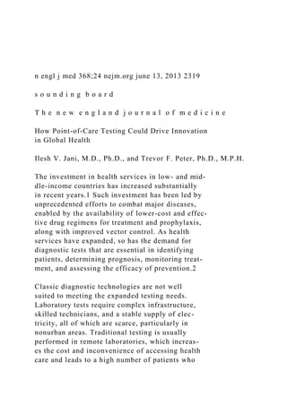
- 2. leave the system before a diagnosis is established.3 These limitations are a critical barrier to equity in health services. Microscopy requires less in- frastructure and is more widely available, but it can be inaccurate (e.g., sputum tests for tubercu- losis) or slow and underutilized (e.g., smear tests for malaria, schistosomiasis, and other parasitic infections).4-6 Many patients with tuberculosis or malaria are simply treated on the basis of a pre- sumptive clinical diagnosis. Although convention- al laboratory testing and microscopy will still be needed, it is expected that faster and more ac- curate point-of-care diagnostic tests that do not require laboratory infrastructure will play an in- creasing role in expanding health care in low- and middle-income countries.7 T h e S h if t t o w a r d P o in t- o f - C a r e T e s t in g Rapid point-of-care testing for diabetes, anemia, pregnancy, human immunodeficiency virus (HIV), and malaria have long been available and have be- come common diagnostic tools in both high- and low-income countries (Fig. 1). The first generation of point-of-care testing relied on easy-to-detect biomarkers, such as antibodies, antigens, and sim- ple biochemical reactions. Such biomarkers are also increasingly used in point-of-care tests for a wide range of infectious diseases (e.g., syphilis, hepatitis, measles, schistosomiasis, and tricho- moniasis) and for applications such as blood typing.8-11 A second generation of point-of-care diagnos-
- 3. tics is now on the horizon, partly because of re- cent industry and donor investment. These tests detect more complex and less accessible biomark- ers, such as nucleic acids and cell-surface markers, and take advantage of advances in microfluidics, microelectronics, optical systems, and laboratory- on-a-chip nucleic acid test (NAT)–based amplifi- cation and detection techniques.12,13 The first applications of these technologies have included enumeration of CD4+ T cells, NAT-based diagno- sis of tuberculosis and drug-resistance screening, and testing of HIV viral load.14-16 Additional ap- plications are in the pipeline for other blood- borne and respiratory infections. A third generation of technologies will enable simultaneous detection of multiple targets (multi- plexing) and will use more accurate biomarkers. Advances in engineering and test chemistry will produce devices that are smaller, simpler to op- erate, and potentially instrument-free,17 enabling reliable home-based testing or self-testing. These technologies will extend a wider range of diag- nostics from the laboratory into clinics and com- munities. Point-of-care testing can have a transforma- tive effect on health care. Rapid HIV tests cata- lyzed increased rates of case finding that have driven global efforts in HIV prevention and treat- ment.18 Malaria rapid tests have been instrumen- tal in raising testing rates in Africa for suspected The New England Journal of Medicine Downloaded from nejm.org by SCOTT BARNUM on February 23, 2016. For personal use only. No other uses without
- 4. permission. Copyright © 2013 Massachusetts Medical Society. All rights reserved. T h e n e w e n g l a n d j o u r n a l o f m e d i c i n e n engl j med 368;24 nejm.org june 13, 20132320 Detection targets Antibodies Antigens Simple biochemical reactions Typical samples Test cartridges Sample (e.g., capillary blood, oral fluid, or urine) is inserted into disposable test cartridge Samples Capillary blood, oral fluid, urine, breath, and other samples Multiple test formats Handheld lab-on-a-chip devices Disposable tests (no instruments) Doctor’s office desk-based devices Devices will fully automate testing and analysis or display of results
- 5. Transmission of results Devices are likely to have wireless connectivity to transmit result data Small instruments process and read results Common test formats First generation of POC diagnostic testing Examples Rapid test strips and dipsticks (HIV antibody and antigen, malaria antigen, urine biochemistry, and pregnancy tests) Simple instruments (glucometers and hemoglobin meters) Detection targets Whole cells DNA or RNA using PCR or other nucleic acid detection method Examples CD4-cell count HIV viral load Tuberculosis diagnosis and potential drug resistance Potential detection targets Nucleic acid sequencing Advanced protein analysis (proteomics) Examples Antiviral and antibiotic drug-resistance screening
- 6. Differential diagnosis (e.g., viral rash and fever, childhood diseases, antenatal tests) Home-based self-testing A Second generation of POC diagnostic testingB Next generation of POC diagnostic testingC Urine Automated reading Manually read cartridge- based strips Manually read dipsticks Oral fluid Capillary blood Lateral-flow test Vertical-flow test 05/29/13 AUTHOR PLEASE NOTE: Figure has been redrawn and type has been reset Please check carefully
- 7. Author Fig # Title ME DE Artist Issue date COLOR FIGURE Rev5 Dr. Jani 06/13/2013 1 Daniel MullerThe New England Journal of Medicine Downloaded from nejm.org by SCOTT BARNUM on February 23, 2016. For personal use only. No other uses without permission. Copyright © 2013 Massachusetts Medical Society. All rights reserved. n engl j med 368;24 nejm.org june 13, 2013 2321 sounding board
- 8. cases from below 5% in 2000 to 45% in 2010,19 thereby reducing inappropriate antimalarial treat- ment and improving community-based manage- ment of fever and health outcomes.20 New point- of-care tests also show promise. In Mozambique, the use of such testing for CD4+ T cells at primary health care clinics doubled the rate of initiation of antiretroviral therapy and halved the time until treatment initiation.21 Rapid, cost-effective NAT- based testing for tuberculosis increased the rate of case detection by up to 50% and reduced the time until treatment initiation by a factor of 10.22,23 Other opportunities exist for point-of-care testing to improve access to appropriate medical services and thus patient outcomes. An accurate test for preeclampsia may enable earlier diagno- sis and appropriate care for a major cause of ma- ternal death,24 and NAT or antigen testing for HIV may improve the rate of pediatric case detec- tion and treatment coverage, which is currently below 50% in many low- and middle-income countries.25 However, weak health systems limit the effect of such testing programs. The initial adoption of promising new diagnostics is hampered by slow regulatory approval and uncertainty over how to deploy new tests relative to existing technology. This may lead to either inappropriate use or over- use. After deployment, inventories of testing sup- plies often run low, and the reliability of point- of-care testing in real-life nonlaboratory settings may be low.26 In addition, the use of such tests has not always improved patient outcomes. For example, rapid antenatal syphilis testing reduced
- 9. treatment delays but did not lead to higher treat- ment rates or a reduction in perinatal mortality.27 The use of rapid tests for malaria has not always improved the prescribing behavior of clinicians.28 Access to rapid tests for HIV did not improve the uptake of same-day testing at antenatal clinics.29 Despite widespread use of rapid tests for HIV, only 40% of HIV-infected persons know their sta- tus, and 40% of those with positive test results do not access follow-up care and may be at increased risk for death or complications because of delayed therapy.3 Study data suggest similar challenges with new point-of-care technologies. Despite the avail- ability of point-of-care testing for CD4+ T cells at primary health care clinics in Mozambique, only 30% of patients underwent same-day test- ing, and 20% were not tested at all.21 Of those tested and eligible for antiretroviral therapy, 40% were lost to follow-up while undergoing addi- tional testing and counseling before treatment. The benefit of new point-of-care tests cannot be taken for granted. N e e d e d C h a n g e s in H e a lt h S y s t e m s Health systems have been designed around ei- ther syndromic management or diagnostic test- ing performed in the laboratory and are not well adapted to the use of point-of-care testing. The coming wave of such technologies demands changes to health systems. We propose four key areas where change is needed (Fig. 2).
- 10. First, testing policies need to be updated. The World Health Organization (WHO) and other normative bodies should provide recom- mendations on how to use point-of-care tests (including guidance on risks, benefits, and cost- effectiveness), how to select the right products, and where and how to deploy new technologies in relation to existing tests. Even if such tests are cost-effective, their use may incur additional costs to health budgets, especially for new and more sophisticated tests, and these implications Figure 1 (facing page). Evolution of Point-of-Care (POC) Diagnostic Testing. Improvements in POC technology will lead to increas- ingly complex tests run on devices that are smaller and easier to use than the current generation of devices. Panel A shows first-generation POC tests, which are conducted with the use of simple chemical analyses and devices. Most tests are lateral- or vertical-flow devices that allow the specimen to flow across or through the solid surface of the test strip past a reac- tion area, resulting in a visual signal. Both manual and automated readings of test results are common. Panel B shows second-generation POC tests, which detect more difficult diagnostic targets with the use of more complex chemical analyses. Sophisticated, disposable microfluidic test cartridges automate sample prepara- tion and test processing. Cartridges are inserted into small, portable instruments that automatically process and read the results, which are displayed digitally. Pan- el C shows the next generation of POC tests, which will probably include more complex diagnostics for the si- multaneous targeting of multiple diseases with the use of instruments that are smaller and easier to use and
- 11. disposable devices that fully automate sample process- ing, testing, and reporting of results. HIV denotes hu- man immunodeficiency virus, and PCR polymerase chain reaction. The New England Journal of Medicine Downloaded from nejm.org by SCOTT BARNUM on February 23, 2016. For personal use only. No other uses without permission. Copyright © 2013 Massachusetts Medical Society. All rights reserved. T h e n e w e n g l a n d j o u r n a l o f m e d i c i n e n engl j med 368;24 nejm.org june 13, 20132322 need to be made clear. For example, use of new NAT-based testing for tuberculosis in South Af- rica increased the cost of diagnosis by 55% and the cost of treatment by 8%.30 Guidance on clini- cal-management algorithms will also be needed. Point-of-care tests will provide health care work- ers, especially those in primary care and com- munity-based settings, with unprecedented diag- nostic information for guiding clinical decisions. Governments, with guidance from the WHO, have developed and implemented practical disease- management algorithms that are based on clini- cal judgment for primary health care settings in which routine laboratory diagnostics were not feasible or reliable. The increased availability of accurate test data in these settings will necessi- tate new clinical algorithms and guidance on how
- 12. to interpret and use diagnostic information. In addition, point-of-care testing will increasingly be used in the private health sector and in less formal settings, such as pharmacies, retail outlets, and homes. The increased availability raises con- cern about product quality and testing perfor- mance, and supportive but firm regulations on the use of such tests in these settings will be needed. Second, innovation will be needed in the de- sign, operation, and workflow of clinics to ensure that testing is accessible and results are used in real time to guide treatment. Point-of-care test- ing may lengthen clinic visits and place extra de- mands on staffing and space. Bottlenecks at any stage can increase waiting times and result in extra visits by patients, and the benefits of on- site testing may be lost. Clinics may need to hire additional staff in key cadres, extend clinic hours or work shifts, and change the scheduling of pa- tients, clinic flow, and use of space in order to facilitate onsite testing and immediate delivery of follow-up care. Improved medical-record sys- tems that capture test results and make them available across different service departments may improve the tracking and follow-up of patients.31 Steps should also be taken to increase testing rates and reduce the effect of shortages in space or test operators (e.g., use of multiplex or parallel testing). The implementation of many of these initiatives in public health systems will require changes in government policy and resource al- location — for example, to facilitate the exten- sion of clinic operating times, hiring of addition- al staff, and improvements in data-management
- 13. systems. Third, systematic steps should be taken to effectively decentralize point-of-care testing and to improve the retention of patients both before and after testing. Policies that enable new models for expanded community-based testing and that facilitate safe and reliable self-diagnosis provide opportunities to better exploit such testing, as well as drive technology innovation. Addressing weaknesses in retention that persist despite on- site testing will require initiatives both upstream and downstream of the test to improve access to testing and ensure appropriate linkage to follow- up care. Interventions such as transportation and food allowances for clinic visits and mobile-phone reminders32 can help ensure that patients com- plete their treatment as well as promote adherence to clinical protocols among health care workers.33 Streamlining and integrating testing and related services can improve access to treatment. For Revised policy and normative guidance Cost and cost-effectiveness of POC testing Decentralization of services Testing guidelines Clinical algorithms Community-based testing and self-testing Improved operational systems Product regulation Supply chain Training Quality assurance
- 14. Maintenance Streamlined clinic services New staff cadres and shifts Space reassignment Patient scheduling Improved medical records Bundled procedures Decentralization and retention initiatives Community-based testing Self-testing Linkage to care Integrated services Patient-centric services Adherence tools Figure 2. Health-System Improvements to Support Expanded POC Testing. Shown are four key areas of improvement in health systems — revised policy and normative guidance, improved operational systems, streamlined clinic ser- vices, and decentralization and retention initiatives — that will require strengthening in order to increase the effect of POC testing. In these areas, the use of POC testing may prompt system improvements that may eventually extend beyond diagnostics to other areas of health care. The New England Journal of Medicine Downloaded from nejm.org by SCOTT BARNUM on February
- 15. 23, 2016. For personal use only. No other uses without permission. Copyright © 2013 Massachusetts Medical Society. All rights reserved. n engl j med 368;24 nejm.org june 13, 2013 2323 sounding board example, the integration of outpatient services and HIV therapy in Zambia resulted in increased rates of case finding,31 and point-of-care testing for CD4+ T cells in HIV screening clinics in South Africa increased linkage to care.34 Other opportunities exist across disease programs, such as point-of-care tuberculosis testing in HIV screening clinics or therapy centers.35 However, the feasibility and cost-effectiveness of these in- terventions need further investigation. Many health-system innovations exist only in pilot form. Translating useful pilots into policy and routine practice is a challenge that requires in- creased attention from both governments and development partners. Fourth, operational challenges to implement- ing point-of-care testing need to be overcome. Weaknesses in supportive services — including product regulation, supply chain, human resourc- es and training, quality assurance, and equipment maintenance — are widespread and systemic.36 In particular, new in-service and preservice ini- tiatives in training and retention of clinic staff
- 16. will be needed to ensure that new technologies are used appropriately. Initiatives to improve these areas are under way and can benefit other areas of health care delivery.37 In addition, rational planning for product uptake is necessary to en- sure that the investment in point-of-care diag- nostics is cost-effective and sustainable and that tests are widely accessible. D r i v in g t h e In n o v at i o n in H e a lt h S y s t e m s The rise of point-of-care testing is expected to expand access to medical services, improve health outcomes, and facilitate the sustainability of dis- ease-control programs in low- and middle-income countries. Although such technologies were ini- tially focused on HIV, tuberculosis, and malaria, they will be used in the diagnosis and treatment of other diseases, and their deployment at scale will require substantial investment. However, such testing may not be cost-effective if the diagnos- tic innovation is not matched with innovation in health systems. As point-of-care testing becomes more com- mon in diagnostic medicine, it could drive this innovation in health systems in at least three ways. First, the supply of point-of-care tests will directly induce changes, such as improved patient flow within clinics. Second, the new technologies tend to increase testing rates substantially, and as more patients are tested, the demand for as- sociated services will increase and existing sys- temic weaknesses will be highlighted. This in-
- 17. creased pressure on health services will motivate local and international initiatives to seek ways to address such limitations. Finally, the enthusi- asm for new point-of-care technologies among public health practitioners, scientists, and the pri- vate sector should elicit proactive efforts in re- solving health-system bottlenecks so that the tests can be successfully used. There are many examples of system innovation that have been prompted by the use of point-of- care testing, such as the use of provider-initiated HIV testing to increase diagnostic rates and im- prove patient retention,38 wireless networks that capture test data from remote sites and monitor quality,39 and the “Test, Track, and Treat” pro- gram for malaria, an international initiative of the WHO designed to scale up malaria testing linked to treatment and disease surveillance.19 However, more is needed to address the chal- lenges described above. Commitment from gov- ernments and global-health actors is necessary, and strengthening of initiatives should be evi- dence-based, drawing on operational research to identify high-priority and cost-effective interven- tions.40 The investment in developing new point- of-care diagnostics has started to yield fruit. Now health systems need to evolve to reap the benefits. Disclosure forms provided by the authors are available with the full text of this article at NEJM.org. From the Instituto Nacional da Saúde, Maputo, Mozambique (I.V.J.); and Clinton Health Access Initiative, Boston (T.F.P.). 1. McCoy D, Chand S, Sridhar D. Global health funding: how
- 18. much, where it comes from and where it goes. Health Policy Plan 2009;24:407-17. 2. Nkengasong JN, Nsubuga P, Nwanyanwu O, et al. Laboratory systems and services are critical in global health: time to end the neglect? Am J Clin Pathol 2010;134:368-73. 3. Rosen S, Fox MP. Retention in HIV care between testing and treatment in sub-Saharan Africa: a systematic review. PLoS Med 2011;8(7):e1001056. 4. Parsons LM, Somoskövi A, Gutierrez C, et al. Laboratory diagnosis of tuberculosis in resource-poor countries: challenges and opportunities. Clin Microbiol Rev 2011;24:314-50. 5. Zurovac D, Midia B, Ochola SA, English M, Snow RW. Mi- croscopy and outpatient malaria case management among older children and adults in Kenya. Trop Med Int Health 2006;11:432- 40. 6. Gray DJ, Ross AG, Li YS, McManus DP. Diagnosis and man- agement of schistosomiasis. BMJ 2011;342:d2651. The New England Journal of Medicine Downloaded from nejm.org by SCOTT BARNUM on February 23, 2016. For personal use only. No other uses without permission. Copyright © 2013 Massachusetts Medical Society. All rights reserved. n engl j med 368;24 nejm.org june 13, 20132324 sounding board 7. Urdea M, Penny LA, Olmstead SS, et al. Requirements for
- 19. high impact diagnostics in the developing world. Nature 2006;444:Suppl 1:73-9. 8. Huppert JS, Hesse E, Kim G, et al. Adolescent women can perform a point-of-care test for trichomoniasis as accurately as clinicians. Sex Transm Infect 2010;86:514-9. 9. Xu J, Feng T, Lin DD, et al. Performance of a dipstick dye immunoassay for rapid screening of Schistosoma japonicum in- fection in areas of low endemicity. Parasit Vectors 2011;4:87. 10. Mabey DC, Sollis KA, Kelly HA, et al. Point-of-care tests to strengthen health systems and save newborn lives: the case of syphilis. PLoS Med 2012;9(6):e1001233. 11. Warrener L, Slibinskas R, Chua KB, et al. A point-of-care test for measles diagnosis: detection of measles-specific IgM anti- bodies and viral nucleic acid. Bull World Health Organ 2011;89:675-82. 12. Yager P, Domingo GJ, Gerdes J. Point-of-care diagnostics for global health. Annu Rev Biomed Eng 2008;10:107-44. 13. Chin CD, Linder VB, Sia SK, et al. Lab-on-a-chip devices for global health: past studies and future opportunities. Lab Chip 2007;7:41-57. 14. Niemz A, Ferguson TM, Boyle DS. Point-of-care nucleic acid testing for infectious diseases. Trends Biotechnol 2011;29:240- 50. 15. Murtagh M. HIV/AIDS diagnostic technology landscape. 2nd ed. UNITAID technical report. Geneva: UNITAID, 2012 (http://www.unitaid.eu/images/marketdynamics/publications/ UNITAID-HIV_Diagnostics_Landscape-2nd_edition.pdf ). 16. Pai NP, Pai M. Point-of-care diagnostics for HIV and tuber- culosis: landscape, pipeline, and unmet needs. Discov Med 2012;13:35-45. 17. LaBarre P, Boyle D, Hawkins K, Weigl B. Instrument-free
- 20. nucleic acid amplification assays for global health settings. Proc SPIE 2011;8029:1-15. 18. De Cock KM, Bunnell R, Mermin J. Unfinished business — expanding HIV testing in developing countries. N Engl J Med 2006;354:440-2. 19. Test treat track scaling up diagnostic testing, treatment and surveillance for malaria. 2012 (http://www.who.int/malaria/ test_treat_track/en/index.html). Geneva: World Health Organi- zation. 20. Yeboah-Antwi K, Pilingana P, Macleod WB, et al. Commu- nity case management of fever due to malaria and pneumonia in children under five in Zambia: a cluster randomized controlled trial. PLoS Med 2010;7(9):e1000340. 21. Jani IV, Sitoe NE, Alfai ER, et al. Effect of point-of-care CD4 cell count tests on retention of patients and rates of antiretrovi- ral therapy initiation in primary health clinics: an observational cohort study. Lancet 2011;378:1572-9. 22. Boehme CC, Nicol MP, Nabeta P, et al. Feasibility, diagnostic accuracy, and effectiveness of decentralised use of the Xpert MTB/RIF test for diagnosis of tuberculosis and multidrug resis- tance: a multicentre implementation study. Lancet 2011;377:1495- 505. 23. Vassall A, van Kampen S, Sohn H, et al. Rapid diagnosis of tuberculosis with the Xpert MTB/RIF assay in high burden coun- tries: a cost-effectiveness analysis. PLoS Med 2011;8(11):e1001120. 24. Gullai N, Stenczer B, Molvarec A, et al. Evaluation of a rapid and simple placental growth factor test in hypertensive disor- ders of pregnancy. Hypertens Res 2013;36:457-62. 25. Parpia ZA, Elghanian R, Nabatiyan A, Hardie DR, Kelso
- 21. DM. p24 Antigen rapid test for diagnosis of acute pediatric HIV infec- tion. J Acquir Immune Defic Syndr 2010;55:413-9. 26. Wolpaw BJ, Mathews C, Chopra M, et al. The failure of rou- tine rapid HIV testing: a case study of improving low sensitivity in the field. BMC Health Serv Res 2010;10:73-6. 27. Myer L, Wilkinson D, Lombard C, Zuma K, Rotchford K, Karim SS. Impact of on-site testing for maternal syphilis on treatment delays, treatment rates, and perinatal mortality in ru- ral South Africa: a randomised controlled trial. Sex Transm In- fect 2003;79:208-13. 28. Ansah EK, Narh-Bana S, Epokor M, et al. Rapid testing for malaria in settings where microscopy is available and peripheral clinics where only presumptive treatment is available: a random- ised controlled trial in Ghana. BMJ 2010;340:c930. 29. Mkwanazi NB, Patel D, Newell ML, et al. Rapid testing may not improve uptake of HIV testing and same day results in a rural South African community: a cohort study of 12,000 women. PLoS One 2008;3(10):e3501. 30. Meyer-Rath G, Schnippel K, Long L, et al. The impact and cost of scaling up GeneXpert MTB/RIF in South Africa. PLoS One 2012;7(5):e36966. 31. Topp SM, Chipukuma JM, Giganti M, et al. Strengthening health systems at facility-level: feasibility of integrating antiret- roviral therapy into primary health care services in Lusaka, Zambia. PLoS One 2010;5(7):e11522. 32. Bärnighausen T, Chaiyachati K, Chimbindi N, Peoples A, Haberer J, Newell ML. Interventions to increase antiretroviral adherence in sub-Saharan Africa: a systematic review of evalua- tion studies. Lancet Infect Dis 2011;11:942-51. 33. Zurovac D, Sudoi RK, Akhwale WS, et al. The effect of mo-
- 22. bile phone text-message reminders on Kenyan health workers’ adherence to malaria treatment guidelines: a cluster randomised trial. Lancet 2011;378:795-803. 34. Larson BA, Schnippel K, Ndibongo B, et al. Rapid point-of- care CD4 testing at mobile HIV testing sites to increase linkage to care: an evaluation of a pilot program in South Africa. J Ac- quir Immune Defic Syndr 2012;61(2):e13-e17. 35. Lawn SD, Wood R. Tuberculosis in antiretroviral treatment services in resource-limited settings: addressing the challenges of screening and diagnosis. J Infect Dis 2011;204:Suppl 4:S1159- S1167. 36. Schito ML, Peter TF, Cavanaugh S, et al. Opportunities and challenges for cost-efficient implementation of new point-of- care diagnostics for HIV and tuberculosis. J Infect Dis 2012;205:Suppl 2:S169-S180. 37. Nkengasong JN. A shifting paradigm in strengthening labo- ratory health systems for global health: acting now, acting col- lectively, but acting differently. Am J Clin Pathol 2010;134:359- 60. 38. Dalal S, Lee CW, Farirai T, et al. Provider-initiated HIV test- ing and counseling: increased uptake in two public community health centers in South Africa and Implications for scale-up. PLoS One 2011;6(11):e27293. 39. Tobaiwa O, Bollinger T, Sitoe N, et al. Implementation of a wireless GPRS-based monitoring system for point-of-care CD4 testing at rural primary health facilities in Mozambique. Gene- va: International AIDS Society, 2012 (http://www.iasociety.org/ Default.aspx?pageId=11&abstractId=200747646). 40. Schwartländer B, Stover J, Hallett T, et al. Towards an im- proved investment approach for an effective response to HIV/ AIDS. Lancet 2011;377:2031-41. DOI: 10.1056/NEJMsb1214197
- 23. Copyright © 2013 Massachusetts Medical Society. The New England Journal of Medicine Downloaded from nejm.org by SCOTT BARNUM on February 23, 2016. For personal use only. No other uses without permission. Copyright © 2013 Massachusetts Medical Society. All rights reserved. Final Applied Lab Project (1 credit Lab Component) Addresses course outcomes 1-5: · apply the scientific method to scientific investigations · state a scientific hypothesis and design a basic experiment · conduct an experiment, make observations, and collect data · use knowledge of biological principles to correctly interpret qualitative and quantitative information · use critical analysis to draw conclusions This is the culminating assessment in BIOL 102. It is designed to assess your ability to apply the principles of the scientific method. For this project, you will complete the activity below. Make sure to address all points (questions) associated with the activity. The Effect of low pH on Enzyme Activity Design an experiment in which you will test the effect of an acidic fluid on enzymatic activity. (Recall: enzymes are proteins.) To complete this project, it may be useful for you to review the Scientific Method Tutorial (found the Course Content section of the classroom under the Science Learning Center link) and the Scientific Method lab (Lab 1), so that you can better understand how to design an experiment. It may also be helpful for you to review your textbook and Lab 4 (Enzymes). As you review Lab 4, you will be reminded that
- 24. there are several factors that impact enzymatic activity: pH, temperature, and amount of reagent. Feel free to refer to observations and information from Lab 4 as you complete the Final Applied Project (see the questions below). Or in other words, it is OK to use the same enzyme/subtrate/method as you did in lab 4 (but modify the treatment), or you can search on- line to find a different enzyme/subtrate/method for measuring enzyme activity. As you design your experiment for this project, please remember that you are trying to examine how an acidic fluid will modify the outcome of an enzymatic reaction. To successfully complete this project, you will need to identify the question(s) being asked in your experiment and the hypothesis that you are testing. In your experimental design, you must clearly explain what you are doing. That means that you will need to identify the enzyme and the acid, as well as explain your experimental protocol (this information will help you to answer question 2). You must also thoroughly explain how the addition of the acidic fluid impacted the overall reaction process (this information will help you to answer question 4). Hint: Keep in mind that the acid will change the environmental conditions of the experiment (for example, a low pH value could change the shape of the active site on the enzyme protein), without directly participating in the reaction. Lab Materials You may need all or some of the following, depending on your experimental design: Materials from your lab kit: · pH paper · hydrogen peroxide solution (you can purchase this at a pharmacy if you have used up the bottle that came with the lab kit) · plastic beakers or cups · vinegar · yeast (can be purchased at grocery store if you need more) · balloons
- 25. · plastic bottle · marker for labeling of beakers You may choose to use additional materials (different acidic solutions and/or different organisms and/or differnet subtrate(s) if you chose to look at an enzyme other than catalase). Outline submit in assignment folder in week 7. I suggest you include the following in your outline: · Name of enzyme you will use, and source (organism) · The substrate · How you will measure enzyme activity (method) · What type of treatment you will you; type of solution(s), pH, length of exposure, how you will treat your samples · The control(s) in the experiment · Sample size · Maybe how you plan to present your data (table and/or type of graph) · Anything else you would like to get feedback on before you start your experiment. Write a paper that includes the following: 1. Title page: title, your name, course name, semester 2. Introduction: introduce your project, include needed background information, the question(s) that you are asking and a clear hypothesis for your experiment. 3. Design an experiment. Provide a detailed account of the materials and methods used to conduct the experiment. Also include the methods for data collection and analysis. 4. Conduct the experiment and record your results. What did you observe? Present your data in table and/or graph . Remember to include the unit of measure. 5. Use your knowledge about enzymes and acids to interpret and discuss your results. It may be necessary for you to refer to your textbook and/or use other information resources. What effect does the acidic treatement have on the enzyme activity? Looking back, how could you have improved your experiment? 6. What is your conclusion? Was your hypothesis supported? 7. Cite all reference sources used (including text book) and
- 26. provide a reference section with citations in APA format Submission Submit your final applied lab project as an attached Word document in the assignment folder by the due date specified in the course schedule THE LANCET 1886 Vol 349 • June 28, 1997 (seven partial or complete responses, four stable disease for more than 6 months), while 12 (52%) did not. The median time to progression for responding patients was 11 months (range 5–29) and for non-responding patients 3 months (range 0–6). The rate of response did not depend on the type of chemotherapy given. There was no difference between responders and non-responders in age, lymph node status, or recurrence-free period. Two of eight (25%) postmenopausal patients responded, compared with nine of 15 (60%) premenopausal patients. The incidence of MRP-positive tumours was not different for patients with soft tissue (one of three), bone (two of eight), or visceral metastases (five of twelve) as predominant site of relapse. In patients receiving first-line chemotherapy, MRP was more often positive in non-responding tumours (50%) than in responding tumours (18%). Only one of eight (13%) MRP-positive tumours had an objective response (5 months), compared with six of 15 (40%) MRP-negative tumours. Analysing for overall response, including stable disease, two of eight (25%) MRP- positive tumours responded, compared with nine of 15 (60%) MRP-negative tumours (odds ratio: 0·22; 95% CI 0·03–1·49). Patients with MRP-positive tumours showed a shorter time to progression on first-line chemotherapy than
- 27. those with MRP-negative tumours (Cox proportional hazard model, p=0·006, figure). The relative hazard rate for time to progression in patients with MRP-positive tumours, compared with MRP-negative tumours, was 4·08 (95% CI 1·50–11·12). At 9 months, all eight patients with MRP- positive tumours showed disease progression, while seven of 15 of those with MRP-negative tumours did not (four objective responses, three stable disease). In Cox multivariate analysis for time to progression, MRP was the only significant variable in the model. Of the 41 patients who received chemotherapy after one or more lines of hormonal therapy, 19 (46%) responded (seven partial responses, 12 stable disease), whereas 22 (54%) did not. In these patients, there was no significant difference in the rate or duration of response, or in the time to progression between patients with MRP-positive and MRP-negative tumours, suggesting differences in tumour cell biology. Metastatic breast cancer patients who receive chemotherapy as the first choice of treatment usually are premenopausal, are oestrogen-receptor and progesterone- receptor negative, and may have visceral metastases. These are all unfavourable prognostic factors. Women first treated with hormonal therapy are usually postmenopausal, have receptor positive tumours, and have bone rather than visceral metastases. We conclude that MRP expression is an important predictor of poor prognosis in patients with breast cancer who were treated with chemotherapy as first-line systemic therapy for recurrence. We thank Maxime Look for statistical analysis. This work was supported by the Dutch Cancer Society (Grants DDHK95-1051, DDHK96- 1236).
- 28. 1 Cole SPC, Bhardwaj G, Gerlach JH, et al. Overexpression of a transporter gene in a multidrug-resistant human lung cancer cell line. Science 1992; 258: 1650–54. 2 Flens MJ, Zaman GJR, van der Valk P, et al. Tissue distribution of the multidrug resistance protein. Am J Pathol 1996; 148: 1237–47. 3 Nooter K, Westerman AM, Flens MJ, et al. Expression of the multidrug resistance-associated protein (MRP) gene in human cancers. Clin Cancer Res 1995; 1: 1301–10. Departments of Medical Oncology (K Nooter) and Clinical Pathology and Division of Endocrine Oncology, University Hospital Rotterdam and Rotterdam Cancer Institute, PO Box 2040, Rotterdam, Netherlands 100 80 60 40 20
- 29. 0P ro g re ss io n -f re e s u rv iv a l (% ) 0 3 6 9 12 15 18 21 24 27 30 15 12 7 6 5 2 1 1 1 1 0 Time (months) p=0·006 RHR (95% CI) 4·08 (1·50–11·12)
- 30. MRP- positive (n=8) MRP-negative (n=15) MRP-negative 8 3 1 0 0 0 0 0 0 0 0MRP-positive Time to progression for patients treated with first-line chemotherapy for recurrence as a function of MRP status. Patients at risk at start and at every 3 months are indicated RHE=relative hazard rate. Complement C3 and factor B cerebrospinal fluid concentrations in bacterial and aseptic meningitis Philip F Stahel, David Nadal, Hans-Walter Pfister, P Maria Paradisis, Scott R Barnum Establishing a diagnosis is difficult in most cases of acute meningitis, since its clinical signs are non-specific, and laboratory examination of cerebrospinal fluid (CSF) often does not accurately differentiate between bacterial and aseptic meningitis.1,2 Therefore the identification of a discriminating parameter, which might lead to a rapid and accurate clinical test, would be of value in the differential diagnosis of acute meningitis. Several studies have suggested that the complement system contributes to intrathecal inflammation in bacterial meningitis.3 In a retrospective analysis, we measured the concentrations of the alternative pathway complement components C3 and factor B in CSF samples obtained by lumbar puncture from 39 patients with clinically suspected
- 31. acute infectious meningitis, and from 64 controls without central nervous system infection, with an ELISA developed in our laboratory. 18 patients (median age 40 years; range 14–67 years; 9 female) were diagnosed as having bacterial meningitis, on the basis of positive bacterial culture or on detection of bacterial antigen in CSF. The pathogens were Streptococcus pneumoniae (n=10), Haemophilus influenzae (3), Neisseria meningitidis (3), Listeria monocytogenes (1), and Streptococcus bovis (1). 21 patients were diagnosed as having aseptic meningitis (median age 8 years; range 2 months to 13 years; 6 female) on the basis of CSF pleocytosis with a predominance of mononuclear cells, negative bacterial and fungal CSF and blood cultures, negative results on CSF antigen detection tests for S pneumoniae, H influenzae, and N meningitidis, and full recovery without antibiotic treatment. No patient had received antibiotics or steroids before diagnostic lumbar puncture, and all CSF samples were obtained on admission. The mean C3 concentration in the CSF of patients with bacterial meningitis (48·32 [SD 50·74] µg/mL) was significantly higher than in aseptic meningitis (2·16 [1·82] µg/mL; p<0·001, Wilcoxon rank sum test) or in controls (2·49 [2·18] µg/mL; p<0·001). Similarly, factor B CSF concentrations were significantly raised in patients with bacterial meningitis (15·89 [17·36] µg/mL) compared with those with aseptic meningitis (0·25 [0·20] µg/mL; p<0·001) or controls (0·29 [0·26] µg/mL; p<0·001). C3 and factor B CSF concentrations in bacterial meningitis did not correlate THE LANCET Vol 349 • June 28, 1997 1887 Nonsense mutation of prostacyclin synthase gene in a family
- 32. Tomohiro Nakayama, Masayoshi Soma, Dolkun Rahmutula, Yoichi Izumi, Katsuo Kanmatsuse We found a nonsense mutation in exon 2 of the human prostacyclin-synthase gene in a family with essential hypertension and cerebral infarction. Prostacyclin (PGI2) is an inhibitor of platelet aggregation, smooth muscle cell proliferation, and vasoconstriction. Prostacyclin synthase (PGIS), which catalyses the formation of PGI2 from prostaglandin H2, is widely distributed, predominantly in vascular endothelial and smooth muscle cells. We have reported the organisation of this gene.1 We searched for possible point mutations in the exons using peripheral blood from 100 patients with essential hypertension by PCR and single strand conformation polymorphism (PCR-SSCP) analysis. One patient had an abnormally migrating band on exon 2. Sequencing of this exon showed a nonsense mutation in codon 26 (CGA/TGA). This nucleotide change makes Bst EII the restriction site. 300 people (150 with essential hypertension and 150 healthy controls) were screened by PCR and Bst EII digestion. The mutation was found in one patient with essential hypertension and in none of the controls. The patient was shown to be heterozygous for this mutation. This mutation of the stop codon is 76 bp downstream from ATG, the start codon in cDNA, thus a large part of mRNA, Male Female Dead HT
- 33. CI Hypertension Cerebral infarction Patient first detected 66y HT 69y HT 66y HT 61y HT 68y Apoplexy HT 63y HT CI 57y
- 34. HT CI – The 247-bp fragment digested with Bst EII will give fragments of 189 bps, 58bps – + +– + + 247bp 189bp 58bp Mutation Family tree and PCR with CSF total white blood cell counts or CSF protein concentrations (r<0·6, Spearman’s rank correlation coefficient). We found that complement concentrations in the CSF may be of clinical value in distinguishing bacterial from aseptic meningitis. With cut-off levels of the mean value +2 SD for the aseptic meningitis population (5·8 µg/mL for C3 and 0·65 µg/mL for factor B; figure), C3 and factor B CSF concentrations were highly sensitive (both 100%) and highly specific (95·2% and 100%, respectively) tests for the diagnosis of bacterial meningitis, and associated with a negative predictive value of 100%, and a positive predictive value of 94·7% (C3) and 100% (factor B). Quantification of
- 35. C3 and factor B CSF concentrations can be completed within 3–4 hours. We plan to test these preliminary results in a multicentre prospective study. 1 Lindquist L, Linné T, Hansson L-O, Kalin M, Axelsson G. Value of cerebrospinal fluid analysis in the differential diagnosis of meningitis: 1000 100 10 1 0·1 0·01 1000 100 10 1
- 37. Aseptic meningitis Controls Mean value Cut-off Complement C3 and factor B concentrations in the CSF of patients with infectious meningitis and controls Each point represents the mean of duplicate sample analysis. Cut-off level=mean of aseptic meningitis group+2 SD for differentiation between bacterial and aseptic meningitis. a study in 710 patients with suspected central nervous system infection. Eur J Clin Microbiol Infect Dis 1988; 7: 374–80. 2 Rodewald LE, Woodin KA, Szilagyi PG, Arvan DA, Raubertas RF, Powell KR. Relevance of common tests of cerebrospinal fluid in screening for bacterial meningitis. J Pediatr 1991; 119: 363–69. 3 Stahel PF, Barnum SR. Bacterial meningitis: complement gene expression in the central nervous system. Immunopharmacology. (In press) Department of Microbiology, University of Alabama at Birmingham, Birmingham, AL 35294, USA (S Barnum); Unit of Infectious Diseases,
- 38. University Children’s Hospital, Zürich, Switzerland; and Department of Neurology, Klinikum Grosshadern,University of Munich, Munich, Germany THE LANCET 1888 Vol 349 • June 28, 1997 Colonic perforation and serosal tears associated with colonoscopy Yoshiharu Uno, Takayuki Morita Serosal tears with no mucosal damage are a complication of colonoscopy.1–4 These tears have been attributed to the pressure of the air introduced through the colonoscope or to stretching the wall of the colon. A colonoscope (CF230I, Olympus Inc, Japan) was pressed against the mucosa of a piece of sigmoid colon removed during surgery for rectal cancer at a continuous pressure of 2–3 kg/cm2 (figure, a). First, the muscularis propria ruptured (figure, b), and then the serosa tore (figure, c), before the mucosa ruptured and the colonoscope penetrated the wall (figure, d). We repeated this experiment at 40 different points (ten points in each of four segments: sigmoid, descending, transverse, and ascending colon) of resected colons from eight patients with mean age 62 (SD 11) years. Perforation occurred in the above order, regardless of age, colonic segment, or sex. We asked 60 doctors in our hospital how injury occurred when the tip of a colonoscope was pressed against the colonic wall. They all replied that perforation started from
- 39. the mucosa, proceeded to the muscularis propria and then the serosa. Our results suggest that, when the tip or a bend of a colonoscope is pressed hard against the colonic mucosa, a seromuscular tear will probably occur, even when there is no mucosal injury. If the mucosa does not rupture, peritonitis is unlikely. 1 Livstone EM, Cohen GM, Troncale FJ, Touloukian RJ. Diastatic serosal lacerations. Gastroenterology 1974; 67: 1245–47. 2 Livstone EM, Kerstein MD. Serosal tears following colonoscopy. Arch Surg 1976; 111: 88. 3 Kozarek RA, Earnest DL, Silverstein ME, Smith RG. Air- pressure- induced colon injury during diagnostic colonoscopy. Gastroenterology 1980; 78: 7–14. 4 Ehrlich CP, Hall FM, Joff N. Postendoscopic perforation of normal colon in an area remote from instrumentation. Gastrointest Endosc 1984; 30: 190–91. First Department of Internal Medicine and Second Department of Surgery, Hirosaki University School of Medicine, Aomori 036, Japan (Y Uno) Perforation of colon by colonoscope a, b, c, d depict how serosal tears occur as complication of colonoscopy.
- 40. 6 kbp,2 can not be translated. Consequently the activity of PGIS may be decreased. The patient is a 57-year-old woman with essential hypertension who presented with a blood pressure of 177/113 mm Hg. Her electrocardiogram showed left ventricular hypertrophy. Although she had never smoked and rarely consumed alcohol, at the age of 50 she had a transient ischaemic attack. We looked for this mutation in her family. Her father died of stroke aged 70. Her mother was healthy until her death at age of 92. She had eight siblings including two elder brothers one of whom died in the war and the other of chronic renal failure. Three of the five living siblings had the mutation (figure); all were hypertensive. One with the mutation had had a cerebral infarction. As essential hypertension is thought to be a multifactorial disorder, PGIS may be one of the genes involved. 1 Nakayama T, Soma M, Izumi Y, Kanmatsuse K. Organization of the human prostacyclin synthase gene. Biochem Biophys Res Commun 1996; 221: 803–06. 2 Miyata A, Hara S,Yokoyama C, Inoue H, Ullrich V, Tanabe T. Molecular cloning and expression of human prostacyclin synthase. Biochem Biophys Res Commun 1994; 200: 1728–34. Second Department of internal Medicine, Nihon University School of Medicine, Ooyaguchikamimachi 30–1, Tokyo 173, Japan (T Nakayama)