SlideShare una empresa de Scribd logo
1 de 85
1 
FAMILY 
FAMILY is the basic social institution and the primary group in society. 
 According to Murdok, family is a social group characterized by common 
residence, economic cooperation and reproduction. 
 According to Burges and Locke, family is a group of persons united by ties of 
marriage, blood or adoption, constituting a single household, interacting and 
communicating with each other in their respective social roles of husband and 
wife, mother and father, son and daughter, brother and sister, and creating and 
maintaining a common culture. 
 According to Murray and Zentner (1997) is a social system and primary reference 
group made up of two or more persons living together who are related by blood, 
marriage or adoption or who are living together by arrangement over a period of 
time. 
CHARACTERISTICS OF A FAMILY 
 The family as a social group is universal and is significant element in man’s 
social life 
 It is the first social group to which the individual is exposed. Family contact and 
relationships are repetitive and continuous 
 The family is very close and intimate group. It is setting of the most intense 
emotional experiences during the life time of the individual.The family affects the 
individual social values, disposition and outlook in life 
 The family has the unique position of serving as a link between the individual and 
the larger society 
 The family is also unique in providing continuity of social life. 
Functions of Family 
 Regulates sexual behavior and reproduction 
 Biological maintenance function
2 
 Socialization function 
 Provide legitimate children with a status 
 Social control function 
 Economic function 
 Educational, recreational, religious and political functions 
Classification of Family Structure 
Based on Descent 
 Patrilineal- affiliates a person with a group of relatives through his or her father. 
 Matrilineal- affiliates a person with a group of relatives through his or her mother. 
 Bilateral- affiliates a person with a group of relatives related through both his or 
her parents. 
Based on Authority 
 Patriarchial- authority is vested on the oldest male in the family, often the father. 
 Matriarchial- authority is vested in the mother or mother’s kin 
 Matricentric- prolonged absence of the father gives the mother a dominant 
position in the family, although the father may also share with the mother in 
decision making. 
Based on Internal Organization and Membership 
 Nuclear Family. Also known as primary or elementary family. 
 Extended Family. Extensions maybe through the parent-child relationship or 
husband-wife relationship, as in polygamous marriage. 
Based on Place of Residence 
 Patrilocal- requires the newly wed to reside near the groom’s parents. 
 Matrilocal- near the bride’s parents. 
 Bilocal- provides the couple the choice to reside on either parents. 
 Neolocal- permits the couple to reside independently of their parents
 Avunculocal- prescribes the newly wed couple to reside with or near the maternal 
3 
uncle of the groom. 
Families consist of special functional subsystems: 
 marital subsystem (parents) 
 sibling subsytem (children) 
 individual parent –child system subsystem 
View of the person: The person is a member of the family where each new member 
adds to the complexity of the interaction within the family. 
Stages of Family 
1. Beginning *Establishing a mutually satisfying Family marriage *Planning to have 
or not to have children. 
2. Child- *Having and adjusting to infant bearing family *Supporting the needs of all 
three members *Renegotiating marital relationships. 
3. Family with *adjusting to cost of family life a pre-school *adapting to the needs of 
pre-school children *Coping with parental loss of energy and privacy 
4. Family with *Adjusting to the activity of the growing school age children 
*Promoting joint decisions between children and parents *Encouraging and 
supporting children’s educational achievements 
5. Family with *Maintaining open communication among teenagers and members 
young adult *Supporting ethical and moral values within the family *Balancing 
freedom with responsibility of teenagers *Releasing young adults with 
appropriate rituals and assistance 
6. Post- *Strengthening marital relationships parental *Maintaining supportive home 
base family *Preparing for retirement 
7. Aging *Maintaining ties with younger and Family older generations *Adjusting for 
retirement *Adjusting to loss of spouse *Closing family house
4 
FETAL AND MATERNAL HEALTH PRENATAL CARE 
 Ensures the overall health of newborns and their mothers. 
OB 
Follow-up visits 
 Every 4 weeks through the 28th week 
 Every 2 weeks through the 36th weeks 
 Every week until delivery 
The health care given to a woman and her family during pregnancy. 
The earliest stage at which fetuses could survive if they were born at that time. 
Any pregnancy terminated before the age of viability. 
An infant born at the end of 37 weeks to the end of 42 weeks. 
An infant born before 37 weeks. 
An infant born beyond 42 weeks. 
A woman in labor. 
A woman who has just given birth. 
A pregnant woman or refers to the present pregnancy. 
The number of pregnancy that reached viability. 
A woman who is pregnant the first time. 
A woman with a second or later pregnancy. 
A woman who has never been pregnant and is nor currently pregnant. 
A woman who has completed one pregnancy of viability. 
A woman who has carried two or more pregnancies of viability. 
A woman who had 6 or more viable pregnancies. 
History of past pregnancies 
Gravida (G) 
 A number of times the woman has been pregnant. 
Para (P) 
 A number of children above viability the woman has previously borne. 
 Multigestation is considered as one para 
GTPAL 
G- number of times the woman has been pregnant.
5 
P- is broken down into: 
T- term BIRTH 
P- preterm BIRTH 
A- Abortion 
L- living children 
GTPALM 
G- number of times the woman has been pregnant 
P- is broken down into: 
T- term INFANT 
P- preterm INFANT 
A- abortion 
L- living children 
M- multiple pregnancies 
History of present pregnancy 
Estimate date of birth/ date of delivery 
Naegele’s rule 
 standard method used to predict the length of a pregnancy 
 approximates that pregnancy last 40 weeks from LMP 
 To get EDB/ EDD 
 1st day of LMP 
Estimate fetal growth 
McDonald’s Rule 
 Common method of determining fetal growth in utero. 
 Symphisis-fundal height measurement 
 The distance from the symphisis pubis to the uterine fundus in cm es equal to the 
week of gestation between the 20th and 31st weeks of preganancy 
 To estimate AOG 
 Fundic height in cm x 2/7= AOG in moths
6 
 Fundic height in cm x 8/7= AOG in weeks 
Leopold’s Maneuver 
 A systematic method of observation and palpation of the abdomen to determine 
fetal presentation and position. 
Preparation 
 Explain the procedure 
 Ask to void 
 Supine with knees slightly flexed 
 Wash hands with warm water 
Purpose Procedure Findings 
First Maneuver: 
Fundal Grip 
To determine fetal 
part lying in the 
fundus. 
To determine 
presentation. 
Using both hands, 
feel for the fetal 
part lying in the 
fundus. 
Head is more firm, 
hard and round 
that moves 
independently of 
the body. 
Breech is less well 
defined that moves 
only in conjunction 
with the body. 
Second 
Maneuver: 
Umbilical Grip 
To identify 
location of fetal 
back. 
To determine 
position. 
One hand is used 
to steady the 
uterus on one 
side of the 
abdomen while 
the other hand 
moves slightly on 
a circular motion 
from top to the 
lower segment of 
the uterus to feel 
for the fetal back 
and small fetal 
parts. 
Use gentle but 
deep pressure. 
Fetal back is 
smooth, hard, and 
resistant surface 
Knees and 
elbows of fetus 
feel with 
a number of 
angular nodulation 
Third Maneuver: 
Pawlik’s Grip 
To determine 
engagement of 
presenting part. 
Using thumb and 
finger, grasp the 
lower portion of 
the abdomen 
above symphisis 
pubis, press in 
slightly and make 
The presenting 
part is notengaged 
if it is not movable. 
It is not yet 
engaged if it is still 
movable.
7 
gentle movements 
from side to side. 
Fourth 
Maneuver: 
Pelvic Grip 
To determine the 
degree of flexion 
of fetal head. 
To determine 
attitude or habitus. 
Facing foot part of 
the woman, 
palpate fetal head 
pressing 
downward about 2 
inches above the 
inguinal ligament. 
Use both hands 
Good attitude – if 
brow correspond to 
the side (2nd 
maneuver) that 
contained the 
elbows and knees. 
Poor atitude – if 
examining fingers 
will meet an 
obstruction on the 
same side as fetal 
back 
(hyperextended 
head) 
Also palpates 
infant’s 
anteroposterior 
position. If brow is 
very easily 
palpated, fetus is 
at posterior 
position (occiput 
pointing towards 
woman’s back) 
PRENATAL VISIT 
8-12 weeks: Initial prenatal visit. The plan for your prenatal care at the practice you 
have chosen will be explained. A health history is taken, and a physical exam, including 
a pelvic exam is done. Lab work is completed, including your blood type and 
hemoglobin, sexually transmitted infection screening, a urine test, and a PAP test if you 
are due for one. You may be able to hear the baby's heart beat at this visit. If you
cannot say with accuracy when you had your last period, an ultrasound might be 
scheduled to help determine how far along you are. 
Optional genetic counseling visit: Early in your pregnancy, you may be offered 
genetic screening. This is commonly offered to women over the age of 35, or women 
who have a family history of genetic problems, but it is increasingly being offered to 
every woman. If you choose this screening, your care provider and/or genetic counselor 
may suggest additional genetic screening or diagnostic tests, including blood tests, 
chorionic villus sampling, ultrasound, and/or amniocentesis. These tests are done at 
specific times during pregnancy. 
First two trimesters: Prenatal visits continue every 4-6 weeks through the first two 
trimesters, or until you are 28 weeks along. At each appointment, your care provider will 
weigh you and take your blood pressure, listen to the baby's heartbeat, and measure 
the growth of your uterus and baby. Some providers check your urine for protein and 
sugar at each visit. 
15 to 20 weeks: At one of your appointments within this period, you will be offered the 
Quad Screen test, which screens for genetic and spinal cord abnormalities. You may 
also be offered an ultrasound between 18 and 20 weeks to view the baby's organs, and 
measure the growth of the baby and the placenta. 
27 or 28 weeks: At an appointment within this period, you will be encouraged to take a 
glucose challenge test to screen for gestational diabetes. Your hemoglobin may be 
rechecked. Some providers do a pelvic exam. Expect to review warning signs of late 
pregnancy that would alert you to preterm labor or high blood pressure. You may be 
encouraged to sign up for prenatal classes, find a doctor or nurse-practitioner who will 
provide well-child care for your baby, and information may be provided about making 
plans for labor. 
28 to 36 weeks: After 28 weeks, prenatal visits continue every 2-3 weeks until 36 
weeks. Your doctor or midwife will continue to record the growth of the baby, listen to 
the baby's heartbeat, and will check the position of the baby. 
8
36 weeks: At this visit, your midwife or doctor will do a pelvic exam, and encourage you 
to have a Group B Strep test. Screening tests for sexually transmitted infections may be 
repeated at this visit. The position and size of the baby are estimated. If your baby is not 
head down, your provider may suggest exercises to encourage the baby to turn, or 
suggest a physical manipulation called external version. The risks and benefits of this 
procedure should be carefully explained. 
36 to 40 weeks: The usual monitoring of your weight and blood pressure, and the 
baby's size, position, and heart rate are done. Your care provider may offer to check 
your cervix for dilation. 
40+ weeks: After your due date, your care provider may offer what is called "post-dates" 
testing, including nonstress tests, ultrasound, and biophysical profiles. Some 
9 
providers start at 40 weeks, others not until 10 days past your due date. 
Safe Medications to Take During Pregnancy 
Allergy 
 Benadryl (diphenhydramine) 
 Claritin 
Check with your doctor before taking these in the first trimester. 
Cold and Flu 
 Tylenol (acetaminophen) 
 Saline nasal drops or spray 
 Warm salt/water gargle 
Check with your doctor before taking any other medications, especially in the first 
trimester. 
Constipation 
 Colace 
 Metamucil 
First Aid Ointment
10 
 Bacitracin 
 J&J First-Aid Cream 
 Neosporin 
 Polysporin 
Rashes 
 Benadryl cream 
 Caladryl lotion or cream 
 Hydrocortisone cream or ointment 
 Oatmeal bath (Aveeno) 
*Note: No drug can be considered 100% safe to use during pregnancy. 
What Alternative Therapies Are Considered Safe During Pregnancy? 
Some alternative therapies have been shown to be safe and effective for pregnant 
women to relieve some of the uncomfortable side effects of pregnancy. Talk it over with 
your doctor first before using any of them. And remember, “Natural” doesn’t always 
equal “safe” when you’re pregnant. 
Nausea in early pregnancy: Acupuncture, acupressure, ginger root (250 milligram 
capsules 4 times a day), and vitamin B6 (pyridoxine, 25 milligrams two or three times a 
day) work well. Sipping the thick syrup from inside a can of peaches, pears, mixed 
fruits, pineapples, or orange slices may also help. 
Backache: Chiropractic manipulation holds the best track record. Another option is 
massage but it is important to make sure your massage therapist is adequately trained 
in pre-natal massage. 
Turning a breech baby: Exercise and hypnosis may help. 
Pain relief in labor: Epidurals are most effective, but immersion in a warm bath can 
also relieve tension. Relaxation and breathing techniques, emotional support, and self-hypnosis 
are widely used in labor. Acupuncture can also work for some women.
11 
Common Discomforts During Pregnancy 
Symptoms of discomfort due to pregnancy vary from woman-to-woman. The following 
are some common discomforts. However, each mother-to-be may experience 
symptoms differently or not at all: 
 Nausea and vomiting 
About half of all pregnant women experience nausea and sometimes vomiting in the 
first trimester--also called morning sickness because symptoms are most severe in the 
morning. Some women may have nausea and vomiting throughout the 
pregnancy. Morning sickness may be due to the changes in hormone levels during 
pregnancy. 
Morning sickness seems to be aggravated by stress, traveling, and certain foods, such 
as spicy or fatty foods. Eating small meals several times a day may help lessen the 
symptoms. A diet high in protein and complex carbohydrates (such as whole wheat 
bread, pasta, bananas, and green, leafy vegetables) may also help reduce the severity 
of the nausea. 
If vomiting is severe, causing a woman to lose fluids and weight, it may indicate a 
condition called hyperemesis gravidarum. Hyperemesis can lead to dehydration and 
may require hospitalization for intravenous fluids and nutrition. Call your physician or 
midwife if you are having constant or severe nausea and vomiting. 
 Fatigue 
As the body works overtime to provide a nourishing environment for the fetus, it is no 
wonder a pregnant woman often feels tired. In the first trimester, her blood volume and 
other fluids increase as her body adjusts to the pregnancy. Sometimes anemia is the 
underlying cause of the fatigue. Anemia is a reduction in the oxygen-carrying capability 
of red blood cells, and is usually due to low iron levels. A simple blood test performed at 
a prenatal visit will check for anemia. 
 Hemorrhoids 
Because of increased pressure on the rectum and perineum, the increased blood 
volume, and the increased likelihood of becoming constipated as the pregnancy
progresses, hemorrhoids are common in late pregnancy. Avoiding constipation and 
straining may help to prevent hemorrhoids. Always check with your physician or midwife 
before using any medication to treat this condition. 
12 
 Varicose veins 
Varicose veins--swollen, purple veins--are common in the legs and around the vaginal 
opening during late pregnancy. In most cases, varicose veins are caused by the 
increased pressure on the legs and the pelvic veins, and by the increased blood 
volume. 
 Heartburn and indigestion 
Heartburn and indigestion, caused by pressure on the intestines and stomach (which, in 
turn, pushes stomach contents back up into the esophagus), can be prevented or 
reduced by eating smaller meals throughout the day and by avoiding lying down shortly 
after eating. 
 Bleeding gums 
Gums may become more spongy as blood flow increases during pregnancy, causing 
them to bleed easily. A pregnant woman should continue to take care of her teeth and 
gums and go to the dentist for regular checkups. This symptom usually disappears after 
pregnancy. 
 Pica 
Pica is a rare craving to eat substances other than food, such as dirt, clay, or coal. The 
craving may indicate a nutritional deficiency. 
 Swelling/fluid retention 
Mild swelling is common during pregnancy but severe swelling that persists may 
indicate preeclampsia (abnormal condition marked by high blood pressure). Lying on 
the left side, elevating the legs, and wearing support hose and comfortable shoes may 
help to relieve the swelling. Be sure to notify your physician or midwife about sudden 
swelling, especially in the hands or face, or rapid weight gain. 
 Skin changes 
Due to fluctuations in hormone levels, including hormones that stimulate pigmentation of
13 
the skin, brown, blotchy patches may occur on the face, forehead, and/or cheeks. This 
is often called the mask of pregnancy, orchloasma, and often disappears soon after 
delivery. Using sunscreen when outside can reduce the amount of darkening that 
occurs. 
Pigmentation may also increase in the skin surrounding the nipples, called the areola. In 
addition, a dark line frequently appears down the middle of the abdomen. Freckles may 
darken, and moles may grow. 
 Stretch marks 
Pinkish stretch marks may appear on the abdomen, breasts, thighs, or buttocks. Stretch 
marks are generally caused by a rapid increase in weight, and the marks usually fade 
after pregnancy. 
 Yeast infections 
Due to hormone changes and increased vaginal discharge, also called leukorrhea, a 
pregnant woman is more susceptible to yeast infections. Yeast infections are 
characterized by a thick, whitish discharge from the vagina and itching. Yeast infections 
are highly treatable. Always consult your physician or midwife before taking any 
medication for this condition. 
 Congested or bloody nose 
During pregnancy, the lining of the respiratory tract receives more blood, often making it 
more congested. This congestion can also cause stuffiness in the nose or nosebleeds. 
In addition, small blood vessels in the nose are easily damaged due to the increased 
blood volume, causing nosebleeds. 
 Constipation 
Increased pressure from the pregnancy on the rectum and intestines can interfere with 
digestion and subsequent bowel movements. In addition, hormone changes may slow 
down the food being processed by the body. Increasing fluids, regular exercise, and 
increasing the fiber in your diet are some of the ways to prevent constipation. Always 
check with your physician or midwife before taking any medication for this condition.
14 
 Backache 
As a woman's weight increases, her balance changes, and her center of gravity is 
pulled forward, straining her back. Pelvic joints that begin to loosen in preparation for 
childbirth also contribute to this back strain. Proper posture and proper lifting techniques 
throughout the pregnancy can help reduce the strain on the back. 
 Dizziness 
Dizziness during pregnancy is a common symptom, which may be caused by: 
o Low blood pressure due to the uterus compressing major arteries 
o Low blood sugar 
o Low iron 
o Quickly moving from a sitting position to a standing position 
o Dehydration 
To prevent injury from falling during episodes of dizziness, a pregnant woman should 
stand up slowly and hold on to the walls and other stable structures for support and 
balance. 
 Headaches 
Hormonal changes may be the cause of headaches during pregnancy, especially during 
the first trimester. Rest, proper nutrition, and adequate fluid intake may help alleviate 
headache symptoms. Always consult your physician or midwife before taking any 
medication for this condition. If you have a severe headache or a headache that does 
not resolve, call your health care provider. It may be a sign of preeclampsia. 
Danger signs of Pregnancy 
1. Decreased fetal movement 
If you notice that your baby isn't moving around as much as she normally does, stop 
exercising and take a minute to pay attention to what she's doing. Remember that 
sometimes it's hard to tell if your baby is moving around when you're moving around,
too. Also, be sure to eat and drink water before your workout because that may affect 
your baby's movements. 
Call your healthcare provider: if your baby isn't moving around as much as normal or 
you notice a sudden decrease in your baby's movement. 
15 
2. Dizziness 
Is It Safe During Pregnancy? 
Persistent dizziness together with fatigue and headaches can be symptoms of severe 
anemia or another serious condition. 
Call your healthcare provider: if you're still dizzy after you've cooled down, rested, 
and had some water. 
3. Overheating 
If you feel faint or dizzy, or if you develop a headache, nausea, cramps, or a racing 
heart, your body's telling you that it's having a hard time regulating your internal 
temperature, which can be harmful to your baby. The baby can get overheated just as 
you do. When your body overheats, blood flowing to the uterus is diverted to the skin to 
help the body cool itself off, putting the baby in jeopardy. 
It's unusual to overheat from exercise alone, but if it's hot outside or in the gym where 
you exercise, you could run into trouble. If you're exercising indoors, it's best to do so in 
a well-ventilated room with fans. If you're exercising outdoors, avoid the sun when it's 
strongest in the middle of the day. Consider staying inside if it's especially hot out. 
Call your healthcare provider: if you feel very hot and you have symptoms of 
overheating, like profuse sweating, dizziness or lightheadedness, a headache, nausea, 
cramps, or an irregular heartbeat. 
4. Heart palpitations 
If your heart is pounding and you can't carry on a conversation without being out of 
breath, or if you sweat buckets while you exercise, you're probably working too hard. 
Heart palpitations may be a sign of dehydration, severe anemia, thyroid disease, or a 
heart problem. 
Call your healthcare provider: if your heart continues to race after you've cooled 
down, rested, and had some water. 
5. Swelling in your calf
Your feet and hands may puff up a little after you exercise, but if you notice calf pain or 
swelling, it could be a sign of deep vein thrombosis (DVT), a potentially life-threatening 
condition caused by a type of blood clot. DVT usually affects veins deep in the lower leg 
and thigh and occurs on one side of the body. You may experience redness and skin 
that feels warm to the touch. 
Also, sudden swelling in your legs (and face and hands), along with high blood 
pressure, may be a sign of preeclampsia. 
Call your healthcare provider: immediately if you think you have DVT. If you have 
DVT and you experience chest pain, difficulty breathing, fainting, or any other serious 
condition, go to the emergency room right away. 
6. Vaginal bleeding 
Some women do experience light spotting throughout their pregnancy, but vaginal 
bleeding during pregnancy is always a cause for concern. Early in your pregnancy, it 
could signal a miscarriage. In the second and third trimesters, bleeding is associated 
with premature labor and complications with the placenta, such as placenta 
previa or placenta abruption. All require immediate medical attention. 
Call your healthcare provider: immediately if you have vaginal bleeding. If you 
can't reach anyone, go to the emergency room. 
16 
7. Blurred vision 
It's common for blood pressure to drop during the first 6 months of pregnancy. But 
combine low blood pressure with exercise and dehydration, and you may be headed for 
trouble. 
If your eyesight gets hazy in the middle of your workout, you may be dehydrated. That 
alone is enough to send your blood pressure plummeting and your heart into overdrive. 
As a result, not enough blood may be getting to your developing baby's vital organs. 
Blurred vision may also be a sign of preeclampsia. This condition can be dangerous for 
your baby because preeclampsia can severely restrict the flow of blood to the placenta. 
Call your healthcare provider: immediately. If you can't reach anyone, go to the 
emergency room. 
8. Fainting 
Fainting during pregnancy shouldn't be taken lightly. It could signal something as simple 
as dehydration or something serious like major circulatory or heart problems. You may
not be getting enough oxygen to your brain, which means your baby may not be getting 
enough either. 
Call your healthcare provider: immediately. If you can't reach anyone, go to the 
emergency room. 
17 
9. Recurring pain in abdomen or chest 
It may just be your ligaments stretching, but you could also be having contractions – 
especially if the pain recurs at somewhat regular intervals. 
Women experience labor pain differently: For some the pain is similar to a severe 
menstrual cramp. For others, the pain is sharp and comes in waves or feels like a 
recurring pain in the back. Abdominal pain accompanied by bleeding might be a sign 
of placental abruption. You may need to be hooked up to a fetal monitor so your 
healthcare provider can determine whether you're in labor. 
Life-threatening chest pain most likely signals a serious problem with your heart or 
lungs. If you're pregnant and have chest pain while exercising, the American College of 
Obstetricians and Gynecologists recommends that you stop what you're doing right 
away. 
Call your healthcare provider: immediately. If you can't reach anyone, go to the 
emergency room. 
10. Fluid leaking from your vagina 
If your underpants are constantly wet or if you feel watery fluid leaking (or gushing) from 
your vagina, it could mean premature rupture of the membranes. That can be a signal 
that your body is about to go into labor. 
Theories of Labor Onset 
Labor is a coordinated sequence of involuntary, intermittent uterine contractions. It is 
the series of events that expels the fetus and placenta out of the mother’s body. This is 
made possible by the presence of uterine contractions and abdominal pressure that 
push the fetus out during the expulsion period of delivery. Regular contractions result to 
gradual cervical effacement and dilatation. Adequate pressure from abdominal 
muscles allows the baby to be pushed outside the mother’s womb.
Labor and delivery require a woman to utilize her coping methods psychologically and 
physiologically. Normally, labor begins when the fetus reaches a mature age (38-42 
weeks age of gestation). This is to ensure survival of the fetus with the extrauterine life. 
The mechanism that converts Braxton Hicks Contractions (painless contractions) to 
strong and coordinated uterine contractions is unknown. In some cases, labor occurs 
before the fetus reaches the mature age (preterm birth) while in others it is delayed 
(postterm birth). 
Although the exact mechanism that initiates labor is unknown. Theories have been 
proposed to explain how and why labor occurs. 
Uterine Stretch theory 
The idea is based on the concept that any hollow body organ when stretched to its 
capacity will inevitably contract to expel its contents. The uterus, which is a hollow 
muscular organ, becomes stretched due to the growing fetal structures. In return, the 
pressure increases causing physiologic changes (uterine contractions) that initiate 
labor. 
Oxytocin theory 
Pressure on the cervix stimulates the hypophysis to release oxytocin from the maternal 
posterior pituitary gland. As pregnancy advances, the uterus becomes more sensitive to 
oxytocin. Presence of this hormone causes the initiation of contraction of the smooth 
muscles of the body (uterus is composed of smooth muscles). 
Progesterone deprivation theory 
Progesterone is the hormone designed to promote pregnancy. It is believed that 
presence of this hormone inhibits uterine motility. As pregnancy advances, changes in 
the relative effects estrogen and progesterone encourage the onset of labor. A marked 
increase in estrogen level is noted in relation to progesterone, making the latter 
hormone less effective in controlling rhythmic uterine contractions. Also, in later 
18
pregnancy, rising fetal cortisol levels inhibit progesterone production from the placenta. 
Reduce progesterone formation initiates labor. 
Prostaglandin theory 
In the latter part of pregnancy, fetal membranes and uterine decidua increase 
prostaglandin levels. This hormone is secreted from the lower area of the fetal 
membrane (forebag). A decrease in progesterone amount also elevates the 
prostaglandin level. Synthesis of prostaglandin, in return, causes uterine contraction 
thus, labor is initiated. 
Theory of Aging Placenta 
Advance placental age decreases blood supply to the uterus. This event triggers uterine 
contractions, thereby, starting the labor. 
19 
Signs of Labor 
A common concern of women as they near the end of pregnancy is how they will know 
if they are beginning labor 
Preliminary Signs of Labor 
Before labor , a woman often experiences subtle signs that signal labor is imminent. It is 
important to review these with women during the last trimester of pregnancy so they can 
more easily recognize beginning signs . 
Lightening 
In primiparas, lightening or descent of the fetal presenting part into the pelvis, occurs 
approximately 10 to 14 days before labor begins. This fetal descent changes a woman’s 
abdominal contour , because it positions the uterus lower and more anterior in the 
abdomen. Lightening gives a woman relief from the diaphragmatic pressure and 
shortness of breath that she has been experiencing and in this way “lightens” her load. 
Lightening probably occurs early in primiparas because of tight abdominal muscles. In 
multiparas, it is not as dramatic and usually occurs on the day of labor or even after
labor has begun. As the fetus sinks lower into the pelvis, a woman may experience 
shooting leg pains from the increased pressure on her sciatic nerve, increased amounts 
of vaginal discharge, and urinary frequency from pressure on her bladder. 
20 
Increase in level of Activity 
A woman may awaken on the morning of labor full of energy, in contrast to the feeling of 
chronic fatigue she felt during the previous month. This increase in activity is related to 
increase in epinephrine release initiated by decrease in progesterone produced by 
placenta . This additional epinephrine prepares a woman’s body for the work of labor 
ahead. 
Slight loss of weight 
As progesterone level falls body fluid is more easily excreted from the body. This 
increase in urine production can lead to a weightloss between 1 and 3 pounds. 
Braxton Hicks Contraction 
Braxton Hicks contractions are sporadic uterine contractions that start about 6 weeks 
into your pregnancy, although you won't be able to feel them that early. You probably 
won't start to notice them until sometime after mid-pregnancy, if you notice them at all. 
(Some women don't.) They get their name from John Braxton Hicks, an English doctor 
who first described them in 1872. 
As your pregnancy progresses, Braxton Hicks contractions tend to come somewhat 
more often, but until you get to your last few weeks, they'll probably remain infrequent, 
irregular, and essentially painless. Sometimes, though, Braxton Hicks contractions are 
hard to distinguish from early signs of preterm labor. 
Play it safe and don't try to make the diagnosis yourself. If you haven't hit 37 weeks yet 
and you're having more than four contractions in an hour — or you have any other signs 
of preterm labor
By the time you're within a couple of weeks of your due date, your cervix has likely 
begun to "ripen" or gradually soften up in preparation for labor. Your contractions may 
get more intense and more frequent, and they may cause some discomfort. Unlike the 
earlier painless and sporadic Braxton Hicks contractions, which caused no obvious 
cervical changes, these contractions may help your cervix thin out (efface) and maybe 
even open up (dilate) a bit. This period is sometimes referred to as pre-labor. 
21 
True labor vs. false labor 
True Labor or False Labor 
“How will I know when it is real labor?” This is a question you may have as you near the 
end of your pregnancy. 
Many women have periods of “false” labor late in their pregnancy. During false labor, 
you have contractions that seem to come and go. False labor pains are called “Braxton 
Hicks” contractions. These contractions help soften and thin your cervix. They tend to 
happen more often as you get closer to your due date (2 to 4 weeks before birth). 
Sometimes it is hard to tell the difference between false labor and true labor. Don’t be 
upset or embarrassed if you think labor is beginning when it is actually a false alarm. 
Differences between false labor and true labor 
There are several ways to tell the difference between true and false labor. 
Timing of contractions 
 False labor: Contractions are often irregular. They don’t get closer together over time. 
 True labor: Contractions come regularly and get closer together. Each contraction lasts 
about 30 to 60 seconds.
22 
Strength of contractions 
 False labor: Contractions are often weak and do not get stronger. 
 True labor: Contractions get stronger as time goes on. 
Change with movement 
 False labor: Contractions may stop or slow down when you walk, lie down, or change 
positions. 
 True labor: Contractions continue no matter what you do. 
Pain with contractions 
 False labor: Discomfort is usually felt in the front, like menstrual cramps. 
 True labor: Discomfort or pressure starts in the back and moves to the front. 
If your water breaks 
Sometimes labor begins when the bag of waters, or membranes, breaks. This may 
happen with your early contractions. Or your water may not break until later into your 
labor. 
If your water breaks, you may notice a near constant trickle of fluid from the vagina or a 
sudden gush of fluid. 
If you think your bag of waters is leaking or broken, call your doctor right away. 
Other physical changes 
You also may have physical changes that occur as your body gets ready for labor. It is 
normal to have a slight increase of thin, white discharge at the end of pregnancy. 
Activities like coughing, sneezing, or laughing may cause leaking of urine. 
You also may notice a change in appetite, nausea, diarrhea, or constipation. The loss of 
your mucus plug often precedes labor by a few days. Mucus may be present 2 to 14 
days before true labor begins.
23 
Everyone experiences labor in a different way. Call your doctor if you think you are in 
labor. 
True Labor vs. False Labor 
One of the biggest confusions many expectant mothers face is telling the difference 
between true labor and false labor. The chart below describes the specific 
characteristics of both true and false labor contractions. 
Characteristic True Labor False Labor 
Frequency Regular, usually happens 4-6 
minutes apart as they become 
closer together. Lasts 30-70 
seconds 
Irregular, don’t show signs of 
consistency or becoming 
closer together 
Strength Consistently increase in 
strength as time goes on, 
vaginal pressure likely to 
increase 
Weak, usually do not gain 
strength as time goes on, can 
begin strong then weaken as 
time passes 
Amount of Pain Starts in the back and moves 
forward 
Usually only felt in the front of 
the abdomen
24 
Changing Positions Changing positions has no 
effect on pain, strength or 
frequency 
Inconsistent, may stop or slow 
down when you walk, lie 
down, increase fluids or 
change positions in any other 
way 
False contractions 
 Begin and remain irregular 
 Felt first abdominally and remain confined to abdomen and groin 
 Often disappear with ambulation or sleep 
 Do not increase in duration, frequency, or intensity 
 Do not achieve cervical dilation 
True Contraction 
 Begin irregularly but become regular and predictable 
 Felt first in lower back and sweep around to the abdomen in a wave 
 Continue no matter what the woman’s level of activity 
 Increase in duration, frequency, and intensity 
 Achieve cervical dilation. 
5 P’s Components of labor 
 Passenger (fetus) 
 Powers (uterine contractions) 
 Passage (the pelvis & maternal soft parts) 
 Position (maternal) 
 Psyche (maternal psychological status)
25 
PASSENGER (FETUS): 
 Biological influences 
A pregnancy that terminates during the 38-42 week gestation is likely to indicate a 
healthy fetus. 
 Mechanical influences 
 Fetal head 
 Fetopelvic relationships 
 Cardinal movements 
 Fetal Head: ( a mechanical influence) 
 Bones: The head is the largest portion of the fetal body, & because it is a firm, 
noncompliant bony structure, it is the fetal component that is of most significance 
(from an obstetrical perspective). 
 Sutures & Fontanelles: Between the bones of the fetal head are membranous 
spaces called sutures. The fontanelles are areas of the head where suture lines 
intersect. 
 Landmarks: Head is divided into designated areas (1) the sinciput or brow 
portion; (2) the vertex, or top of the head between the 2 fontanelles; (3) the 
occiput or back of the head over the occipital bone. 
 Diameters: During birth it is desirable that the smallest diameter of the fetal head 
move through the maternal bony pelvis. The diameter tht presents through the 
pelvis depends on the amount of flexion or extension of the head (attitude). 
 Fetopelvic Relationships: 
 Fetal Lie: refers to the relationship of the long axis of the fetus, as related to the 
spinal column, to the long axis of the mother. (vertical lie = most common). 
 Fetal Attitude: refers to the relationship of the fetal parts to one another. Fetus is 
described as being in a state of flexion or extension. 
 Fetal Presentation: The part of the fetal body that enters (or presents to) the 
maternal pelvis. Most common = cephalic presentation (head first).
 Fetal Position: refers to the relationship of an assigned area of the presenting 
26 
part (often called the fetal denominator) to the maternal pelvis. 
 Determine the fetal denominator. 
 Mentally divide the maternal pelvis into 4 quadrants (R&L anterior, R&L 
posterior). 
 Assign a standard abbreviation indicating the fetal position based on 
findings of vaginal exam. 
 Synclitism & Asynclitism: Asynclitic refers to a fetal head that is not parallel to 
the anteroposterior plane of the pelvis. The head is synclitic when the sagittal 
suture lies midway between the symphysis pubis and the sacral promontory. 
 Cardinal Movements: 
 Also called the “mechanisms of labor”. 
 A series of adaptations the fetus makes as it moves through the maternal bony 
pelvis during the process of lavor & birth. 
 Influenced by the size and position of the fetus, the powers of labor, the size and 
shape of the maternal pelvis, and the mother’s position. 
 8 Cardinal Movements: (in an anterior occiput position) 
 Engagement 
 Descent 
 Flexion 
 Internal rotation 
 Extension 
 Restitution 
 External rotation of the shoulders 
 Expulsion 
 Engagement: the mechanism by which the fetus nestles into the pelvis. 
 Also referred to as “dropping” or “lightening”.
 A fetus is engaged when the biparietal diameter of the fetal head 
reached the level of the maternal ischial spines; known as zero station. 
 Leopold’s maneuvers: the head is more difficult to move and less of 
27 
the head is able to be palpated abdominally after engagement. 
 Descent: describes the process that the fetal head undergoes as it 
begins its journey through the pelvis. 
 Pressure from uterine ctx, hydrostatic forces, abdominal muscles, and 
gravity promote descent of the fetus through the pelvic inlet and 
midplane. 
 Descent is continuous from the time of engagement until birth. 
 Assessed by measurements called stations. 
 Ranges from –3 to +3 station. 
 Flexion: the process of the fetal head’s nodding forward toward the fetal chest 
and occurs as a result of descent, the thickening of the uterine fundus, & 
increased resistance of the soft tissues. 
o Engagement, descent and flexion tend to occur simultaneously. 
 Internal Rotation: most commonly the fetus rotates internally from the occiput 
transverse position assumed at engagement into the pelvis to an occiput anterior 
position while continuously descending. 
 Extension: enables the head to be born when the fetus is in a cephalic position. 
Results from the downward forces of the uterine contractions and the resistance 
of the pelvic floor muscles. 
o Begins after the head has crowned and is complete when the head 
passes under the symphysis pubis and the occiput, anterior fontanelle, 
brow, face, and chin pass over the sacrum & coccyx and are born over the 
perineum. 
 Restitution: results in a realignment of the fetal head with the body, after the 
head is born. 
 It is common that as the head internally rotates to an anterior position 
before its birth, the shoulders may enter the pelvis in the oblique diameter.
28 
 This allows the head to turn, but as a result, the neck twists. 
 Restitution occurs when the head is free of pelvic resistance, allowing the 
head to turn back until it is again at right angles to the shoulders. 
 External Rotation: After the head is born & restitution occurs, the shoulders 
externally rotate so that they are in the anteroposterior diameter of the pelvis. 
 This is the largest diameter of the outlet, it easily allows the birth of the 
broad shoulders. 
 Shoulders are born by first delivering the anterior shoulder from under the 
symphysis pubis and then the posterior shoulder from over the perineum. 
 Expulsion: the last cardinal movement; consists of the birth of the entire body. 
 The body usually follows easily after the birth of the head and shoulders. 
 The time of birth is often documented at the moment of expulsion. 
PASSAGE: “P” # 2 
 Major pelvic bones include the innominate bones (formed by the fusion of the 
ilium, ischium, and pubis around the acetabulum), the sacrum, and the coccyx. 
DIVISIONS: 
 Pelvis is arbitrarily divided into halves – the false pelvis and the true pelvis. 
 False pelvis: wide broad area btw. the iliac crests & has no major clinical 
significance for L&D. 
 True Pelvis: the actual bony passage that the fetus must traverse during labor 
and birth. Shape is a curved axis, not a straight passage , d/t the diameters & 
planes of the pelvis.
29 
PLANES: 
 3 common planes of the pelvis are the inlet (the pelvic brim), midpelvis, and 
outlet. 
 A pelvis with an adequate inlet & midplane rarely if ever has reduced diameters 
for the outlet. 
 The coccyx also has slight mobility, which increases the available space in the 
outlet. 
PRENATAL ASSESSMENT OF PELVIS: 
 Clinical pelvimetry reassures both the health care provider & the woman about 
the normalcy of the pelvis. 
 When any variation exists in the pelvic structures, it can be discussed & 
anticipatory guidance given (ex- how to cope with back aches, back labor, etc.) 
 Rarely an abnormal pelvis such as true android, guidance may include the 
planning for a C/S. 
SOFT PASSAGE THROUGH MATERNAL SOFT TISSUE STRUCTURES: 
 Soft tissues of the cervix, vagina, and perineum must stretch to allow passage of 
the fetus through the axis of the birth canal. 
 Progesterone & relaxin help facilitate the softening & increase the elasticity of 
muscles & ligaments. 
POWERS: “P” # 3 
 Uterine labor ctx. of the myometrium. 
 Ctx.phase consists of a descending gradient: 
o The wave begins in the fundus (greatest # myometrial cells). 
o Then moves downward through the corpus of the uterus. 
o Intensity of ctx.diminishes from fundus to cervix.
30 
 Retraction phase. 
EFFACEMENT & DILATATION: 
 The purpose of uterine ctx. 
o Accomplish the effacement and dilation of the cervix. 
o Facilitate the descent & rotation of the fetus through the passages. 
o Facilitate the separation & expulsion of the placenta. 
o Control bleeding after delivery by compressing blood vessels. 
 Effacement= the thinning or shortening of the cervix. 
 Dilatation = the gradual opening of th cervix and is a continued extension of the 
contraction-retraction process already described. 
 Dilatation and effacement take place concurrently throughout labor. 
 Dilatation is assessed by vaginal examination, and is recorded in centimeters 
from 0-10 cm. 
 Hydrostatic Force = another power that facilitates the process of labor and birth. 
 Includes the pressure of the fetus within the amniotic sac. 
 As ctx. occur, the membranes and amniotic fluid facilitates dilation and 
effacement. 
 Since the lower uterine segment and cervix are regions of lesser resistance, the 
additional pressure of the amniotic sac is of great importance in promoting the 
birth process. 
 Abdominal Force = the final power for labor & birth. Intra-abdominal force. 
 This power is reserved for the 2nd stage of labor, after effacement & dilation are 
complete. 
 Maternal pushing, or bearing down effort. 
 In the expulsion stage, the ctx.change in character, & many women begin to 
experience an involuntary urge to push.
31 
POSITION: “P” # 4 
 In the last half of the 20th century, the position used most frequently for labor in 
the US has supine in a hospital bed. 
 The most common position for birth has been a lithotomy position. 
 Limited ambulation of laboring women resulted from use of continuous fetal 
monitoring, routine use of IV hydration, epidural anesthesia and use of analgesia 
PSYCHOLOGY OF BIRTH: “P” # 5 
 The progress of labor and birth can be adversely affected maternal fear and 
tension. 
 Norepinephrine and epinephrine may stimulate both alpha and beta receptors of 
the myometrium and interfere with the rhythmic nature of labor. 
 Anxiety can also increase pain perception and lead to an increased need for 
analgesia & anesthesia. 
STAGES OF LABOR 
Childbirth usually occurs in three stages: 
First stage: The time of the onset of true labor unti l the cervix is completely 
di lated to 10 cm. 
Second stage: The period after the cervix is di lated to 10 cm unti l the baby is 
delivered. 
Third stage: Delivery of the placenta. 
First Stage 
The first stage of labor is the longest and involves three phases: 
 Early Labor Phase -The time of the onset of labor until the cervix is dilated to 3 
cm.
32 
 Active Labor Phase - Continues from 3 cm. until the cervix is dilated to 7 cm. 
 Transition Phase - Continues from 7 cm. until the cervix is fully dilated to 10 cm. 
Each phase is characterized by di fferent emotions and physical 
challenges. Think of i t as a big adventure wi th some important guidelines. 
Early Labor Phase 
What to do: 
During this phase you should just try to relax. It is not necessary to rush to the 
hospi tal or bi rth center. Try to enjoy the comfort of the fami liar surroundings at 
home. If early labor occurs during the day, do some simple routines around the 
house. Keep yourself occupied whi le conserving your energy. Drink ple nty of 
water and eat small snacks. Keep track of the time of your contractions. If early 
labor begins during the night i t is a good idea to try to get some sleep. If you are 
unable to fall asleep, focus on doing some light activities like cleaning out your 
closet, packing your bag, or making sack lunches for the next day. 
What to expect: 
 Early labor will last approximately 8-12 hours 
 Your cervix will efface and dilate to 3 cm 
 Contractions will last about 30-45 seconds, giving you 5-30 minutes of rest 
between contractions 
 Contractions are typically mild, somewhat irregular, but become progressively 
stronger and more frequent 
 Contractions can feel like aching in your lower back, menstrual cramps, and 
pressure or tightening in the pelvic area 
 Your water might break. This is known as amniotic sac rupture and can happen 
any time within the first stage of labor. 
 When experiencing contractions, ask if they are:
33 
 Growing more intense 
 Following a regular pattern 
 Lasting longer 
 Becoming closer together 
 When your water breaks (amniotic sac rupture) note the following: 
 Color of fluid 
 Odor of fluid 
 Time rupture occurred 
 Tips for the support person: 
 Practice timing contractions 
 Be a calming influence 
 Offer comfort, reassurance, and support 
 Suggest simple activities to draw her focus from the labor 
 Keep up your own strength. You will need it! 
 Active Labor Phase: 
 What to do: 
 Now is time for you to head to the hospi tal or bi rth center.Your 
contractions wi ll be stronger, longer and closer together. It is very 
important that you have plenty of support. It is also a good time to start 
your breathing techniques and try a few relaxation exercises for use 
between contractions. You should swi tch posi tions often during this time. 
You might want to try walking or taking a warm bath. Continue to drink 
plenty of water and urinate periodically. 
 What to expect: 
 Active labor will last about 3-5 hours 
 Your cervix will dilate from 4cm to 7cm
 Contractions during this phase will last about 45-60 seconds with3-5 
34 
minutes rest in between 
 Contractions will feel stronger and longer 
 This is usually the time to head to the hospital or birth center 
 Tips for the support person: 
 Give the mother your undivided attention 
 Offer her verbal reassurance and encouragement 
 Massage her abdomen and lower back 
 Keep track of the contractions (if she is being monitored, find out how the 
machine works) 
 Go through the breathing techniques with her 
 Help make her comfortable (prop pillows, get her water, apply touch) 
 Remind her to change positions frequently (take her for a walk or offer her a 
bath) 
 Continue with distractions from labor such as music, reading a book, or playing a 
simple card game 
 Don’t think that there is something wrong if she is not responding to you 
 Transition Phase 
 What to do: 
 During this phase the mother wi ll rely heavily on her support person. This 
is the most challenging phase but i t is also the shortest. Try to thi nk “one 
contracti on at a time” (Thi s may be hard to do i f the contractions are very 
close together). Remember how far you have already come, and when 
you feel an urge to push, tell your health care provider. 
 What to expect: 
 Transition will last about 30 min-2 hrs 
 Your cervix will dilate from 8cm to 10cm
 Contractions during this phase will last about 60-90 seconds with a 30 second- 
35 
2 minute rest in between 
 Contractions are long, strong, intense, and can overlap 
 This is the hardest phase but also the shortest 
 You might experience hot flashes, chills, nausea, vomiting, or gas 
 Tips for the support person: 
 Offer lots of encouragement and praise 
 Avoid small talk 
 Continue breathing with her 
 Help guide her through her contractions with encouragement 
 Encourage her to relax between contractions 
 Don’t think that there is something wrong if she seems to be angry. It is a normal 
part of transition. 
Non stress test 
This simple, painless procedure is done during pregnancy to evaluate your baby's 
condition. During the test, your healthcare practitioner or a technician monitors your 
baby's heartbeat, first while the baby is resting and then while he's moving. Just as your 
heart beats faster when you're active, your baby's heart rate should go up while he's 
moving or kicking. 
The test is typically done if you've gone past your due date, or in the month or two 
leading up to your due date if you're having a high-risk pregnancy. Here are some 
reasons you might have a nonstress test: 
 You have diabetes that's treated with medication, high blood pressure, or some 
other medical condition that could affect your pregnancy. 
 You have gestational hypertension. 
 Your baby appears to be small or not growing properly. 
 Your baby is less active than normal.
36 
 You have too much or too little amniotic fluid. 
 You've had a procedure such as an external cephalic version (to turn a breech 
baby) or third trimester amniocentesis (to determine whether your baby's lungs are 
mature enough for birth or to rule out a uterine infection). Afterward, your practitioner 
will order a nonstress test to make sure that your baby's doing well. 
 You're past your due date and your practitioner wants to see how your baby is 
holding up during his extended stay in the womb. 
 You've previously lost a baby in the second half of pregnancy, for an unknown 
reason or because of a problem that might happen again in this pregnancy. In this 
case, nonstress testing may start as early as 28 weeks. 
 You have a medical problem that may jeopardize your baby's health. 
 Your baby has been diagnosed with an abnormality or birth defect and needs 
to be monitored. 
How Is A NST Performed? 
The test i nvolves attachi ng one belt to the mother’ s abdomen to measure fetal 
heart rate and another belt to measure contractions. Movement, heart rate and 
“reacti vi ty” of heart rate to movement i s measured for 20-30 minutes. If the baby 
does not move, i t does not necessari ly indicate that there is a problem; the baby 
could just be asleep. A nurse may use a small “buzzer” to wake the baby for the 
remainder of the test. 
Why Would A NST Be Performed? 
A NST may be performed if: 
 You sense that the baby is not moving as frequently as usual 
 You are overdue 
 There is any reason to suspect that the placenta is not functioning adequately 
 You are high risk for any other reason
The test can indicate i f the baby is not receiving enough oxygen because of 
placental or umbi lical cord problems; i t can also indicate other types of fetal 
distress. 
37 
When Is A NST Performed? 
NSTs are generally performed after 28 weeks of gestation. Before 28 weeks, the 
fetus is not developed enough to respond to the test proto col. 
What Does The NST Look For? 
The primary goal of the test is to measure the heart rate of the fetus in response 
to i ts own movements. Healthy babies wi ll respond wi th an increased heart rate 
during times of movement, and the heart rate wi ll decrease at rest. The concept 
behind a non-stress test is that adequate oxygen is requi red for fetal activity and 
heart rate to be wi thin normal ranges. When oxygen levels are low, the fetus 
may not respond normally. Low oxygen levels can often be caused by problems 
wi th the placenta or umbi lical cord. 
What Do The NST Results Mean And What Are The Reasons For Further Testing? 
A reactive non-stress result indicates that blood flow (and oxygen) to the fetus is 
adequate. A nonreactive non-stress result requi res addi tional testing to 
determine whether the result is truly due to poor oxygenation, or whether there 
are other reasons for fetal non reactivity. 
Contraction Stress Test 
What is a contraction stress test? 
A contraction stress test uses fetal monitoring to check the health of an unborn baby. An 
external fetal monitor is attached with belts to the mother's abdomen (belly) to record 
the baby's heart rate during contractions. If you are not having contractions, you may be
given the medicine oxytocin to cause contractions. During the test, the baby's heart rate 
and the mother's contractions are recorded. 
Most contractions decrease the flow of blood and oxygen to the baby for a short time. 
By seeing how the baby's heart rate reacts to contractions, your healthcare provider can 
tell if the baby will be able to handle the stress of the contractions that occur during 
labor. Normally, a healthy baby's heart rate does not change during contractions. 
38 
This test is also called an oxytocin challenge test. 
When is it used? 
A contraction stress test is usually done the 32nd week of pregnancy or later. If a 
pregnancy is high risk, the first test may be done at 26 to 28 weeks. 
This test may be done if: 
 During a nonstress test the baby's heart rate did not rise enough during 
movements to be considered a reactive result. (A nonstress test uses an external 
fetal monitor to look at how the baby's heart rate changes when the baby moves.) 
 The biophysical profile score is low. (For a biophysical profile, an ultrasound scan 
is done with a nonstress test.) 
 Your healthcare provider knows or thinks that your baby or you have a high-risk 
condition. 
Often a biophysical profile may be done instead of a contraction stress test. 
How do I prepare for this test? 
 Pregnant women should not smoke, but if do you smoke, do not smoke for at 
least 4 hours before the test. Smoking can decrease your baby's movements. 
 Ask your provider if you need to avoid eating for 4 to 8 hours before the test. 
What happens during the test? 
You will lie on your left side with a strap and pressure gauge around your abdomen. The 
gauge measures contractions of the uterus. An ultrasound transducer will be placed on
your abdomen over the baby's heart to measure the baby's heart rate. Your blood 
pressure, the baby's heart rate, and contractions of the uterus will be recorded for 
several minutes. 
For the test to be valid, you must have contractions that are strong and frequent enough 
to be similar to the first phase of labor. Your healthcare provider may give you a very 
small amount of oxytocin through a vein (IV) to start contractions if you are not having 
contractions that are strong and frequent enough. The oxytocin may be given until you 
have 3 contractions, each lasting 40 seconds, in 10 minutes. 
39 
How is the test interpreted? 
Your provider will look at when and how often the baby's heart rate slows. 
 If the baby's heart rate does not slow during the contractions, the result of the 
test is normal. (A normal result is called negative.) This is a reassuring result that 
suggests that the baby is healthy. 
 If the baby's heart rate slows during a contraction, the result of the test may be 
interpreted as abnormal and nonreassuring. (An abnormal result is called 
positive.) This may mean that the baby may be having some problems and there 
is a chance the baby may have more serious problems during labor. It is 
important to remember that not all slowing of the heart rate during a contraction 
means the baby may have a problem. The test has to be read by your healthcare 
provider based on the situation and circumstances of the test. 
What happens after the test? 
A result that is not clearly positive or negative may be repeated in 24 hours. Your 
healthcare provider may want to repeat the contraction stress test weekly if your 
pregnancy is high risk. 
If the result of the stress test is positive and nonreassuring, you may be admitted to the 
hospital. If your baby is having a problem that cannot be corrected, your healthcare 
provider may want to deliver the baby early by inducing labor, or with a cesarean 
section (C-section).
What are the benefits of this procedure? 
The contraction stress test allows your healthcare provider to check the baby's 
response to contractions. If the baby is not doing well, steps may be taken to help the 
baby. If the monitoring shows a normal pattern, it is reassuring to the mother and her 
provider. 
What are the risks of a contraction stress test? 
The stress test could cause too many uterine contractions, especially if you are given 
oxytocin to stimulate the contractions. It could make you go into labor. For this reason 
the oxytocin is given slowly and carefully. It is stopped if it is causing too many 
contractions. 
40 
Your healthcare provider may not use this test if: 
 You are at risk of going into premature labor. 
 You had a previous C-section with a vertical incision of the uterus. 
 There is a chance that the placenta will separate from the uterus early (placental 
abruption). 
 You have more than 1 baby in the uterus. 
 The placenta is low in the uterus (placenta previa). 
 You have early rupture of your membranes (your bag of waters has broken). 
Disclaimer: This content is reviewed periodically and is subject to change as new health 
information becomes available. The information provided is intended to be informative 
and educational and is not a replacement for professional medical evaluation, advice, 
diagnosis or treatment by a healthcare professional.
41 
Signs of Impending Labor 
1. Lightening: You can breathe again! This is an indication that the baby has 
dropped, settling deeper into your pelvis and relieving some of the pressure on 
yourdiaphragm helping you to not be so short of breath. You may feel increased 
pressure on your bladder, meaning more trips to the bathroom. People may 
comment on your changed appearance even though you might not recognize the 
changes. 
2. Bloody show: Loss of mucus plug. During pregnancy, a thick plug of mucus 
protects the cervical opening from bacteria entering the uterus. When your 
cervix begins to thin and relax, this plug is expelled. Some women think the 
plug wi ll look solid like a cork, but i t is actually stringy mucus or discharge. It 
can be clear, pink or blood tinged and can appear minutes, hours or even days 
before the onset of labor. Not all women notice this sign. 
3. Rupture of membranes: Your water breaks! Only 1 in 10 woman experience 
a dramatic gush of amniotic fluid. This event usually happens at home, often 
when you are in bed. Sometimes the amniotic sac breaks or leaks prior to labor 
and because your uterus is resting di rectly on top o f your bladder, i t can cause 
you to leak urine. Sometimes i t can be di fficult to distinguish the urine from 
amniotic fluid. 
If your membranes have ruptured and you are leaking amniotic fluid, i t wi ll be an 
odorless fluid. The discharge can be a sudden gush or a constant trickle. If you 
notice fluid leaking, you should try to determine i f i t smells like urine or i f i t is 
odorless. If i t does not seem to be urine, you should contact your health care 
provider. 
Unti l you see your physician or midwi fe do not use tampons, have sexual 
intercourse or do anything that would introduce bacteria into your vagina. Let 
your health care provider know i f the fluid is anything other than clear and 
odorless, especially i f i t is green in color or foul smelling which can indi cate the 
presence of infection.
4. Nesting: Spurt of energy. For most of your pregnancy you have probably 
been fighting the urge to take a nap, so you should easi ly recognize this 
symptom. A day wi ll come when you wi ll wake up feeling full of energy! You wi ll 
be motivated to make lists of things to do, things to clean, things to buy, etc., 
and you wi ll feel a sense of urgency about everythi ng you’ ve put off doi ng. 
D espi te these urges, remember that “Labor D ay” may be just around the corner, 
so try to conserve your energy. 
5. Effacement: Thinning of the cervix. In the last month of pregnancy the 
cervix wi ll begin to stretch and thin. This is an indication that the lower p ortion of 
the uterus is getting prepared for delivery because a thinner cervices di late more 
easi ly. 
Your health care provider can check for effacement in the final 2 months of 
pregnancy. Effacement is measured in percentages. You might hear your health 
care provi der say,“You are 25% effaced, 50% effaced, 75%…” TheBraxton 
Hicks contracti ons or “practi ce contracti ons” you have been experi enci ng may 
play a part in the effacement process. You wi ll not have the abi li ty to evaluate 
your degree of effacement. It can only be determined by a health care provi der’ s 
exam. 
6. Dilation: Opening of the cervix. Di lation is the process of the cervix opening 
in preparation for chi ldbi rth. Di lation is measured in centimeters or, less 
accurately, i n “fi ngers” duri ng an i nternal (manual) pelvi c exam. “ Fully di lated” 
means you’ re at 10 centimeters and are ready to gi ve bi rth. Your health care 
provider can tell you how many centimeters your cervix has di lated. 
42
43 
Fontanelle 
The anterior fontanelle (bregmatic fontanelle, frontal fontanelle) is the 
largest fontanelle, and is placed at the junction of the sagittal suture, coronal suture, 
and frontal suture; it is lozenge-shaped, and measures about 4 cm in its antero-posterior 
and 2.5 cm in its transverse diameter. The fontanelle allows the skull to deform 
during birth to ease its passage through the birth canal and for expansion of the brain 
after birth. 
While the posterior and lateral fontanelles are obliterated by about six months after 
birth, the anterior is not completely closed until about the middle of the second year. Full 
ossification starts in the late twenties and finishes before the age of 50 . 
The posterior fontanelle (lambdoid fontanelle, occipital fontanelle) is a gap 
between bones in the human skull (known asfontanelle), triangular in form and situated 
at the junction of the sagittal suture and lambdoidal suture. It generally closes in 6–8 
weeks from birth. A delay in closure is associated with congenital hypothyroidism.
44 
Fetal presentation 
In obstetrics, the presentation of a fetus about to be born refers to which anatomical 
part of the fetus is leading, that is, is closest to the pelvic inlet of the birth canal. 
According to the leading part, this is identified as a cephalic, breech, 
or shoulder presentation. A malpresentation is any presentation other than a vertex 
presentation (with the top of the head first). 
Classification 
Thus the various presentations are: 
 cephalic presentation (head first): 
 vertex (crown) — the most common and associated with the fewest 
complications 
 occiput (forehead) 
 brow (eyebrows) 
 face 
 chin 
 breech presentation (buttocks or feet first): 
 complete breech 
 footling breech
45 
 frank breech 
 shoulder presentation: 
 arm 
 shoulder 
 trunk 
Related obstetrical terms 
Attitude 
 Definition: Relationship of fetal head to spine: 
 flexed, (this is the normal situation) 
 neutral (“military”), 
 extended. 
Position 
 Definition: Relationship of presenting part to maternal pelvis: and based on 
presentation: 
 Cephalic presentation 
 Vertex presentation with longitudinal lie:[1] 
 Left occipitoanterior (LOA)—the occiput is close to the vagina (hence 
known as vertex presentation) faces anteriorly (forward with mother 
standing) and towards left. This is the most common position and lie. 
 Right occipitoanterior (ROA)—the occiput faces anteriorly and towards 
right. Less common than LOA, but not associated with labor 
complications. 
 Left occipitoposterior (LOP)—the occiput faces posteriorly (behind) and 
towards left. 
 Right occipitoposterior (ROP)—the occiput faces posteriorly and towards 
right. 
 Occipitoanterior —the occiput faces anteriorly (absolutely straight without 
any turning to any of the sides)
 Occipitoposterior —the occiput faces posteriorly (absolutely straight 
46 
without any turning to any of the sides) 
 Face presentation 
 Breech presentation with longitudinal lie:[1] 
 Left sacrum anterior (LSA)—the buttocks, as against the occiput of the vertex 
presentation, like close to the vagina (hence known as breech presentation), 
which like anteriorly and towards the left. 
 Right sacrum anterior (RSA)—the buttocks face anteriorly and towards the 
right. 
 Left sacrum posterior (LSP)—the buttocks face posteriorly and towards the 
left. 
 Right sacrum posterior (RSP)—the buttocks face posteriorly and towards 
right. 
 Sacrum anterior(SA)—the buttocks face anteriorly. 
 Sacrum posterior (SP)—the buttocks face posteriorly. 
 Shoulder presentation with transverse lie are classified into four types, based on 
the location of the scapula (shoulderblade); note: this presentation needs to be 
delivered bycesarean section. 
 Left scapula-anterior (LSA) 
 Right scapula-anterior (RSA) 
 Left scapula-posterior (LSP) 
 Right scapula-posterior (RSP) 
Lie 
 Definition: Relationship between the longitudinal axis of fetus and mother: 
 longitudinal, (resulting in either cephalic or breech presentation) 
 oskie, (cephalic presentation, fetus legs straight along frontal axis of mother) 
 oblique, (unstable, will eventually become either transverse or longitudinal)
47 
 transverse (resulting in shoulder presentation). 
THE THREE STAGES OF LABOR 
Labor is described in three stages, and together these stages complete the delivery and 
the passage of the placenta. 
Stage One 
The first stage is the process of reaching full cervical dilatation. This begins with 
the onset of uterine labor contractions, and it is the longest phase of labor. The first 
stage is divided into three phases: latent, active, and deceleration. 
 In the latent phase, the contractions become more frequent, stronger, and gain 
regularity, and most of the change of the cervix involves thinning, or effacement. The 
latent phase is the most variable from woman to woman, and from labor to labor. It 
may take a few days, or be as short as a few hours. We typically expect the latent 
phase to be 10 to 12 hours for a woman who has had children. For first pregnancies, 
it may last closer to 20 hours. For many women, the latent phase of labor can be 
confused with Braxton Hicks contractions. Membranes may spontaneously rupture in 
the early- to mid-portion of the first stage of labor. If they rupture, the labor process 
usually speeds up. 
 The next portion of the first stage of labor is the active phase, which is the phase of 
the most rapid cervical dilatation. For most women this is from 3 to 4 centimeters of 
dilatation until 8 to 9 centimeters of dilatation. The active phase is the most 
predictable, lasting an average of five hours in first-time mothers and two hours in 
mothers who have birthed before. 
 Finally, there is the deceleration phase, during which the cervical dilation continues, 
but at a slower pace, until full dilation. In some women the deceleration phase is not 
really noticeable, blending into the active phase. This is also a phase of more rapid 
descent, when the baby is passing lower into the pelvis and deeper into the birth 
canal. The deceleration phase is also called transition, and, in mothers with no 
anesthesia, it’s often punctuated by vomiting and uncontrollable shaking. These 
symptoms can be frightening to watch, but they’re a part of normal birth, and they 
signal that the first state is almost completed.
48 
Stage Two 
The second stage is the delivery of the infant. During the second stage, mom 
actively pushes out the baby. For first time mothers, this can take two to three hours, so 
it’s important to save your energy and pace yourself. For second babies and beyond, 
the second stage often lasts less than an hour – and sometimes, only a few minutes. 
Stage Three 
The third stage of labor is the passage of the placenta, which can be 
immediate, or take up to thirty minutes. The process may be sped up naturally by 
breastfeeding (which releases oxytocin), or medically by administering a drug called 
pitocin. 
FETAL HEART TONE MONITORING 
The picture below is known as an “early decelerations”. The top line is monitoring 
the baby’s heart rate and the bottom line is monitoring mom’s contractions. On the 
bottom line (mom’s contraction), you can see that the line start to go up and then down. 
This means mom is having a contraction. The top line (baby’s heart rate) then responds 
to this contraction and notice that it slightly dips down while mom is having her 
contraction. 
The key to remembering if this 
an early deceleration is to see if the 
baby’s heart rate mirrors moms 
contraction and it does here. Plus look 
to see if the baby’s heart rate is staying 
within normal limits of 110-160 beats 
per minutes. The baby’s heart rate dips
slightly at the same time the contraction starts and recovers to a normal range after 
49 
mom’s contraction is over. 
Early decelerations are nothing to be alarmed about. The reason the 
baby’s heart rate starts to slightly decrease is due to head compression (probably from 
the baby’s head being in the birth canal) causing the vagus nerve to be compressed 
which in turn decreases the heart rate. 
This crazy looking strip is called 
“variable decelerations“. I 
remember it because the dips in the 
fetal heart tones look like V’s. The v’s 
remind me that this is a “variable 
deceleration”. Variable decelerations 
are NOT good! Notice that every time 
mom has a contraction the baby’s heart 
rate majorly decreases. Remember a 
normal fetal heart rate is 110-160 
bpms. 
The cause of the decrease fetal heart rate is due to umbilical cord compression. 
So if you are presented with this type of strip on NCLEX or HESI some of the answers 
you would need to pick would be change mom’s position (moving her around could help 
relieve cord compression), administer Oxygen usually 10 L (because cord is being 
compressed which in turn is causing the baby to not receive enough Oxygen), stop
Picotin infusion if running, and contact the doctor. Plus you may be asked on the exam 
50 
what is causing this strip to look like this and the answer would be cord compression. 
The picture beside is known as “late 
decelerations“. The name tells you 
exactly what should be presented on the 
strip. Late decelerations are NOT good 
either just like variable decelerations. 
Notice that when mom has a contraction 
the baby’s heart rate goes down long after 
the beginning of mom’s contraction and 
recovers way after the contraction is over. 
Umbilical Cord Compression 
Because the fetus moves and kicks inside the uterus, the umbilical cord can wrap 
and unwrap itself around the baby many times throughout pregnancy. While there are 
"cord accidents" in which the cord gets twisted around and harms the baby, this is 
extremely rare and usually can't be prevented. 
Sometimes the umbilical cord gets stretched and compressed during labor, 
leading to a brief decrease in blood flow to the fetus. This can cause sudden, short 
drops in fetal heart rate, called variable decelerations, which are usually picked up by
monitors during labor. Cord compression happens in about one in 10 deliveries. In most 
cases, these heart rate changes are of no major concern, and the birth proceeds 
normally. But a C-section may be necessary if the baby's heart rate worsens or the baby 
51 
shows other signs of distress. 
Fetal tachycardia 
It is an abnormal increase in fetal heart rate and is variably defined as a heart 
rate above 160-180 beats per minute (bpm). The rate typically ranges between 170-220 
bpm (higher rates can however occur with tachyarrhythmias). 
Epidemiology: The estimated prevalence is at around 0.4-1 of pregnancies. 
Pathology: In the majority of cases, the abnormal electrical impulses origniate from the 
atria. 
Classification: A fetal tachycardia can range from a simple sinus tachycardia to 
various fetal tachyarrhythmia’s. In a sinus tachycardia there is a one to one conduction 
from the atria through to the ventricles. 
Associations: A fetal tachycardia can be associated with many maternal as well as 
fetal conditions which include 
 maternal 
o maternal hyperthyroidism 
o maternal medications 
 fetal 
o in utero infection 
o in utero hypoxia 
o fetal anaemia 
o chromosomal anomalies 
 trisomy 13
52 
 Turner syndrome 
Radiographic assessment 
Ultrasound 
Fetal echocardiography 
An M-mode Doppler study is best for assessment. It is recommended that the sampling 
line intercepts both the atrial and ventricular walls thereby allowing simultaneous 
assessment of both ventricular and atrial contractility. 
Ancilliary features 
Ulrasound may also show evidence of development complications such as signs 
ofhydrops fetalis. 
Treatment and prognosis 
The long-term prognosis for most fetuses diagnosed with a sinus tachycardia is 
generally good, with the abnormal rhythm resolving spontaneously during the first year 
of life in the majority of cases. Treatment options (if at all required) include 
transplacental administration of antiarrhythmic drugs. 
Fetal Bradycardia 
It refers to an abnormally low fetal heart rate. It is regarded as a sustained first 
trimester heart rate below 100 beats per minute (bpm). The average fetal rate however 
changes during pregnancy and some consider the lower limit of normal at. 
 100 bpm up to 6.2 weeks of gestation 
 120 bpm at 6.3 - 7.0 weeks
53 
Pathophysiology 
A fetal bradycardia can arise from a number of causes which include 
 underlying conduction abnormality 
 following cordocentesis 
 vagal cardiovascular reflex (especially if transient during 2nd trimester): this may 
occur from 
o fetal head compression 
o umbilical cord occlusion / compression 
o maternal exertion : possibly from indequate maternal gas exchange 
o hypoxia caused by myocardial depression 
o stimulation of the stretch receptors in aortic arch and / or carotid sinus 
walls 
Classification 
 fetal sinus bradycardia 
 fetal bradyarrhythmia(s) 
o fetal partial atrioventricular block (PAVB) 
o fetal complete atrioventricular block (CAVB) : commonest type of 
bradyarrhythmia 
o blocked premature atrial contractions 
Associations 
 increased risk of chromosomal anomalies especially trisomy 
 maternal connective tissue disease : particularly with bradyarrhythmias
54 
Treatment and prognosis 
The lower the rate the worse the prognosis and heart rates of less than 90 bpm in the 
first trimester are considered to have dismal prognosis. Transfer to a tertiary centre with 
cardiology support is often recommended. 
Differential diagnosis 
General considerations include 
 transient sinus bradycardia from excessive transducer pressure 
Laceration Classified by Degrees : 
 First-degree tear: 
Laceration is limited to the fourchette and superficial perineal skin or vaginal 
mucosa. 
 Second degree tear: 
Laceration extends beyond fourchette, perineal skin and vaginal mucosa to 
perineal muscles and fascia, but not the anal sphincter. 
 Third-degree tear: 
Fourchette, perineal skin, vginal mucosa, muscles, and anal sphincter are torn 
Third-degree teas maybe further subdivided into three subcategories. 
3a: Partial tear of the external anal sphincter involving less than 50% 
thickness 
3b: Greater than 50% tear of the external sphincter 
3c: Internal sphincter is torn 
 Fourth-degree tear: 
Fourchette, perineal skin, vaginal mucosa, muscles, anal sphincter, and rectal 
mucosa are torn. 
POSTPARTUM ASSESSMENT AND PATIENT EDUCATION
55 
Primary responsibilities of nurses in postpartum settings are to assess postpartum patients, provide 
care and teaching, and if necessary, report any significant findings. Postpartum nurses are essentially 
detectives searching for findings that might lead to negative outcomes for patients if left unattended. 
Many nurses find it useful to use the acronym BUBBLE-LE to remember the necessary components of 
the postpartum assessment and teaching topics. These include: 
 Breasts 
 Uterus 
 Bowel function 
 Bladder 
 Lochia 
 Episiotomy/perineum 
Breasts 
Assess the breasts for: 
 Signs of engorgement, including fullness, around postpartum days 3 and 4 
 Hot, red, painful, and edematous areas, which could indicate mastitis 
 Nipple condition and latch-on technique of women who are breastfeeding 
Breastfeeding women should wear a comfortable, well-fitted support bra. Instruct them to gently rub 
colostrum or breast milk into their nipples and allow the nipples to air dry after each feeding to “condition” the 
nipples. Mothers can prevent drying by avoiding soap when washing the nipples. 
It is also extremely important to teach patients proper breastfeeding techniques to ensure a positive 
experience for mothers and their infants. Teaching proper latch-on techniques and how to break the infant’s 
suction after feeding can have a positive and lasting effect upon mothers’ breastfeeding experiences. 
Otherwise, mothers may develop sore, cracked, and sometimes bleeding nipples, which can discourage the 
continuation of breastfeeding. 
Uterus 
Assess the fundus: 
 By approximately one hour post delivery, the fundus is firm and at the level of the umbilicus. 
 The fundus continues to descend into the pelvis at the rate of approximately 1 cm or finger-breadth 
per day and should be nonpalpable by 10 days postpartum. 
 In addition, assess patients for uterine cramping and treat for pain as needed. 
 Patients or a family member can be taught to assess the firmness of the fundus and to 
provide massage in the event of a boggy uterus or excessive bleeding. Encourage patients to 
void before palpation of the uterine fundus because a full bladder displaces the uterus and 
can lead to excessive bleeding.
56 
Bowel Function 
Assessment of the bowel is important in all postpartum patients. It is especially vital for patients following C-sections. 
Assess for the following: 
 Bowel sounds 
 Return of bowel function 
 Flatus 
 Color and consistency of stool 
Administer prescribed stool softeners or laxatives as needed to treat constipation and ease perineal 
discomfort during defecation. 
Encourage patients to ambulate soon after delivery. Teach the need to eat fruits, vegetables, and other 
high-fiber foods daily. Postpartum patients should consume at least 2,000 mL/day of fluid. While patients may 
consider 2,000 mL a lot to drink in one day, consumption can be spread out throughout the day. 
Bladder 
Assess urination and bladder function for the following: 
 Return of urination, which should occur within 6 to 8 hours of delivery 
 For approximately 8 hours after delivery, amount of urine at each void. Patients should void a minimum 
of 150 mL per void; less than 150 mL per void could indicate urinary retention due to decreased 
bladder tone post delivery (in the absence of preeclampsia or other significant health problems). 
 Signs and symptoms of a urinary tract infection (UTI) 
 The bladder should be nonpalpable above the symphysis pubis. 
 Encourage patients to drink adequate fluid each day and to report signs and symptoms of a urinary 
tract infection, including frequency, urgency, painful urination, and hematuria. 
Lochia 
Assess lochia during the postpartum period: 
 Saturating one pad in less than an hour, a constant trickle of lochia, or the presence of large blood 
clots is indicative of more serious complications and should be investigated immediately. 
 Foul-smelling lochia typically indicates an infection . 
 Lochia should progress from rubra to serosa to alba. Any changes in this progression could be 
considered abnormal and should be reported. Lochia rubra is present on days 1–3, lochia serosa on 
days 4–10, and lochia alba on days 11–21.
 It is important to note that patients who had a C-section will typically have less lochia than patients who 
57 
delivered vaginally; however, some lochia should be present. 
 After discharge, patients should report any abnormal progressions of lochia, excessive bleeding, foul - 
smelling lochia, or large blood clots to their physician immediately. Instruct patients to avoid sexual 
activity until lochial flow has ceased. 
Episiotomy / Perineum 
The acronym REEDA is often used to assess an episiotomy or laceration of the perineum. REEDA stands for: 
 Redness 
 Edema 
 Ecchymosis 
 Discharge 
 Approximation 
Redness is considered normal with episiotomies and lacerations; however, if there is significant pain present, 
further assessment is necessary. Furthermore, excessive edema can delay wound healing. The use of ice 
packs during the immediate postpartum period is generally indicated. 
Signs of Hypovolemic Shocks: 
 Hypertension 
 Cold Clummy skin 
 Tachycardia 
Types of Forceps Assisted Delivery 
1.Outlet = Crowning 
2.Low Forceps = + 2 station 
3.Mid Forceps = + 1 station 
Indication 
1.Maternal Exhaustion 
2.Fetal Distress 
3.Arrested rotaion 
4.Cardiac Disease 
Requirements in Forceps
58 
1.Full effacement 
2.Engagement 
3.Cphalic Presentation 
4.Ruptured membreane 
5.Empty bowel and bladder 
6.No CPD – Cephalo Pelvic Disproportion 
7.Fetal Heart Rate is stable 
Vacuum Assissted Delivery 
1.Crowning 
2.A cap is connected to occiput station 
Cesarean Delivery 
Indication: 
1.Fetal Distressed 
2.CPD 
3.Placenta Previa 
4.Abruptio Placenta 
5.Pregnancy induce 
6.Multiple Gestation 
7.Breech presentation , Transverse presentation 
8.Hydrocephalus 
9.Genital Herpes 
Types of Caesarean incisions 
There are 3 types of incisions that can be used to perform a Caesarean birth. These are: 
 A low, transverse or horizontal incision, in the lower segment of the uterus. This is referred to a Lower 
Segment Caesarean Section (or LSCS). 
 A vertical incision made higher up the middle of the uterus into the upper segment of the uterus. This is 
referred to as a Classical Caesarean (or CS). 
 A low vertical incision, in the lower segment of the uterus, which can be extended into a classical 
incision into the upper segment if necessary.
59 
Informed Consent 
 Risk 
 Disadvantage 
 Advantage 
 Complications 
Maylard Incision 
 The Maylard incision is an abdominal incision that can afford extensive exposure to the 
pelvic organs when this is needed. Although it can be used for most gynecologic procedures, 
it is not particularly useful in such upper abdominal surgeries as ovarian cancer that may be 
associated with tumor in and around the liver or spleen. 
 Its main disadvantage is that it is a more painful incision for the patient during the first 
postoperative week. This may be weighed against its reduced rate of incisional hernia and 
the fact that it is cosmetically advantageous, since it does not scar the midabdomen. 
Induction of Labor 
Description: 
1. The deliberate initiation of labor before spontaneous contractions begin may be 
either mechanical (amniotomy ie, rupture of amniotic membranes]), physiologic 
(ambulation and nipple stimulation), or chemical (prostaglandins and oxytocin). 
2. Artificial rupture of membranes (AROM) may be adequate stimulation to initiate 
contractions, or AROM may be done after oxytocin administration establishes 
effective contractions. 
3. Induction and AROM are initiated when the cervix is soft, partially effaced, and 
slightly dilated, preferably when the fetal presenting part is engaged.
4. Oxytocin-induced labor must be done with careful, ongoing monitoring; oxytocin 
is a powerful drug. Hyper stimulation of the uterus may result in tonic 
contractions prolonged to more than 90 seconds, which could cause fetal 
compromise due to impaired uteroplacental perfusion, abruption placentae, and 
laceration of the cervix, uterine rupture, and neonatal trauma. 
60 
Indications : 
Commonly accepted medical reasons for induction include: 
 Post terrm pregnancy, i.e. if the pregnancy has gone past the 41st week. 
 Intrauterine fetal growth retardation (IUGR). 
 There are health risks to the woman in continuing the pregnancy (e.g. she 
has pre-eclampsia). 
 Premature rupture of the membranes (PROM); this is when the membranes have 
ruptured, but labor does not start within a specific amount of time.[1] 
 Premature termination of the pregnancy (abortion). 
 Fetal death in utero.
61 
 Twin pregnancy continuing beyond 38 weeks. 
Methods of induction: 
 Methods of inducing labor include both pharmacological medication and 
mechanical or physical approaches. 
 Mechanical and physical approaches can include artificial rupture of membranes 
or membrane sweeping. The use of intrauterine catheters are also indicated. 
These work by compressing the cervix mechanically to generate release on 
prostaglandins in local tissues. There is no direct effect on the uterus. 
 Pharmacological methods are mainly using either dinoprostone (prostaglandin 
E2) or misoprostol (a prostaglandin E1 analogue) 
Medication 
 Intravaginal, endocervical or extra-amniotic administration of prostaglandin, such 
as dinoprostone or misoprostol.[2] Prostaglandin E2 is the most studied compound 
and with most evidence behind it. A range of different dosage forms are available 
with a variety of routes possible. The use of misoprostol has been extensively 
studied but normally in small, poorly defined studies. Only a very few countries have 
approved misoprostol for use in induction of labour. 
 Intravenous administration of synthetic oxytocin preparations, such as Pitocin. 
 Use of mifepristone has been described but is rarely used in practice.[3] 
 Relaxin has been investigated,[4] but is not currently commonly used. 
Post mature pregnancy 
Post maturity is the condition of a baby that has not yet been born after 42 weeks of 
gestation, two weeks beyond the normal 40. Post-term, post maturity, prolonged 
pregnancy, and post-dates pregnancy all refer to post mature birth. Post-mature births 
can carry risks for both the mother and the infant, including fetal malnutrition. After the 
42nd week of gestation, the placenta, which supplies the baby with nutrients and
oxygen from the mother, starts aging and will eventually fail. If the fetus passes fecal 
matter, which is not typical until after birth, and the child breathes it in, then the baby 
could become sick with Meconium aspiration syndrome. Post term pregnancy may be a 
reason to induce labor. 
62 
Causes 
The causes of post-term births are unknown, but post-mature births are more likely 
when the mother has experienced a previous post-mature birth. Due dates are easily 
miscalculated when the mother is unsure of her last menstrual period. When there is a 
miscalculation, the baby could be delivered before or after the expected due date.Post-mature 
births can also be attributed to irregular menstrual cycles. When the menstrual 
period is irregular it is very difficult to judge when the ovaries would be available for 
fertilization and subsequently result in pregnancy. Some post-mature pregnancies are 
because the mother is not certain of her last period, so in reality the baby is not 
technically post-mature However in most first world countries where gestation is 
measured by ultrasound scan technology, this is less likely. 
Symptoms 
Post maturity symptoms vary. The most common are dry skin, overgrown nails, creases 
on the baby's palms and soles of their feet, minimal fat, a lot of hair on their head, and 
either a brown, green, or yellow discoloration of their skin. Doctors diagnose post-mature 
birth based on the baby's physical appearance and the length of the mother's 
pregnancy.[4] Some post mature babies will show no or little sign of post maturity. 
Complications 
Fetal and Neonatal Risks 
 Reduced placental perfusion—Once a pregnancy has surpassed the 40 week 
gestation period, doctors closely monitor the mother for signs of placental 
deterioration. Toward the end of pregnancy, calcium is deposited on the walls of 
blood vessels, and proteins are deposited on the surface of the placenta, which
changes the placenta. This limits the blood flow through the placenta and ultimately 
leads to placental insufficiency, and the baby is no longer properly nourished. 
Induced labor is strongly encouraged if this happens 
63 
 Oligohydramnios 
 Meconium aspiration 
Maternal Risks 
 Large for gestational age: 
 Increased incidence of forceps assisted, vacuum assisted or cesarean birth—Post-term 
babies may be larger than an average baby, thus increasing the length of 
labor. The labor is increased because the baby's head is too big to pass through the 
mother's pelvis. This is called cephalopelvic disproportion. Caesarean sections are 
encouraged if this happens. When post-mature babies are larger than average, 
forceps or vacuum delivery may be used to resolve the difficulties at the delivery 
time. Difficulty in delivering the shoulders, shoulder dystocia, becomes an increased 
risk. 
 Increased psychological stress 
 Probable labour induction 
Methods of Monitoring Post-Mature Babies 
Once a baby is diagnosed post-mature, the mother should be offered additional 
monitoring as this can provide valuable clues that the baby's health is being maintained. 
Fetal movement recording 
Regular movements of the baby is the best sign indicating that it is still in good health. 
The mother should keep a "kick-chart" to record the movements of her baby. Less than 
10 movements in 2 hours is not a good sign, and a doctor should be contacted. If there 
is a reduction in the number of movements it could indicate placental deterioration. 
Electronic fetal monitoring
Electronic fetal monitoring uses a cardiotocograph to check the baby's heartbeat and is 
typically monitored over a 30-minute period. If the heartbeat proves to be normal, the 
doctor will not usually suggest induced labor. 
64 
Ultrasound scan 
An ultrasound scan evaluates the amount of amniotic fluid around the baby. If the 
placenta is deteriorating, then the amount of fluid will be low, and induced labor is highly 
recommended. However, ultra sounds are not always accurate since they also monitor 
the fetus's development, and if the fetus is smaller than normal, the doctor's guess at 
the age can be quite off. The actual placenta won't start to deteriorate until about 48 
weeks. Because of the risks, doctors favour induction by 42 weeks. 
Biophysical profile 
A biophysical profile checks for the baby's heart rate, muscle tone, movement, 
breathing, and the amount of amniotic fluid surrounding the baby. 
Doppler flow study 
Doppler flow study is a type of ultrasound that measures the amount of blood flowing in 
and out of the placenta. 
Fetal Macrosomia 
Large for gestational age : 
Large for gestational age (LGA) is an indication of high prenatal growth rate. LGA is 
often defined as a weight, length, or head circumference that lies above the 90th 
percentile for that gestational age. However, it has been suggested that the definition be 
restricted to infants with birth weights greater than the 97th percentile (2 standard 
deviations above the mean) as this more accurately describes infants who are at 
greatest risk for perinatal morbidity and mortality. Macrosomia, which literally means 
"big body," is sometimes confused with LGA. Some experts consider a baby to be big 
when it weighs more than 8 pounds 13 ounces (4,000 g) at birth, and others say a baby
is big if it weighs more than 9 pounds 15 ounces (4,500 g). A baby is also called “large 
for gestational age” if its weight is greater than the 90th percentile at birth. 
65 
Diagnosis 
It's important to note that LGA and macrosomia cannot be diagnosed until after birth, as 
it is impossible to accurately estimate the size and weight of a child in the womb.Babies 
that are large for gestational age throughout the pregnancy may be suspected because 
of an ultrasound, but fetal weight estimations in pregnancy are quite imprecise. For non-diabetic 
women, ultrasounds and care providers are equally inaccurate at predicting 
whether or not a baby will be big. If an ultrasound or a care provider predicts a big baby, 
they will be wrong half the time. 
Although big babies are only born to 1 out of 10 women, the 2013 Listening to Mothers 
Survey found that 1 out of 3 American women were told that their babies were too big. 
In the end, the average birth weight of these suspected “big babies” was only 7 pounds 
13 ounces (3,500 g). In the end, care provider concerns about a suspected big baby 
were the 4th most common reason for an induction (16% of all inductions), and the 5th 
most common reason for a C-section (9% of all C-sections). Unfortunately, this 
treatment is not based on current best evidence. 
In fact, research has consistently shown that the care provider’s perception that a baby 
is big is more harmful than an actual big baby by itself. In a very important 2008 study, 
researchers compared what happened to women who were suspected of having a big 
baby to what happened to women who were not suspected of having a big baby—but 
who ended up having one. In the end, women who were suspected of having a big baby 
(and actually ended up having one) had a triple in the induction rate; more than triple 
the C-section rate, and a quadrupling of the maternal complication rate, compared to 
women who were not suspected of having a big baby but who had one anyway. 
Complications were most often due to C-sections and included bleeding (hemorrhage), 
wound infection, wound separation, fever, and need for antibiotics. There were no 
differences in shoulder dystocia between the two groups. In other words, when a care 
provider “suspected” a big baby (as compared to not knowing the baby was going to be
Maternal and Child Nursing Lecture

Más contenido relacionado

La actualidad más candente

NCP Ineffective Infant Feeding Pattern
NCP Ineffective Infant Feeding PatternNCP Ineffective Infant Feeding Pattern
NCP Ineffective Infant Feeding PatternPaula Sarmiento
 
Nursing Informatics historical perspective
Nursing Informatics historical perspectiveNursing Informatics historical perspective
Nursing Informatics historical perspectivejhonee balmeo
 
Essential intrapartum-newborn-care
Essential intrapartum-newborn-careEssential intrapartum-newborn-care
Essential intrapartum-newborn-careDR MUKESH SAH
 
Drug study- Paracetamol and Cefuroxime Na
Drug study- Paracetamol and Cefuroxime NaDrug study- Paracetamol and Cefuroxime Na
Drug study- Paracetamol and Cefuroxime NaMj Hernandez
 
Nursing care plan (hypertension)
Nursing care plan (hypertension)Nursing care plan (hypertension)
Nursing care plan (hypertension)Faye Austero
 
F-Dar, Focus Charting
F-Dar, Focus ChartingF-Dar, Focus Charting
F-Dar, Focus ChartingJack Frost
 
Essential newborn care
Essential newborn careEssential newborn care
Essential newborn careReynel Dan
 
Typology of nursing problems in family health care
Typology of nursing problems in family health careTypology of nursing problems in family health care
Typology of nursing problems in family health careNova Leon
 
For delivery fdar charting
For delivery fdar chartingFor delivery fdar charting
For delivery fdar chartingLyca Mae
 
DOH National Immunization Program
DOH National Immunization ProgramDOH National Immunization Program
DOH National Immunization ProgramWilma Beralde
 
Community Health Nursing (complete)
Community Health Nursing (complete)Community Health Nursing (complete)
Community Health Nursing (complete)MarkFredderickAbejo
 
Family health nursing
Family health   nursingFamily health   nursing
Family health nursingkunal770909
 
Imci answer key
Imci answer keyImci answer key
Imci answer keyChisupa
 
Fdar charting
Fdar chartingFdar charting
Fdar chartingkataliya
 
Infant Physical Assessment
Infant Physical AssessmentInfant Physical Assessment
Infant Physical AssessmentMonique Trejeros
 
Family Case Study.pdf
Family Case Study.pdfFamily Case Study.pdf
Family Case Study.pdfMixcyMabatid
 

La actualidad más candente (20)

Perineal care
Perineal carePerineal care
Perineal care
 
NCP Ineffective Infant Feeding Pattern
NCP Ineffective Infant Feeding PatternNCP Ineffective Infant Feeding Pattern
NCP Ineffective Infant Feeding Pattern
 
Diary
DiaryDiary
Diary
 
Handout Prenatal
Handout PrenatalHandout Prenatal
Handout Prenatal
 
Nursing Informatics historical perspective
Nursing Informatics historical perspectiveNursing Informatics historical perspective
Nursing Informatics historical perspective
 
Essential intrapartum-newborn-care
Essential intrapartum-newborn-careEssential intrapartum-newborn-care
Essential intrapartum-newborn-care
 
Family Diagnosis *CHN
Family Diagnosis *CHNFamily Diagnosis *CHN
Family Diagnosis *CHN
 
Drug study- Paracetamol and Cefuroxime Na
Drug study- Paracetamol and Cefuroxime NaDrug study- Paracetamol and Cefuroxime Na
Drug study- Paracetamol and Cefuroxime Na
 
Nursing care plan (hypertension)
Nursing care plan (hypertension)Nursing care plan (hypertension)
Nursing care plan (hypertension)
 
F-Dar, Focus Charting
F-Dar, Focus ChartingF-Dar, Focus Charting
F-Dar, Focus Charting
 
Essential newborn care
Essential newborn careEssential newborn care
Essential newborn care
 
Typology of nursing problems in family health care
Typology of nursing problems in family health careTypology of nursing problems in family health care
Typology of nursing problems in family health care
 
For delivery fdar charting
For delivery fdar chartingFor delivery fdar charting
For delivery fdar charting
 
DOH National Immunization Program
DOH National Immunization ProgramDOH National Immunization Program
DOH National Immunization Program
 
Community Health Nursing (complete)
Community Health Nursing (complete)Community Health Nursing (complete)
Community Health Nursing (complete)
 
Family health nursing
Family health   nursingFamily health   nursing
Family health nursing
 
Imci answer key
Imci answer keyImci answer key
Imci answer key
 
Fdar charting
Fdar chartingFdar charting
Fdar charting
 
Infant Physical Assessment
Infant Physical AssessmentInfant Physical Assessment
Infant Physical Assessment
 
Family Case Study.pdf
Family Case Study.pdfFamily Case Study.pdf
Family Case Study.pdf
 

Destacado

Maternal and child health care
Maternal and child health careMaternal and child health care
Maternal and child health careSabeena Sasidharan
 
Maternal and child health programme
Maternal and child health programmeMaternal and child health programme
Maternal and child health programmeIndra Mani Mishra
 
Maternal and child health care services
Maternal and child health care servicesMaternal and child health care services
Maternal and child health care servicesKailash Nagar
 
National health programmes related to child health and welfare
National health programmes related to child health and welfareNational health programmes related to child health and welfare
National health programmes related to child health and welfareUniversity of Hyderabad
 
Maternal and Child Health Care Nursing
Maternal and Child Health Care NursingMaternal and Child Health Care Nursing
Maternal and Child Health Care NursingAnnalyn Corpuz
 
Maternal, Newborn and Child Health: A Global Perspective
Maternal, Newborn and Child Health: A Global PerspectiveMaternal, Newborn and Child Health: A Global Perspective
Maternal, Newborn and Child Health: A Global PerspectiveMichelle Avelino
 
High risk approach in maternal and child health
High risk approach in maternal and child healthHigh risk approach in maternal and child health
High risk approach in maternal and child healthShrooti Shah
 
Reproductive and child health programme
Reproductive and child health programmeReproductive and child health programme
Reproductive and child health programmeThomaskutty Saji
 
The gynaecological examination ppt
The gynaecological examination pptThe gynaecological examination ppt
The gynaecological examination pptReina Ramesh
 
Nursing care plan chronic renal failure
Nursing care plan   chronic renal failureNursing care plan   chronic renal failure
Nursing care plan chronic renal failureReynel Dan
 

Destacado (20)

Maternal and child health care
Maternal and child health careMaternal and child health care
Maternal and child health care
 
Maternal and child health programme
Maternal and child health programmeMaternal and child health programme
Maternal and child health programme
 
Maternal and child health care services
Maternal and child health care servicesMaternal and child health care services
Maternal and child health care services
 
Care of the mother, child and family (NCM 101)
Care of the mother, child and family (NCM 101)Care of the mother, child and family (NCM 101)
Care of the mother, child and family (NCM 101)
 
Mch and rch programmes
Mch and rch  programmesMch and rch  programmes
Mch and rch programmes
 
OB Nursing Bullets
OB Nursing BulletsOB Nursing Bullets
OB Nursing Bullets
 
NCM 102
NCM 102NCM 102
NCM 102
 
National health programmes related to child health and welfare
National health programmes related to child health and welfareNational health programmes related to child health and welfare
National health programmes related to child health and welfare
 
Gtpal questions
Gtpal questionsGtpal questions
Gtpal questions
 
Dec 2012 NLE TIPS MS (A)
Dec 2012 NLE TIPS MS (A)Dec 2012 NLE TIPS MS (A)
Dec 2012 NLE TIPS MS (A)
 
Maternal and Child Health Care Nursing
Maternal and Child Health Care NursingMaternal and Child Health Care Nursing
Maternal and Child Health Care Nursing
 
NCM 100 *LECTURES*
NCM 100  *LECTURES*NCM 100  *LECTURES*
NCM 100 *LECTURES*
 
Maternal, Newborn and Child Health: A Global Perspective
Maternal, Newborn and Child Health: A Global PerspectiveMaternal, Newborn and Child Health: A Global Perspective
Maternal, Newborn and Child Health: A Global Perspective
 
Exam 1
Exam 1Exam 1
Exam 1
 
High risk approach in maternal and child health
High risk approach in maternal and child healthHigh risk approach in maternal and child health
High risk approach in maternal and child health
 
Reproductive and child health programme
Reproductive and child health programmeReproductive and child health programme
Reproductive and child health programme
 
The gynaecological examination ppt
The gynaecological examination pptThe gynaecological examination ppt
The gynaecological examination ppt
 
Nursing care plan chronic renal failure
Nursing care plan   chronic renal failureNursing care plan   chronic renal failure
Nursing care plan chronic renal failure
 
Extreme prematurity
Extreme prematurityExtreme prematurity
Extreme prematurity
 
Rationalize research
Rationalize researchRationalize research
Rationalize research
 

Similar a Maternal and Child Nursing Lecture

Second Quarter Lesson 1 Health in MAPEH.pptx
Second Quarter Lesson 1 Health in MAPEH.pptxSecond Quarter Lesson 1 Health in MAPEH.pptx
Second Quarter Lesson 1 Health in MAPEH.pptxnmagday01
 
Risk factors in pregnancy
Risk factors in pregnancyRisk factors in pregnancy
Risk factors in pregnancynishasaiju
 
Bioethics the Moral Issue of Abortion
Bioethics the Moral Issue of AbortionBioethics the Moral Issue of Abortion
Bioethics the Moral Issue of AbortionJofred Martinez
 
Fetus GROWTH AND ITS INFLUENCE ON DRUGS
Fetus GROWTH AND ITS INFLUENCE ON DRUGSFetus GROWTH AND ITS INFLUENCE ON DRUGS
Fetus GROWTH AND ITS INFLUENCE ON DRUGSKAVIYA AP
 
Developmental psychology from conception to birth and after an overview
Developmental psychology from conception to birth  and after an overviewDevelopmental psychology from conception to birth  and after an overview
Developmental psychology from conception to birth and after an overviewjabberkhan
 
HEALTH 8: Dating, Courtship and Marriage
HEALTH 8: Dating, Courtship and MarriageHEALTH 8: Dating, Courtship and Marriage
HEALTH 8: Dating, Courtship and MarriageKristineRanyah
 
Courtship, Dating, and Marriage MAPEH 8.pptx
Courtship, Dating, and Marriage MAPEH 8.pptxCourtship, Dating, and Marriage MAPEH 8.pptx
Courtship, Dating, and Marriage MAPEH 8.pptxquimcokeichie
 
Growth and Development of Children.ppt
Growth and Development of Children.pptGrowth and Development of Children.ppt
Growth and Development of Children.pptpurvipatel517209
 
introduction to G & D -2020.pptx
introduction to G & D -2020.pptxintroduction to G & D -2020.pptx
introduction to G & D -2020.pptxDeenaDavid4
 
Cafs pre reading biological parents
Cafs pre reading biological parentsCafs pre reading biological parents
Cafs pre reading biological parentsdomsidaros
 
Healthy Women Happy Motherhood :: International Mother's day
Healthy Women Happy Motherhood :: International Mother's dayHealthy Women Happy Motherhood :: International Mother's day
Healthy Women Happy Motherhood :: International Mother's dayNARENDRA MALHOTRA
 
Pregnancy Fetal Development.ppt
Pregnancy Fetal Development.pptPregnancy Fetal Development.ppt
Pregnancy Fetal Development.pptShama
 
Nursing Case study nsvd normal spontaneous delivery
Nursing Case study nsvd normal spontaneous deliveryNursing Case study nsvd normal spontaneous delivery
Nursing Case study nsvd normal spontaneous deliverypinoy nurze
 
Growth and development in children
Growth and development in childrenGrowth and development in children
Growth and development in childrenjohn522129
 

Similar a Maternal and Child Nursing Lecture (20)

Second Quarter Lesson 1 Health in MAPEH.pptx
Second Quarter Lesson 1 Health in MAPEH.pptxSecond Quarter Lesson 1 Health in MAPEH.pptx
Second Quarter Lesson 1 Health in MAPEH.pptx
 
Risk factors in pregnancy
Risk factors in pregnancyRisk factors in pregnancy
Risk factors in pregnancy
 
Bioethics the Moral Issue of Abortion
Bioethics the Moral Issue of AbortionBioethics the Moral Issue of Abortion
Bioethics the Moral Issue of Abortion
 
Fetus GROWTH AND ITS INFLUENCE ON DRUGS
Fetus GROWTH AND ITS INFLUENCE ON DRUGSFetus GROWTH AND ITS INFLUENCE ON DRUGS
Fetus GROWTH AND ITS INFLUENCE ON DRUGS
 
Responsible Parenthood
Responsible ParenthoodResponsible Parenthood
Responsible Parenthood
 
FAMILY HEALTH
FAMILY HEALTHFAMILY HEALTH
FAMILY HEALTH
 
Developmental psychology from conception to birth and after an overview
Developmental psychology from conception to birth  and after an overviewDevelopmental psychology from conception to birth  and after an overview
Developmental psychology from conception to birth and after an overview
 
HEALTH 8: Dating, Courtship and Marriage
HEALTH 8: Dating, Courtship and MarriageHEALTH 8: Dating, Courtship and Marriage
HEALTH 8: Dating, Courtship and Marriage
 
Courtship, Dating, and Marriage MAPEH 8.pptx
Courtship, Dating, and Marriage MAPEH 8.pptxCourtship, Dating, and Marriage MAPEH 8.pptx
Courtship, Dating, and Marriage MAPEH 8.pptx
 
Reproductive health
Reproductive healthReproductive health
Reproductive health
 
Growth and Development of Children.ppt
Growth and Development of Children.pptGrowth and Development of Children.ppt
Growth and Development of Children.ppt
 
introduction to G & D -2020.pptx
introduction to G & D -2020.pptxintroduction to G & D -2020.pptx
introduction to G & D -2020.pptx
 
Cafs pre reading biological parents
Cafs pre reading biological parentsCafs pre reading biological parents
Cafs pre reading biological parents
 
Healthy Women Happy Motherhood :: International Mother's day
Healthy Women Happy Motherhood :: International Mother's dayHealthy Women Happy Motherhood :: International Mother's day
Healthy Women Happy Motherhood :: International Mother's day
 
Sec.3.infancy
Sec.3.infancySec.3.infancy
Sec.3.infancy
 
Pregnancy Fetal Development.ppt
Pregnancy Fetal Development.pptPregnancy Fetal Development.ppt
Pregnancy Fetal Development.ppt
 
Pregnancy
Pregnancy Pregnancy
Pregnancy
 
Nursing Case study nsvd normal spontaneous delivery
Nursing Case study nsvd normal spontaneous deliveryNursing Case study nsvd normal spontaneous delivery
Nursing Case study nsvd normal spontaneous delivery
 
Growth and development in children
Growth and development in childrenGrowth and development in children
Growth and development in children
 
Topic 4.2- GE1.pdf
Topic 4.2- GE1.pdfTopic 4.2- GE1.pdf
Topic 4.2- GE1.pdf
 

Más de Rozelle Mae Birador (20)

Documentation - Case Presentation 2015
Documentation - Case Presentation 2015Documentation - Case Presentation 2015
Documentation - Case Presentation 2015
 
Dengue Hemorrhagic Fever- Case Study
Dengue Hemorrhagic Fever- Case StudyDengue Hemorrhagic Fever- Case Study
Dengue Hemorrhagic Fever- Case Study
 
HUNGTINGTON'S CHOREA
HUNGTINGTON'S CHOREAHUNGTINGTON'S CHOREA
HUNGTINGTON'S CHOREA
 
Head injuries
Head injuriesHead injuries
Head injuries
 
Herpes Zoster (Shigella) NCM 104
Herpes Zoster (Shigella) NCM 104Herpes Zoster (Shigella) NCM 104
Herpes Zoster (Shigella) NCM 104
 
Tetanus ( Lock Jaw) NCM 104
Tetanus ( Lock Jaw) NCM 104Tetanus ( Lock Jaw) NCM 104
Tetanus ( Lock Jaw) NCM 104
 
Infections of the Urinary Tract
Infections of the Urinary TractInfections of the Urinary Tract
Infections of the Urinary Tract
 
Inflammatory Bowel Disease
Inflammatory Bowel DiseaseInflammatory Bowel Disease
Inflammatory Bowel Disease
 
Fecal Incontinence
Fecal IncontinenceFecal Incontinence
Fecal Incontinence
 
Pancreatitis NCM 103
Pancreatitis NCM 103Pancreatitis NCM 103
Pancreatitis NCM 103
 
Hepatic Cirrhosis NCM 103
 Hepatic Cirrhosis NCM 103 Hepatic Cirrhosis NCM 103
Hepatic Cirrhosis NCM 103
 
Gastritis NCM 103
Gastritis NCM 103Gastritis NCM 103
Gastritis NCM 103
 
Peritonitis NCM 103
Peritonitis NCM 103Peritonitis NCM 103
Peritonitis NCM 103
 
Addisons Disease
Addisons DiseaseAddisons Disease
Addisons Disease
 
Cholecystitis NCM 103
Cholecystitis NCM 103Cholecystitis NCM 103
Cholecystitis NCM 103
 
Cholelithiasis NCM 103
Cholelithiasis NCM 103Cholelithiasis NCM 103
Cholelithiasis NCM 103
 
Letter lmc
Letter lmcLetter lmc
Letter lmc
 
Angina Pectoris
Angina PectorisAngina Pectoris
Angina Pectoris
 
Diabetes case-study
Diabetes case-studyDiabetes case-study
Diabetes case-study
 
Nutrition and Diet Theraphy
Nutrition and Diet TheraphyNutrition and Diet Theraphy
Nutrition and Diet Theraphy
 

Último

Python Notes for mca i year students osmania university.docx
Python Notes for mca i year students osmania university.docxPython Notes for mca i year students osmania university.docx
Python Notes for mca i year students osmania university.docxRamakrishna Reddy Bijjam
 
How to setup Pycharm environment for Odoo 17.pptx
How to setup Pycharm environment for Odoo 17.pptxHow to setup Pycharm environment for Odoo 17.pptx
How to setup Pycharm environment for Odoo 17.pptxCeline George
 
Exploring_the_Narrative_Style_of_Amitav_Ghoshs_Gun_Island.pptx
Exploring_the_Narrative_Style_of_Amitav_Ghoshs_Gun_Island.pptxExploring_the_Narrative_Style_of_Amitav_Ghoshs_Gun_Island.pptx
Exploring_the_Narrative_Style_of_Amitav_Ghoshs_Gun_Island.pptxPooja Bhuva
 
HMCS Vancouver Pre-Deployment Brief - May 2024 (Web Version).pptx
HMCS Vancouver Pre-Deployment Brief - May 2024 (Web Version).pptxHMCS Vancouver Pre-Deployment Brief - May 2024 (Web Version).pptx
HMCS Vancouver Pre-Deployment Brief - May 2024 (Web Version).pptxmarlenawright1
 
How to Create and Manage Wizard in Odoo 17
How to Create and Manage Wizard in Odoo 17How to Create and Manage Wizard in Odoo 17
How to Create and Manage Wizard in Odoo 17Celine George
 
How to Add New Custom Addons Path in Odoo 17
How to Add New Custom Addons Path in Odoo 17How to Add New Custom Addons Path in Odoo 17
How to Add New Custom Addons Path in Odoo 17Celine George
 
Interdisciplinary_Insights_Data_Collection_Methods.pptx
Interdisciplinary_Insights_Data_Collection_Methods.pptxInterdisciplinary_Insights_Data_Collection_Methods.pptx
Interdisciplinary_Insights_Data_Collection_Methods.pptxPooja Bhuva
 
80 ĐỀ THI THỬ TUYỂN SINH TIẾNG ANH VÀO 10 SỞ GD – ĐT THÀNH PHỐ HỒ CHÍ MINH NĂ...
80 ĐỀ THI THỬ TUYỂN SINH TIẾNG ANH VÀO 10 SỞ GD – ĐT THÀNH PHỐ HỒ CHÍ MINH NĂ...80 ĐỀ THI THỬ TUYỂN SINH TIẾNG ANH VÀO 10 SỞ GD – ĐT THÀNH PHỐ HỒ CHÍ MINH NĂ...
80 ĐỀ THI THỬ TUYỂN SINH TIẾNG ANH VÀO 10 SỞ GD – ĐT THÀNH PHỐ HỒ CHÍ MINH NĂ...Nguyen Thanh Tu Collection
 
TỔNG ÔN TẬP THI VÀO LỚP 10 MÔN TIẾNG ANH NĂM HỌC 2023 - 2024 CÓ ĐÁP ÁN (NGỮ Â...
TỔNG ÔN TẬP THI VÀO LỚP 10 MÔN TIẾNG ANH NĂM HỌC 2023 - 2024 CÓ ĐÁP ÁN (NGỮ Â...TỔNG ÔN TẬP THI VÀO LỚP 10 MÔN TIẾNG ANH NĂM HỌC 2023 - 2024 CÓ ĐÁP ÁN (NGỮ Â...
TỔNG ÔN TẬP THI VÀO LỚP 10 MÔN TIẾNG ANH NĂM HỌC 2023 - 2024 CÓ ĐÁP ÁN (NGỮ Â...Nguyen Thanh Tu Collection
 
Sensory_Experience_and_Emotional_Resonance_in_Gabriel_Okaras_The_Piano_and_Th...
Sensory_Experience_and_Emotional_Resonance_in_Gabriel_Okaras_The_Piano_and_Th...Sensory_Experience_and_Emotional_Resonance_in_Gabriel_Okaras_The_Piano_and_Th...
Sensory_Experience_and_Emotional_Resonance_in_Gabriel_Okaras_The_Piano_and_Th...Pooja Bhuva
 
2024-NATIONAL-LEARNING-CAMP-AND-OTHER.pptx
2024-NATIONAL-LEARNING-CAMP-AND-OTHER.pptx2024-NATIONAL-LEARNING-CAMP-AND-OTHER.pptx
2024-NATIONAL-LEARNING-CAMP-AND-OTHER.pptxMaritesTamaniVerdade
 
Basic Civil Engineering first year Notes- Chapter 4 Building.pptx
Basic Civil Engineering first year Notes- Chapter 4 Building.pptxBasic Civil Engineering first year Notes- Chapter 4 Building.pptx
Basic Civil Engineering first year Notes- Chapter 4 Building.pptxDenish Jangid
 
REMIFENTANIL: An Ultra short acting opioid.pptx
REMIFENTANIL: An Ultra short acting opioid.pptxREMIFENTANIL: An Ultra short acting opioid.pptx
REMIFENTANIL: An Ultra short acting opioid.pptxDr. Ravikiran H M Gowda
 
Key note speaker Neum_Admir Softic_ENG.pdf
Key note speaker Neum_Admir Softic_ENG.pdfKey note speaker Neum_Admir Softic_ENG.pdf
Key note speaker Neum_Admir Softic_ENG.pdfAdmir Softic
 
FSB Advising Checklist - Orientation 2024
FSB Advising Checklist - Orientation 2024FSB Advising Checklist - Orientation 2024
FSB Advising Checklist - Orientation 2024Elizabeth Walsh
 
Kodo Millet PPT made by Ghanshyam bairwa college of Agriculture kumher bhara...
Kodo Millet  PPT made by Ghanshyam bairwa college of Agriculture kumher bhara...Kodo Millet  PPT made by Ghanshyam bairwa college of Agriculture kumher bhara...
Kodo Millet PPT made by Ghanshyam bairwa college of Agriculture kumher bhara...pradhanghanshyam7136
 
On_Translating_a_Tamil_Poem_by_A_K_Ramanujan.pptx
On_Translating_a_Tamil_Poem_by_A_K_Ramanujan.pptxOn_Translating_a_Tamil_Poem_by_A_K_Ramanujan.pptx
On_Translating_a_Tamil_Poem_by_A_K_Ramanujan.pptxPooja Bhuva
 
Google Gemini An AI Revolution in Education.pptx
Google Gemini An AI Revolution in Education.pptxGoogle Gemini An AI Revolution in Education.pptx
Google Gemini An AI Revolution in Education.pptxDr. Sarita Anand
 
How to Manage Global Discount in Odoo 17 POS
How to Manage Global Discount in Odoo 17 POSHow to Manage Global Discount in Odoo 17 POS
How to Manage Global Discount in Odoo 17 POSCeline George
 
Beyond_Borders_Understanding_Anime_and_Manga_Fandom_A_Comprehensive_Audience_...
Beyond_Borders_Understanding_Anime_and_Manga_Fandom_A_Comprehensive_Audience_...Beyond_Borders_Understanding_Anime_and_Manga_Fandom_A_Comprehensive_Audience_...
Beyond_Borders_Understanding_Anime_and_Manga_Fandom_A_Comprehensive_Audience_...Pooja Bhuva
 

Último (20)

Python Notes for mca i year students osmania university.docx
Python Notes for mca i year students osmania university.docxPython Notes for mca i year students osmania university.docx
Python Notes for mca i year students osmania university.docx
 
How to setup Pycharm environment for Odoo 17.pptx
How to setup Pycharm environment for Odoo 17.pptxHow to setup Pycharm environment for Odoo 17.pptx
How to setup Pycharm environment for Odoo 17.pptx
 
Exploring_the_Narrative_Style_of_Amitav_Ghoshs_Gun_Island.pptx
Exploring_the_Narrative_Style_of_Amitav_Ghoshs_Gun_Island.pptxExploring_the_Narrative_Style_of_Amitav_Ghoshs_Gun_Island.pptx
Exploring_the_Narrative_Style_of_Amitav_Ghoshs_Gun_Island.pptx
 
HMCS Vancouver Pre-Deployment Brief - May 2024 (Web Version).pptx
HMCS Vancouver Pre-Deployment Brief - May 2024 (Web Version).pptxHMCS Vancouver Pre-Deployment Brief - May 2024 (Web Version).pptx
HMCS Vancouver Pre-Deployment Brief - May 2024 (Web Version).pptx
 
How to Create and Manage Wizard in Odoo 17
How to Create and Manage Wizard in Odoo 17How to Create and Manage Wizard in Odoo 17
How to Create and Manage Wizard in Odoo 17
 
How to Add New Custom Addons Path in Odoo 17
How to Add New Custom Addons Path in Odoo 17How to Add New Custom Addons Path in Odoo 17
How to Add New Custom Addons Path in Odoo 17
 
Interdisciplinary_Insights_Data_Collection_Methods.pptx
Interdisciplinary_Insights_Data_Collection_Methods.pptxInterdisciplinary_Insights_Data_Collection_Methods.pptx
Interdisciplinary_Insights_Data_Collection_Methods.pptx
 
80 ĐỀ THI THỬ TUYỂN SINH TIẾNG ANH VÀO 10 SỞ GD – ĐT THÀNH PHỐ HỒ CHÍ MINH NĂ...
80 ĐỀ THI THỬ TUYỂN SINH TIẾNG ANH VÀO 10 SỞ GD – ĐT THÀNH PHỐ HỒ CHÍ MINH NĂ...80 ĐỀ THI THỬ TUYỂN SINH TIẾNG ANH VÀO 10 SỞ GD – ĐT THÀNH PHỐ HỒ CHÍ MINH NĂ...
80 ĐỀ THI THỬ TUYỂN SINH TIẾNG ANH VÀO 10 SỞ GD – ĐT THÀNH PHỐ HỒ CHÍ MINH NĂ...
 
TỔNG ÔN TẬP THI VÀO LỚP 10 MÔN TIẾNG ANH NĂM HỌC 2023 - 2024 CÓ ĐÁP ÁN (NGỮ Â...
TỔNG ÔN TẬP THI VÀO LỚP 10 MÔN TIẾNG ANH NĂM HỌC 2023 - 2024 CÓ ĐÁP ÁN (NGỮ Â...TỔNG ÔN TẬP THI VÀO LỚP 10 MÔN TIẾNG ANH NĂM HỌC 2023 - 2024 CÓ ĐÁP ÁN (NGỮ Â...
TỔNG ÔN TẬP THI VÀO LỚP 10 MÔN TIẾNG ANH NĂM HỌC 2023 - 2024 CÓ ĐÁP ÁN (NGỮ Â...
 
Sensory_Experience_and_Emotional_Resonance_in_Gabriel_Okaras_The_Piano_and_Th...
Sensory_Experience_and_Emotional_Resonance_in_Gabriel_Okaras_The_Piano_and_Th...Sensory_Experience_and_Emotional_Resonance_in_Gabriel_Okaras_The_Piano_and_Th...
Sensory_Experience_and_Emotional_Resonance_in_Gabriel_Okaras_The_Piano_and_Th...
 
2024-NATIONAL-LEARNING-CAMP-AND-OTHER.pptx
2024-NATIONAL-LEARNING-CAMP-AND-OTHER.pptx2024-NATIONAL-LEARNING-CAMP-AND-OTHER.pptx
2024-NATIONAL-LEARNING-CAMP-AND-OTHER.pptx
 
Basic Civil Engineering first year Notes- Chapter 4 Building.pptx
Basic Civil Engineering first year Notes- Chapter 4 Building.pptxBasic Civil Engineering first year Notes- Chapter 4 Building.pptx
Basic Civil Engineering first year Notes- Chapter 4 Building.pptx
 
REMIFENTANIL: An Ultra short acting opioid.pptx
REMIFENTANIL: An Ultra short acting opioid.pptxREMIFENTANIL: An Ultra short acting opioid.pptx
REMIFENTANIL: An Ultra short acting opioid.pptx
 
Key note speaker Neum_Admir Softic_ENG.pdf
Key note speaker Neum_Admir Softic_ENG.pdfKey note speaker Neum_Admir Softic_ENG.pdf
Key note speaker Neum_Admir Softic_ENG.pdf
 
FSB Advising Checklist - Orientation 2024
FSB Advising Checklist - Orientation 2024FSB Advising Checklist - Orientation 2024
FSB Advising Checklist - Orientation 2024
 
Kodo Millet PPT made by Ghanshyam bairwa college of Agriculture kumher bhara...
Kodo Millet  PPT made by Ghanshyam bairwa college of Agriculture kumher bhara...Kodo Millet  PPT made by Ghanshyam bairwa college of Agriculture kumher bhara...
Kodo Millet PPT made by Ghanshyam bairwa college of Agriculture kumher bhara...
 
On_Translating_a_Tamil_Poem_by_A_K_Ramanujan.pptx
On_Translating_a_Tamil_Poem_by_A_K_Ramanujan.pptxOn_Translating_a_Tamil_Poem_by_A_K_Ramanujan.pptx
On_Translating_a_Tamil_Poem_by_A_K_Ramanujan.pptx
 
Google Gemini An AI Revolution in Education.pptx
Google Gemini An AI Revolution in Education.pptxGoogle Gemini An AI Revolution in Education.pptx
Google Gemini An AI Revolution in Education.pptx
 
How to Manage Global Discount in Odoo 17 POS
How to Manage Global Discount in Odoo 17 POSHow to Manage Global Discount in Odoo 17 POS
How to Manage Global Discount in Odoo 17 POS
 
Beyond_Borders_Understanding_Anime_and_Manga_Fandom_A_Comprehensive_Audience_...
Beyond_Borders_Understanding_Anime_and_Manga_Fandom_A_Comprehensive_Audience_...Beyond_Borders_Understanding_Anime_and_Manga_Fandom_A_Comprehensive_Audience_...
Beyond_Borders_Understanding_Anime_and_Manga_Fandom_A_Comprehensive_Audience_...
 

Maternal and Child Nursing Lecture

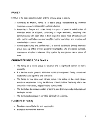
  • 1. 1 FAMILY FAMILY is the basic social institution and the primary group in society.  According to Murdok, family is a social group characterized by common residence, economic cooperation and reproduction.  According to Burges and Locke, family is a group of persons united by ties of marriage, blood or adoption, constituting a single household, interacting and communicating with each other in their respective social roles of husband and wife, mother and father, son and daughter, brother and sister, and creating and maintaining a common culture.  According to Murray and Zentner (1997) is a social system and primary reference group made up of two or more persons living together who are related by blood, marriage or adoption or who are living together by arrangement over a period of time. CHARACTERISTICS OF A FAMILY  The family as a social group is universal and is significant element in man’s social life  It is the first social group to which the individual is exposed. Family contact and relationships are repetitive and continuous  The family is very close and intimate group. It is setting of the most intense emotional experiences during the life time of the individual.The family affects the individual social values, disposition and outlook in life  The family has the unique position of serving as a link between the individual and the larger society  The family is also unique in providing continuity of social life. Functions of Family  Regulates sexual behavior and reproduction  Biological maintenance function
  • 2. 2  Socialization function  Provide legitimate children with a status  Social control function  Economic function  Educational, recreational, religious and political functions Classification of Family Structure Based on Descent  Patrilineal- affiliates a person with a group of relatives through his or her father.  Matrilineal- affiliates a person with a group of relatives through his or her mother.  Bilateral- affiliates a person with a group of relatives related through both his or her parents. Based on Authority  Patriarchial- authority is vested on the oldest male in the family, often the father.  Matriarchial- authority is vested in the mother or mother’s kin  Matricentric- prolonged absence of the father gives the mother a dominant position in the family, although the father may also share with the mother in decision making. Based on Internal Organization and Membership  Nuclear Family. Also known as primary or elementary family.  Extended Family. Extensions maybe through the parent-child relationship or husband-wife relationship, as in polygamous marriage. Based on Place of Residence  Patrilocal- requires the newly wed to reside near the groom’s parents.  Matrilocal- near the bride’s parents.  Bilocal- provides the couple the choice to reside on either parents.  Neolocal- permits the couple to reside independently of their parents
  • 3.  Avunculocal- prescribes the newly wed couple to reside with or near the maternal 3 uncle of the groom. Families consist of special functional subsystems:  marital subsystem (parents)  sibling subsytem (children)  individual parent –child system subsystem View of the person: The person is a member of the family where each new member adds to the complexity of the interaction within the family. Stages of Family 1. Beginning *Establishing a mutually satisfying Family marriage *Planning to have or not to have children. 2. Child- *Having and adjusting to infant bearing family *Supporting the needs of all three members *Renegotiating marital relationships. 3. Family with *adjusting to cost of family life a pre-school *adapting to the needs of pre-school children *Coping with parental loss of energy and privacy 4. Family with *Adjusting to the activity of the growing school age children *Promoting joint decisions between children and parents *Encouraging and supporting children’s educational achievements 5. Family with *Maintaining open communication among teenagers and members young adult *Supporting ethical and moral values within the family *Balancing freedom with responsibility of teenagers *Releasing young adults with appropriate rituals and assistance 6. Post- *Strengthening marital relationships parental *Maintaining supportive home base family *Preparing for retirement 7. Aging *Maintaining ties with younger and Family older generations *Adjusting for retirement *Adjusting to loss of spouse *Closing family house
  • 4. 4 FETAL AND MATERNAL HEALTH PRENATAL CARE  Ensures the overall health of newborns and their mothers. OB Follow-up visits  Every 4 weeks through the 28th week  Every 2 weeks through the 36th weeks  Every week until delivery The health care given to a woman and her family during pregnancy. The earliest stage at which fetuses could survive if they were born at that time. Any pregnancy terminated before the age of viability. An infant born at the end of 37 weeks to the end of 42 weeks. An infant born before 37 weeks. An infant born beyond 42 weeks. A woman in labor. A woman who has just given birth. A pregnant woman or refers to the present pregnancy. The number of pregnancy that reached viability. A woman who is pregnant the first time. A woman with a second or later pregnancy. A woman who has never been pregnant and is nor currently pregnant. A woman who has completed one pregnancy of viability. A woman who has carried two or more pregnancies of viability. A woman who had 6 or more viable pregnancies. History of past pregnancies Gravida (G)  A number of times the woman has been pregnant. Para (P)  A number of children above viability the woman has previously borne.  Multigestation is considered as one para GTPAL G- number of times the woman has been pregnant.
  • 5. 5 P- is broken down into: T- term BIRTH P- preterm BIRTH A- Abortion L- living children GTPALM G- number of times the woman has been pregnant P- is broken down into: T- term INFANT P- preterm INFANT A- abortion L- living children M- multiple pregnancies History of present pregnancy Estimate date of birth/ date of delivery Naegele’s rule  standard method used to predict the length of a pregnancy  approximates that pregnancy last 40 weeks from LMP  To get EDB/ EDD  1st day of LMP Estimate fetal growth McDonald’s Rule  Common method of determining fetal growth in utero.  Symphisis-fundal height measurement  The distance from the symphisis pubis to the uterine fundus in cm es equal to the week of gestation between the 20th and 31st weeks of preganancy  To estimate AOG  Fundic height in cm x 2/7= AOG in moths
  • 6. 6  Fundic height in cm x 8/7= AOG in weeks Leopold’s Maneuver  A systematic method of observation and palpation of the abdomen to determine fetal presentation and position. Preparation  Explain the procedure  Ask to void  Supine with knees slightly flexed  Wash hands with warm water Purpose Procedure Findings First Maneuver: Fundal Grip To determine fetal part lying in the fundus. To determine presentation. Using both hands, feel for the fetal part lying in the fundus. Head is more firm, hard and round that moves independently of the body. Breech is less well defined that moves only in conjunction with the body. Second Maneuver: Umbilical Grip To identify location of fetal back. To determine position. One hand is used to steady the uterus on one side of the abdomen while the other hand moves slightly on a circular motion from top to the lower segment of the uterus to feel for the fetal back and small fetal parts. Use gentle but deep pressure. Fetal back is smooth, hard, and resistant surface Knees and elbows of fetus feel with a number of angular nodulation Third Maneuver: Pawlik’s Grip To determine engagement of presenting part. Using thumb and finger, grasp the lower portion of the abdomen above symphisis pubis, press in slightly and make The presenting part is notengaged if it is not movable. It is not yet engaged if it is still movable.
  • 7. 7 gentle movements from side to side. Fourth Maneuver: Pelvic Grip To determine the degree of flexion of fetal head. To determine attitude or habitus. Facing foot part of the woman, palpate fetal head pressing downward about 2 inches above the inguinal ligament. Use both hands Good attitude – if brow correspond to the side (2nd maneuver) that contained the elbows and knees. Poor atitude – if examining fingers will meet an obstruction on the same side as fetal back (hyperextended head) Also palpates infant’s anteroposterior position. If brow is very easily palpated, fetus is at posterior position (occiput pointing towards woman’s back) PRENATAL VISIT 8-12 weeks: Initial prenatal visit. The plan for your prenatal care at the practice you have chosen will be explained. A health history is taken, and a physical exam, including a pelvic exam is done. Lab work is completed, including your blood type and hemoglobin, sexually transmitted infection screening, a urine test, and a PAP test if you are due for one. You may be able to hear the baby's heart beat at this visit. If you
  • 8. cannot say with accuracy when you had your last period, an ultrasound might be scheduled to help determine how far along you are. Optional genetic counseling visit: Early in your pregnancy, you may be offered genetic screening. This is commonly offered to women over the age of 35, or women who have a family history of genetic problems, but it is increasingly being offered to every woman. If you choose this screening, your care provider and/or genetic counselor may suggest additional genetic screening or diagnostic tests, including blood tests, chorionic villus sampling, ultrasound, and/or amniocentesis. These tests are done at specific times during pregnancy. First two trimesters: Prenatal visits continue every 4-6 weeks through the first two trimesters, or until you are 28 weeks along. At each appointment, your care provider will weigh you and take your blood pressure, listen to the baby's heartbeat, and measure the growth of your uterus and baby. Some providers check your urine for protein and sugar at each visit. 15 to 20 weeks: At one of your appointments within this period, you will be offered the Quad Screen test, which screens for genetic and spinal cord abnormalities. You may also be offered an ultrasound between 18 and 20 weeks to view the baby's organs, and measure the growth of the baby and the placenta. 27 or 28 weeks: At an appointment within this period, you will be encouraged to take a glucose challenge test to screen for gestational diabetes. Your hemoglobin may be rechecked. Some providers do a pelvic exam. Expect to review warning signs of late pregnancy that would alert you to preterm labor or high blood pressure. You may be encouraged to sign up for prenatal classes, find a doctor or nurse-practitioner who will provide well-child care for your baby, and information may be provided about making plans for labor. 28 to 36 weeks: After 28 weeks, prenatal visits continue every 2-3 weeks until 36 weeks. Your doctor or midwife will continue to record the growth of the baby, listen to the baby's heartbeat, and will check the position of the baby. 8
  • 9. 36 weeks: At this visit, your midwife or doctor will do a pelvic exam, and encourage you to have a Group B Strep test. Screening tests for sexually transmitted infections may be repeated at this visit. The position and size of the baby are estimated. If your baby is not head down, your provider may suggest exercises to encourage the baby to turn, or suggest a physical manipulation called external version. The risks and benefits of this procedure should be carefully explained. 36 to 40 weeks: The usual monitoring of your weight and blood pressure, and the baby's size, position, and heart rate are done. Your care provider may offer to check your cervix for dilation. 40+ weeks: After your due date, your care provider may offer what is called "post-dates" testing, including nonstress tests, ultrasound, and biophysical profiles. Some 9 providers start at 40 weeks, others not until 10 days past your due date. Safe Medications to Take During Pregnancy Allergy  Benadryl (diphenhydramine)  Claritin Check with your doctor before taking these in the first trimester. Cold and Flu  Tylenol (acetaminophen)  Saline nasal drops or spray  Warm salt/water gargle Check with your doctor before taking any other medications, especially in the first trimester. Constipation  Colace  Metamucil First Aid Ointment
  • 10. 10  Bacitracin  J&J First-Aid Cream  Neosporin  Polysporin Rashes  Benadryl cream  Caladryl lotion or cream  Hydrocortisone cream or ointment  Oatmeal bath (Aveeno) *Note: No drug can be considered 100% safe to use during pregnancy. What Alternative Therapies Are Considered Safe During Pregnancy? Some alternative therapies have been shown to be safe and effective for pregnant women to relieve some of the uncomfortable side effects of pregnancy. Talk it over with your doctor first before using any of them. And remember, “Natural” doesn’t always equal “safe” when you’re pregnant. Nausea in early pregnancy: Acupuncture, acupressure, ginger root (250 milligram capsules 4 times a day), and vitamin B6 (pyridoxine, 25 milligrams two or three times a day) work well. Sipping the thick syrup from inside a can of peaches, pears, mixed fruits, pineapples, or orange slices may also help. Backache: Chiropractic manipulation holds the best track record. Another option is massage but it is important to make sure your massage therapist is adequately trained in pre-natal massage. Turning a breech baby: Exercise and hypnosis may help. Pain relief in labor: Epidurals are most effective, but immersion in a warm bath can also relieve tension. Relaxation and breathing techniques, emotional support, and self-hypnosis are widely used in labor. Acupuncture can also work for some women.
  • 11. 11 Common Discomforts During Pregnancy Symptoms of discomfort due to pregnancy vary from woman-to-woman. The following are some common discomforts. However, each mother-to-be may experience symptoms differently or not at all:  Nausea and vomiting About half of all pregnant women experience nausea and sometimes vomiting in the first trimester--also called morning sickness because symptoms are most severe in the morning. Some women may have nausea and vomiting throughout the pregnancy. Morning sickness may be due to the changes in hormone levels during pregnancy. Morning sickness seems to be aggravated by stress, traveling, and certain foods, such as spicy or fatty foods. Eating small meals several times a day may help lessen the symptoms. A diet high in protein and complex carbohydrates (such as whole wheat bread, pasta, bananas, and green, leafy vegetables) may also help reduce the severity of the nausea. If vomiting is severe, causing a woman to lose fluids and weight, it may indicate a condition called hyperemesis gravidarum. Hyperemesis can lead to dehydration and may require hospitalization for intravenous fluids and nutrition. Call your physician or midwife if you are having constant or severe nausea and vomiting.  Fatigue As the body works overtime to provide a nourishing environment for the fetus, it is no wonder a pregnant woman often feels tired. In the first trimester, her blood volume and other fluids increase as her body adjusts to the pregnancy. Sometimes anemia is the underlying cause of the fatigue. Anemia is a reduction in the oxygen-carrying capability of red blood cells, and is usually due to low iron levels. A simple blood test performed at a prenatal visit will check for anemia.  Hemorrhoids Because of increased pressure on the rectum and perineum, the increased blood volume, and the increased likelihood of becoming constipated as the pregnancy
  • 12. progresses, hemorrhoids are common in late pregnancy. Avoiding constipation and straining may help to prevent hemorrhoids. Always check with your physician or midwife before using any medication to treat this condition. 12  Varicose veins Varicose veins--swollen, purple veins--are common in the legs and around the vaginal opening during late pregnancy. In most cases, varicose veins are caused by the increased pressure on the legs and the pelvic veins, and by the increased blood volume.  Heartburn and indigestion Heartburn and indigestion, caused by pressure on the intestines and stomach (which, in turn, pushes stomach contents back up into the esophagus), can be prevented or reduced by eating smaller meals throughout the day and by avoiding lying down shortly after eating.  Bleeding gums Gums may become more spongy as blood flow increases during pregnancy, causing them to bleed easily. A pregnant woman should continue to take care of her teeth and gums and go to the dentist for regular checkups. This symptom usually disappears after pregnancy.  Pica Pica is a rare craving to eat substances other than food, such as dirt, clay, or coal. The craving may indicate a nutritional deficiency.  Swelling/fluid retention Mild swelling is common during pregnancy but severe swelling that persists may indicate preeclampsia (abnormal condition marked by high blood pressure). Lying on the left side, elevating the legs, and wearing support hose and comfortable shoes may help to relieve the swelling. Be sure to notify your physician or midwife about sudden swelling, especially in the hands or face, or rapid weight gain.  Skin changes Due to fluctuations in hormone levels, including hormones that stimulate pigmentation of
  • 13. 13 the skin, brown, blotchy patches may occur on the face, forehead, and/or cheeks. This is often called the mask of pregnancy, orchloasma, and often disappears soon after delivery. Using sunscreen when outside can reduce the amount of darkening that occurs. Pigmentation may also increase in the skin surrounding the nipples, called the areola. In addition, a dark line frequently appears down the middle of the abdomen. Freckles may darken, and moles may grow.  Stretch marks Pinkish stretch marks may appear on the abdomen, breasts, thighs, or buttocks. Stretch marks are generally caused by a rapid increase in weight, and the marks usually fade after pregnancy.  Yeast infections Due to hormone changes and increased vaginal discharge, also called leukorrhea, a pregnant woman is more susceptible to yeast infections. Yeast infections are characterized by a thick, whitish discharge from the vagina and itching. Yeast infections are highly treatable. Always consult your physician or midwife before taking any medication for this condition.  Congested or bloody nose During pregnancy, the lining of the respiratory tract receives more blood, often making it more congested. This congestion can also cause stuffiness in the nose or nosebleeds. In addition, small blood vessels in the nose are easily damaged due to the increased blood volume, causing nosebleeds.  Constipation Increased pressure from the pregnancy on the rectum and intestines can interfere with digestion and subsequent bowel movements. In addition, hormone changes may slow down the food being processed by the body. Increasing fluids, regular exercise, and increasing the fiber in your diet are some of the ways to prevent constipation. Always check with your physician or midwife before taking any medication for this condition.
  • 14. 14  Backache As a woman's weight increases, her balance changes, and her center of gravity is pulled forward, straining her back. Pelvic joints that begin to loosen in preparation for childbirth also contribute to this back strain. Proper posture and proper lifting techniques throughout the pregnancy can help reduce the strain on the back.  Dizziness Dizziness during pregnancy is a common symptom, which may be caused by: o Low blood pressure due to the uterus compressing major arteries o Low blood sugar o Low iron o Quickly moving from a sitting position to a standing position o Dehydration To prevent injury from falling during episodes of dizziness, a pregnant woman should stand up slowly and hold on to the walls and other stable structures for support and balance.  Headaches Hormonal changes may be the cause of headaches during pregnancy, especially during the first trimester. Rest, proper nutrition, and adequate fluid intake may help alleviate headache symptoms. Always consult your physician or midwife before taking any medication for this condition. If you have a severe headache or a headache that does not resolve, call your health care provider. It may be a sign of preeclampsia. Danger signs of Pregnancy 1. Decreased fetal movement If you notice that your baby isn't moving around as much as she normally does, stop exercising and take a minute to pay attention to what she's doing. Remember that sometimes it's hard to tell if your baby is moving around when you're moving around,
  • 15. too. Also, be sure to eat and drink water before your workout because that may affect your baby's movements. Call your healthcare provider: if your baby isn't moving around as much as normal or you notice a sudden decrease in your baby's movement. 15 2. Dizziness Is It Safe During Pregnancy? Persistent dizziness together with fatigue and headaches can be symptoms of severe anemia or another serious condition. Call your healthcare provider: if you're still dizzy after you've cooled down, rested, and had some water. 3. Overheating If you feel faint or dizzy, or if you develop a headache, nausea, cramps, or a racing heart, your body's telling you that it's having a hard time regulating your internal temperature, which can be harmful to your baby. The baby can get overheated just as you do. When your body overheats, blood flowing to the uterus is diverted to the skin to help the body cool itself off, putting the baby in jeopardy. It's unusual to overheat from exercise alone, but if it's hot outside or in the gym where you exercise, you could run into trouble. If you're exercising indoors, it's best to do so in a well-ventilated room with fans. If you're exercising outdoors, avoid the sun when it's strongest in the middle of the day. Consider staying inside if it's especially hot out. Call your healthcare provider: if you feel very hot and you have symptoms of overheating, like profuse sweating, dizziness or lightheadedness, a headache, nausea, cramps, or an irregular heartbeat. 4. Heart palpitations If your heart is pounding and you can't carry on a conversation without being out of breath, or if you sweat buckets while you exercise, you're probably working too hard. Heart palpitations may be a sign of dehydration, severe anemia, thyroid disease, or a heart problem. Call your healthcare provider: if your heart continues to race after you've cooled down, rested, and had some water. 5. Swelling in your calf
  • 16. Your feet and hands may puff up a little after you exercise, but if you notice calf pain or swelling, it could be a sign of deep vein thrombosis (DVT), a potentially life-threatening condition caused by a type of blood clot. DVT usually affects veins deep in the lower leg and thigh and occurs on one side of the body. You may experience redness and skin that feels warm to the touch. Also, sudden swelling in your legs (and face and hands), along with high blood pressure, may be a sign of preeclampsia. Call your healthcare provider: immediately if you think you have DVT. If you have DVT and you experience chest pain, difficulty breathing, fainting, or any other serious condition, go to the emergency room right away. 6. Vaginal bleeding Some women do experience light spotting throughout their pregnancy, but vaginal bleeding during pregnancy is always a cause for concern. Early in your pregnancy, it could signal a miscarriage. In the second and third trimesters, bleeding is associated with premature labor and complications with the placenta, such as placenta previa or placenta abruption. All require immediate medical attention. Call your healthcare provider: immediately if you have vaginal bleeding. If you can't reach anyone, go to the emergency room. 16 7. Blurred vision It's common for blood pressure to drop during the first 6 months of pregnancy. But combine low blood pressure with exercise and dehydration, and you may be headed for trouble. If your eyesight gets hazy in the middle of your workout, you may be dehydrated. That alone is enough to send your blood pressure plummeting and your heart into overdrive. As a result, not enough blood may be getting to your developing baby's vital organs. Blurred vision may also be a sign of preeclampsia. This condition can be dangerous for your baby because preeclampsia can severely restrict the flow of blood to the placenta. Call your healthcare provider: immediately. If you can't reach anyone, go to the emergency room. 8. Fainting Fainting during pregnancy shouldn't be taken lightly. It could signal something as simple as dehydration or something serious like major circulatory or heart problems. You may
  • 17. not be getting enough oxygen to your brain, which means your baby may not be getting enough either. Call your healthcare provider: immediately. If you can't reach anyone, go to the emergency room. 17 9. Recurring pain in abdomen or chest It may just be your ligaments stretching, but you could also be having contractions – especially if the pain recurs at somewhat regular intervals. Women experience labor pain differently: For some the pain is similar to a severe menstrual cramp. For others, the pain is sharp and comes in waves or feels like a recurring pain in the back. Abdominal pain accompanied by bleeding might be a sign of placental abruption. You may need to be hooked up to a fetal monitor so your healthcare provider can determine whether you're in labor. Life-threatening chest pain most likely signals a serious problem with your heart or lungs. If you're pregnant and have chest pain while exercising, the American College of Obstetricians and Gynecologists recommends that you stop what you're doing right away. Call your healthcare provider: immediately. If you can't reach anyone, go to the emergency room. 10. Fluid leaking from your vagina If your underpants are constantly wet or if you feel watery fluid leaking (or gushing) from your vagina, it could mean premature rupture of the membranes. That can be a signal that your body is about to go into labor. Theories of Labor Onset Labor is a coordinated sequence of involuntary, intermittent uterine contractions. It is the series of events that expels the fetus and placenta out of the mother’s body. This is made possible by the presence of uterine contractions and abdominal pressure that push the fetus out during the expulsion period of delivery. Regular contractions result to gradual cervical effacement and dilatation. Adequate pressure from abdominal muscles allows the baby to be pushed outside the mother’s womb.
  • 18. Labor and delivery require a woman to utilize her coping methods psychologically and physiologically. Normally, labor begins when the fetus reaches a mature age (38-42 weeks age of gestation). This is to ensure survival of the fetus with the extrauterine life. The mechanism that converts Braxton Hicks Contractions (painless contractions) to strong and coordinated uterine contractions is unknown. In some cases, labor occurs before the fetus reaches the mature age (preterm birth) while in others it is delayed (postterm birth). Although the exact mechanism that initiates labor is unknown. Theories have been proposed to explain how and why labor occurs. Uterine Stretch theory The idea is based on the concept that any hollow body organ when stretched to its capacity will inevitably contract to expel its contents. The uterus, which is a hollow muscular organ, becomes stretched due to the growing fetal structures. In return, the pressure increases causing physiologic changes (uterine contractions) that initiate labor. Oxytocin theory Pressure on the cervix stimulates the hypophysis to release oxytocin from the maternal posterior pituitary gland. As pregnancy advances, the uterus becomes more sensitive to oxytocin. Presence of this hormone causes the initiation of contraction of the smooth muscles of the body (uterus is composed of smooth muscles). Progesterone deprivation theory Progesterone is the hormone designed to promote pregnancy. It is believed that presence of this hormone inhibits uterine motility. As pregnancy advances, changes in the relative effects estrogen and progesterone encourage the onset of labor. A marked increase in estrogen level is noted in relation to progesterone, making the latter hormone less effective in controlling rhythmic uterine contractions. Also, in later 18
  • 19. pregnancy, rising fetal cortisol levels inhibit progesterone production from the placenta. Reduce progesterone formation initiates labor. Prostaglandin theory In the latter part of pregnancy, fetal membranes and uterine decidua increase prostaglandin levels. This hormone is secreted from the lower area of the fetal membrane (forebag). A decrease in progesterone amount also elevates the prostaglandin level. Synthesis of prostaglandin, in return, causes uterine contraction thus, labor is initiated. Theory of Aging Placenta Advance placental age decreases blood supply to the uterus. This event triggers uterine contractions, thereby, starting the labor. 19 Signs of Labor A common concern of women as they near the end of pregnancy is how they will know if they are beginning labor Preliminary Signs of Labor Before labor , a woman often experiences subtle signs that signal labor is imminent. It is important to review these with women during the last trimester of pregnancy so they can more easily recognize beginning signs . Lightening In primiparas, lightening or descent of the fetal presenting part into the pelvis, occurs approximately 10 to 14 days before labor begins. This fetal descent changes a woman’s abdominal contour , because it positions the uterus lower and more anterior in the abdomen. Lightening gives a woman relief from the diaphragmatic pressure and shortness of breath that she has been experiencing and in this way “lightens” her load. Lightening probably occurs early in primiparas because of tight abdominal muscles. In multiparas, it is not as dramatic and usually occurs on the day of labor or even after
  • 20. labor has begun. As the fetus sinks lower into the pelvis, a woman may experience shooting leg pains from the increased pressure on her sciatic nerve, increased amounts of vaginal discharge, and urinary frequency from pressure on her bladder. 20 Increase in level of Activity A woman may awaken on the morning of labor full of energy, in contrast to the feeling of chronic fatigue she felt during the previous month. This increase in activity is related to increase in epinephrine release initiated by decrease in progesterone produced by placenta . This additional epinephrine prepares a woman’s body for the work of labor ahead. Slight loss of weight As progesterone level falls body fluid is more easily excreted from the body. This increase in urine production can lead to a weightloss between 1 and 3 pounds. Braxton Hicks Contraction Braxton Hicks contractions are sporadic uterine contractions that start about 6 weeks into your pregnancy, although you won't be able to feel them that early. You probably won't start to notice them until sometime after mid-pregnancy, if you notice them at all. (Some women don't.) They get their name from John Braxton Hicks, an English doctor who first described them in 1872. As your pregnancy progresses, Braxton Hicks contractions tend to come somewhat more often, but until you get to your last few weeks, they'll probably remain infrequent, irregular, and essentially painless. Sometimes, though, Braxton Hicks contractions are hard to distinguish from early signs of preterm labor. Play it safe and don't try to make the diagnosis yourself. If you haven't hit 37 weeks yet and you're having more than four contractions in an hour — or you have any other signs of preterm labor
  • 21. By the time you're within a couple of weeks of your due date, your cervix has likely begun to "ripen" or gradually soften up in preparation for labor. Your contractions may get more intense and more frequent, and they may cause some discomfort. Unlike the earlier painless and sporadic Braxton Hicks contractions, which caused no obvious cervical changes, these contractions may help your cervix thin out (efface) and maybe even open up (dilate) a bit. This period is sometimes referred to as pre-labor. 21 True labor vs. false labor True Labor or False Labor “How will I know when it is real labor?” This is a question you may have as you near the end of your pregnancy. Many women have periods of “false” labor late in their pregnancy. During false labor, you have contractions that seem to come and go. False labor pains are called “Braxton Hicks” contractions. These contractions help soften and thin your cervix. They tend to happen more often as you get closer to your due date (2 to 4 weeks before birth). Sometimes it is hard to tell the difference between false labor and true labor. Don’t be upset or embarrassed if you think labor is beginning when it is actually a false alarm. Differences between false labor and true labor There are several ways to tell the difference between true and false labor. Timing of contractions  False labor: Contractions are often irregular. They don’t get closer together over time.  True labor: Contractions come regularly and get closer together. Each contraction lasts about 30 to 60 seconds.
  • 22. 22 Strength of contractions  False labor: Contractions are often weak and do not get stronger.  True labor: Contractions get stronger as time goes on. Change with movement  False labor: Contractions may stop or slow down when you walk, lie down, or change positions.  True labor: Contractions continue no matter what you do. Pain with contractions  False labor: Discomfort is usually felt in the front, like menstrual cramps.  True labor: Discomfort or pressure starts in the back and moves to the front. If your water breaks Sometimes labor begins when the bag of waters, or membranes, breaks. This may happen with your early contractions. Or your water may not break until later into your labor. If your water breaks, you may notice a near constant trickle of fluid from the vagina or a sudden gush of fluid. If you think your bag of waters is leaking or broken, call your doctor right away. Other physical changes You also may have physical changes that occur as your body gets ready for labor. It is normal to have a slight increase of thin, white discharge at the end of pregnancy. Activities like coughing, sneezing, or laughing may cause leaking of urine. You also may notice a change in appetite, nausea, diarrhea, or constipation. The loss of your mucus plug often precedes labor by a few days. Mucus may be present 2 to 14 days before true labor begins.
  • 23. 23 Everyone experiences labor in a different way. Call your doctor if you think you are in labor. True Labor vs. False Labor One of the biggest confusions many expectant mothers face is telling the difference between true labor and false labor. The chart below describes the specific characteristics of both true and false labor contractions. Characteristic True Labor False Labor Frequency Regular, usually happens 4-6 minutes apart as they become closer together. Lasts 30-70 seconds Irregular, don’t show signs of consistency or becoming closer together Strength Consistently increase in strength as time goes on, vaginal pressure likely to increase Weak, usually do not gain strength as time goes on, can begin strong then weaken as time passes Amount of Pain Starts in the back and moves forward Usually only felt in the front of the abdomen
  • 24. 24 Changing Positions Changing positions has no effect on pain, strength or frequency Inconsistent, may stop or slow down when you walk, lie down, increase fluids or change positions in any other way False contractions  Begin and remain irregular  Felt first abdominally and remain confined to abdomen and groin  Often disappear with ambulation or sleep  Do not increase in duration, frequency, or intensity  Do not achieve cervical dilation True Contraction  Begin irregularly but become regular and predictable  Felt first in lower back and sweep around to the abdomen in a wave  Continue no matter what the woman’s level of activity  Increase in duration, frequency, and intensity  Achieve cervical dilation. 5 P’s Components of labor  Passenger (fetus)  Powers (uterine contractions)  Passage (the pelvis & maternal soft parts)  Position (maternal)  Psyche (maternal psychological status)
  • 25. 25 PASSENGER (FETUS):  Biological influences A pregnancy that terminates during the 38-42 week gestation is likely to indicate a healthy fetus.  Mechanical influences  Fetal head  Fetopelvic relationships  Cardinal movements  Fetal Head: ( a mechanical influence)  Bones: The head is the largest portion of the fetal body, & because it is a firm, noncompliant bony structure, it is the fetal component that is of most significance (from an obstetrical perspective).  Sutures & Fontanelles: Between the bones of the fetal head are membranous spaces called sutures. The fontanelles are areas of the head where suture lines intersect.  Landmarks: Head is divided into designated areas (1) the sinciput or brow portion; (2) the vertex, or top of the head between the 2 fontanelles; (3) the occiput or back of the head over the occipital bone.  Diameters: During birth it is desirable that the smallest diameter of the fetal head move through the maternal bony pelvis. The diameter tht presents through the pelvis depends on the amount of flexion or extension of the head (attitude).  Fetopelvic Relationships:  Fetal Lie: refers to the relationship of the long axis of the fetus, as related to the spinal column, to the long axis of the mother. (vertical lie = most common).  Fetal Attitude: refers to the relationship of the fetal parts to one another. Fetus is described as being in a state of flexion or extension.  Fetal Presentation: The part of the fetal body that enters (or presents to) the maternal pelvis. Most common = cephalic presentation (head first).
  • 26.  Fetal Position: refers to the relationship of an assigned area of the presenting 26 part (often called the fetal denominator) to the maternal pelvis.  Determine the fetal denominator.  Mentally divide the maternal pelvis into 4 quadrants (R&L anterior, R&L posterior).  Assign a standard abbreviation indicating the fetal position based on findings of vaginal exam.  Synclitism & Asynclitism: Asynclitic refers to a fetal head that is not parallel to the anteroposterior plane of the pelvis. The head is synclitic when the sagittal suture lies midway between the symphysis pubis and the sacral promontory.  Cardinal Movements:  Also called the “mechanisms of labor”.  A series of adaptations the fetus makes as it moves through the maternal bony pelvis during the process of lavor & birth.  Influenced by the size and position of the fetus, the powers of labor, the size and shape of the maternal pelvis, and the mother’s position.  8 Cardinal Movements: (in an anterior occiput position)  Engagement  Descent  Flexion  Internal rotation  Extension  Restitution  External rotation of the shoulders  Expulsion  Engagement: the mechanism by which the fetus nestles into the pelvis.  Also referred to as “dropping” or “lightening”.
  • 27.  A fetus is engaged when the biparietal diameter of the fetal head reached the level of the maternal ischial spines; known as zero station.  Leopold’s maneuvers: the head is more difficult to move and less of 27 the head is able to be palpated abdominally after engagement.  Descent: describes the process that the fetal head undergoes as it begins its journey through the pelvis.  Pressure from uterine ctx, hydrostatic forces, abdominal muscles, and gravity promote descent of the fetus through the pelvic inlet and midplane.  Descent is continuous from the time of engagement until birth.  Assessed by measurements called stations.  Ranges from –3 to +3 station.  Flexion: the process of the fetal head’s nodding forward toward the fetal chest and occurs as a result of descent, the thickening of the uterine fundus, & increased resistance of the soft tissues. o Engagement, descent and flexion tend to occur simultaneously.  Internal Rotation: most commonly the fetus rotates internally from the occiput transverse position assumed at engagement into the pelvis to an occiput anterior position while continuously descending.  Extension: enables the head to be born when the fetus is in a cephalic position. Results from the downward forces of the uterine contractions and the resistance of the pelvic floor muscles. o Begins after the head has crowned and is complete when the head passes under the symphysis pubis and the occiput, anterior fontanelle, brow, face, and chin pass over the sacrum & coccyx and are born over the perineum.  Restitution: results in a realignment of the fetal head with the body, after the head is born.  It is common that as the head internally rotates to an anterior position before its birth, the shoulders may enter the pelvis in the oblique diameter.
  • 28. 28  This allows the head to turn, but as a result, the neck twists.  Restitution occurs when the head is free of pelvic resistance, allowing the head to turn back until it is again at right angles to the shoulders.  External Rotation: After the head is born & restitution occurs, the shoulders externally rotate so that they are in the anteroposterior diameter of the pelvis.  This is the largest diameter of the outlet, it easily allows the birth of the broad shoulders.  Shoulders are born by first delivering the anterior shoulder from under the symphysis pubis and then the posterior shoulder from over the perineum.  Expulsion: the last cardinal movement; consists of the birth of the entire body.  The body usually follows easily after the birth of the head and shoulders.  The time of birth is often documented at the moment of expulsion. PASSAGE: “P” # 2  Major pelvic bones include the innominate bones (formed by the fusion of the ilium, ischium, and pubis around the acetabulum), the sacrum, and the coccyx. DIVISIONS:  Pelvis is arbitrarily divided into halves – the false pelvis and the true pelvis.  False pelvis: wide broad area btw. the iliac crests & has no major clinical significance for L&D.  True Pelvis: the actual bony passage that the fetus must traverse during labor and birth. Shape is a curved axis, not a straight passage , d/t the diameters & planes of the pelvis.
  • 29. 29 PLANES:  3 common planes of the pelvis are the inlet (the pelvic brim), midpelvis, and outlet.  A pelvis with an adequate inlet & midplane rarely if ever has reduced diameters for the outlet.  The coccyx also has slight mobility, which increases the available space in the outlet. PRENATAL ASSESSMENT OF PELVIS:  Clinical pelvimetry reassures both the health care provider & the woman about the normalcy of the pelvis.  When any variation exists in the pelvic structures, it can be discussed & anticipatory guidance given (ex- how to cope with back aches, back labor, etc.)  Rarely an abnormal pelvis such as true android, guidance may include the planning for a C/S. SOFT PASSAGE THROUGH MATERNAL SOFT TISSUE STRUCTURES:  Soft tissues of the cervix, vagina, and perineum must stretch to allow passage of the fetus through the axis of the birth canal.  Progesterone & relaxin help facilitate the softening & increase the elasticity of muscles & ligaments. POWERS: “P” # 3  Uterine labor ctx. of the myometrium.  Ctx.phase consists of a descending gradient: o The wave begins in the fundus (greatest # myometrial cells). o Then moves downward through the corpus of the uterus. o Intensity of ctx.diminishes from fundus to cervix.
  • 30. 30  Retraction phase. EFFACEMENT & DILATATION:  The purpose of uterine ctx. o Accomplish the effacement and dilation of the cervix. o Facilitate the descent & rotation of the fetus through the passages. o Facilitate the separation & expulsion of the placenta. o Control bleeding after delivery by compressing blood vessels.  Effacement= the thinning or shortening of the cervix.  Dilatation = the gradual opening of th cervix and is a continued extension of the contraction-retraction process already described.  Dilatation and effacement take place concurrently throughout labor.  Dilatation is assessed by vaginal examination, and is recorded in centimeters from 0-10 cm.  Hydrostatic Force = another power that facilitates the process of labor and birth.  Includes the pressure of the fetus within the amniotic sac.  As ctx. occur, the membranes and amniotic fluid facilitates dilation and effacement.  Since the lower uterine segment and cervix are regions of lesser resistance, the additional pressure of the amniotic sac is of great importance in promoting the birth process.  Abdominal Force = the final power for labor & birth. Intra-abdominal force.  This power is reserved for the 2nd stage of labor, after effacement & dilation are complete.  Maternal pushing, or bearing down effort.  In the expulsion stage, the ctx.change in character, & many women begin to experience an involuntary urge to push.
  • 31. 31 POSITION: “P” # 4  In the last half of the 20th century, the position used most frequently for labor in the US has supine in a hospital bed.  The most common position for birth has been a lithotomy position.  Limited ambulation of laboring women resulted from use of continuous fetal monitoring, routine use of IV hydration, epidural anesthesia and use of analgesia PSYCHOLOGY OF BIRTH: “P” # 5  The progress of labor and birth can be adversely affected maternal fear and tension.  Norepinephrine and epinephrine may stimulate both alpha and beta receptors of the myometrium and interfere with the rhythmic nature of labor.  Anxiety can also increase pain perception and lead to an increased need for analgesia & anesthesia. STAGES OF LABOR Childbirth usually occurs in three stages: First stage: The time of the onset of true labor unti l the cervix is completely di lated to 10 cm. Second stage: The period after the cervix is di lated to 10 cm unti l the baby is delivered. Third stage: Delivery of the placenta. First Stage The first stage of labor is the longest and involves three phases:  Early Labor Phase -The time of the onset of labor until the cervix is dilated to 3 cm.
  • 32. 32  Active Labor Phase - Continues from 3 cm. until the cervix is dilated to 7 cm.  Transition Phase - Continues from 7 cm. until the cervix is fully dilated to 10 cm. Each phase is characterized by di fferent emotions and physical challenges. Think of i t as a big adventure wi th some important guidelines. Early Labor Phase What to do: During this phase you should just try to relax. It is not necessary to rush to the hospi tal or bi rth center. Try to enjoy the comfort of the fami liar surroundings at home. If early labor occurs during the day, do some simple routines around the house. Keep yourself occupied whi le conserving your energy. Drink ple nty of water and eat small snacks. Keep track of the time of your contractions. If early labor begins during the night i t is a good idea to try to get some sleep. If you are unable to fall asleep, focus on doing some light activities like cleaning out your closet, packing your bag, or making sack lunches for the next day. What to expect:  Early labor will last approximately 8-12 hours  Your cervix will efface and dilate to 3 cm  Contractions will last about 30-45 seconds, giving you 5-30 minutes of rest between contractions  Contractions are typically mild, somewhat irregular, but become progressively stronger and more frequent  Contractions can feel like aching in your lower back, menstrual cramps, and pressure or tightening in the pelvic area  Your water might break. This is known as amniotic sac rupture and can happen any time within the first stage of labor.  When experiencing contractions, ask if they are:
  • 33. 33  Growing more intense  Following a regular pattern  Lasting longer  Becoming closer together  When your water breaks (amniotic sac rupture) note the following:  Color of fluid  Odor of fluid  Time rupture occurred  Tips for the support person:  Practice timing contractions  Be a calming influence  Offer comfort, reassurance, and support  Suggest simple activities to draw her focus from the labor  Keep up your own strength. You will need it!  Active Labor Phase:  What to do:  Now is time for you to head to the hospi tal or bi rth center.Your contractions wi ll be stronger, longer and closer together. It is very important that you have plenty of support. It is also a good time to start your breathing techniques and try a few relaxation exercises for use between contractions. You should swi tch posi tions often during this time. You might want to try walking or taking a warm bath. Continue to drink plenty of water and urinate periodically.  What to expect:  Active labor will last about 3-5 hours  Your cervix will dilate from 4cm to 7cm
  • 34.  Contractions during this phase will last about 45-60 seconds with3-5 34 minutes rest in between  Contractions will feel stronger and longer  This is usually the time to head to the hospital or birth center  Tips for the support person:  Give the mother your undivided attention  Offer her verbal reassurance and encouragement  Massage her abdomen and lower back  Keep track of the contractions (if she is being monitored, find out how the machine works)  Go through the breathing techniques with her  Help make her comfortable (prop pillows, get her water, apply touch)  Remind her to change positions frequently (take her for a walk or offer her a bath)  Continue with distractions from labor such as music, reading a book, or playing a simple card game  Don’t think that there is something wrong if she is not responding to you  Transition Phase  What to do:  During this phase the mother wi ll rely heavily on her support person. This is the most challenging phase but i t is also the shortest. Try to thi nk “one contracti on at a time” (Thi s may be hard to do i f the contractions are very close together). Remember how far you have already come, and when you feel an urge to push, tell your health care provider.  What to expect:  Transition will last about 30 min-2 hrs  Your cervix will dilate from 8cm to 10cm
  • 35.  Contractions during this phase will last about 60-90 seconds with a 30 second- 35 2 minute rest in between  Contractions are long, strong, intense, and can overlap  This is the hardest phase but also the shortest  You might experience hot flashes, chills, nausea, vomiting, or gas  Tips for the support person:  Offer lots of encouragement and praise  Avoid small talk  Continue breathing with her  Help guide her through her contractions with encouragement  Encourage her to relax between contractions  Don’t think that there is something wrong if she seems to be angry. It is a normal part of transition. Non stress test This simple, painless procedure is done during pregnancy to evaluate your baby's condition. During the test, your healthcare practitioner or a technician monitors your baby's heartbeat, first while the baby is resting and then while he's moving. Just as your heart beats faster when you're active, your baby's heart rate should go up while he's moving or kicking. The test is typically done if you've gone past your due date, or in the month or two leading up to your due date if you're having a high-risk pregnancy. Here are some reasons you might have a nonstress test:  You have diabetes that's treated with medication, high blood pressure, or some other medical condition that could affect your pregnancy.  You have gestational hypertension.  Your baby appears to be small or not growing properly.  Your baby is less active than normal.
  • 36. 36  You have too much or too little amniotic fluid.  You've had a procedure such as an external cephalic version (to turn a breech baby) or third trimester amniocentesis (to determine whether your baby's lungs are mature enough for birth or to rule out a uterine infection). Afterward, your practitioner will order a nonstress test to make sure that your baby's doing well.  You're past your due date and your practitioner wants to see how your baby is holding up during his extended stay in the womb.  You've previously lost a baby in the second half of pregnancy, for an unknown reason or because of a problem that might happen again in this pregnancy. In this case, nonstress testing may start as early as 28 weeks.  You have a medical problem that may jeopardize your baby's health.  Your baby has been diagnosed with an abnormality or birth defect and needs to be monitored. How Is A NST Performed? The test i nvolves attachi ng one belt to the mother’ s abdomen to measure fetal heart rate and another belt to measure contractions. Movement, heart rate and “reacti vi ty” of heart rate to movement i s measured for 20-30 minutes. If the baby does not move, i t does not necessari ly indicate that there is a problem; the baby could just be asleep. A nurse may use a small “buzzer” to wake the baby for the remainder of the test. Why Would A NST Be Performed? A NST may be performed if:  You sense that the baby is not moving as frequently as usual  You are overdue  There is any reason to suspect that the placenta is not functioning adequately  You are high risk for any other reason
  • 37. The test can indicate i f the baby is not receiving enough oxygen because of placental or umbi lical cord problems; i t can also indicate other types of fetal distress. 37 When Is A NST Performed? NSTs are generally performed after 28 weeks of gestation. Before 28 weeks, the fetus is not developed enough to respond to the test proto col. What Does The NST Look For? The primary goal of the test is to measure the heart rate of the fetus in response to i ts own movements. Healthy babies wi ll respond wi th an increased heart rate during times of movement, and the heart rate wi ll decrease at rest. The concept behind a non-stress test is that adequate oxygen is requi red for fetal activity and heart rate to be wi thin normal ranges. When oxygen levels are low, the fetus may not respond normally. Low oxygen levels can often be caused by problems wi th the placenta or umbi lical cord. What Do The NST Results Mean And What Are The Reasons For Further Testing? A reactive non-stress result indicates that blood flow (and oxygen) to the fetus is adequate. A nonreactive non-stress result requi res addi tional testing to determine whether the result is truly due to poor oxygenation, or whether there are other reasons for fetal non reactivity. Contraction Stress Test What is a contraction stress test? A contraction stress test uses fetal monitoring to check the health of an unborn baby. An external fetal monitor is attached with belts to the mother's abdomen (belly) to record the baby's heart rate during contractions. If you are not having contractions, you may be
  • 38. given the medicine oxytocin to cause contractions. During the test, the baby's heart rate and the mother's contractions are recorded. Most contractions decrease the flow of blood and oxygen to the baby for a short time. By seeing how the baby's heart rate reacts to contractions, your healthcare provider can tell if the baby will be able to handle the stress of the contractions that occur during labor. Normally, a healthy baby's heart rate does not change during contractions. 38 This test is also called an oxytocin challenge test. When is it used? A contraction stress test is usually done the 32nd week of pregnancy or later. If a pregnancy is high risk, the first test may be done at 26 to 28 weeks. This test may be done if:  During a nonstress test the baby's heart rate did not rise enough during movements to be considered a reactive result. (A nonstress test uses an external fetal monitor to look at how the baby's heart rate changes when the baby moves.)  The biophysical profile score is low. (For a biophysical profile, an ultrasound scan is done with a nonstress test.)  Your healthcare provider knows or thinks that your baby or you have a high-risk condition. Often a biophysical profile may be done instead of a contraction stress test. How do I prepare for this test?  Pregnant women should not smoke, but if do you smoke, do not smoke for at least 4 hours before the test. Smoking can decrease your baby's movements.  Ask your provider if you need to avoid eating for 4 to 8 hours before the test. What happens during the test? You will lie on your left side with a strap and pressure gauge around your abdomen. The gauge measures contractions of the uterus. An ultrasound transducer will be placed on
  • 39. your abdomen over the baby's heart to measure the baby's heart rate. Your blood pressure, the baby's heart rate, and contractions of the uterus will be recorded for several minutes. For the test to be valid, you must have contractions that are strong and frequent enough to be similar to the first phase of labor. Your healthcare provider may give you a very small amount of oxytocin through a vein (IV) to start contractions if you are not having contractions that are strong and frequent enough. The oxytocin may be given until you have 3 contractions, each lasting 40 seconds, in 10 minutes. 39 How is the test interpreted? Your provider will look at when and how often the baby's heart rate slows.  If the baby's heart rate does not slow during the contractions, the result of the test is normal. (A normal result is called negative.) This is a reassuring result that suggests that the baby is healthy.  If the baby's heart rate slows during a contraction, the result of the test may be interpreted as abnormal and nonreassuring. (An abnormal result is called positive.) This may mean that the baby may be having some problems and there is a chance the baby may have more serious problems during labor. It is important to remember that not all slowing of the heart rate during a contraction means the baby may have a problem. The test has to be read by your healthcare provider based on the situation and circumstances of the test. What happens after the test? A result that is not clearly positive or negative may be repeated in 24 hours. Your healthcare provider may want to repeat the contraction stress test weekly if your pregnancy is high risk. If the result of the stress test is positive and nonreassuring, you may be admitted to the hospital. If your baby is having a problem that cannot be corrected, your healthcare provider may want to deliver the baby early by inducing labor, or with a cesarean section (C-section).
  • 40. What are the benefits of this procedure? The contraction stress test allows your healthcare provider to check the baby's response to contractions. If the baby is not doing well, steps may be taken to help the baby. If the monitoring shows a normal pattern, it is reassuring to the mother and her provider. What are the risks of a contraction stress test? The stress test could cause too many uterine contractions, especially if you are given oxytocin to stimulate the contractions. It could make you go into labor. For this reason the oxytocin is given slowly and carefully. It is stopped if it is causing too many contractions. 40 Your healthcare provider may not use this test if:  You are at risk of going into premature labor.  You had a previous C-section with a vertical incision of the uterus.  There is a chance that the placenta will separate from the uterus early (placental abruption).  You have more than 1 baby in the uterus.  The placenta is low in the uterus (placenta previa).  You have early rupture of your membranes (your bag of waters has broken). Disclaimer: This content is reviewed periodically and is subject to change as new health information becomes available. The information provided is intended to be informative and educational and is not a replacement for professional medical evaluation, advice, diagnosis or treatment by a healthcare professional.
  • 41. 41 Signs of Impending Labor 1. Lightening: You can breathe again! This is an indication that the baby has dropped, settling deeper into your pelvis and relieving some of the pressure on yourdiaphragm helping you to not be so short of breath. You may feel increased pressure on your bladder, meaning more trips to the bathroom. People may comment on your changed appearance even though you might not recognize the changes. 2. Bloody show: Loss of mucus plug. During pregnancy, a thick plug of mucus protects the cervical opening from bacteria entering the uterus. When your cervix begins to thin and relax, this plug is expelled. Some women think the plug wi ll look solid like a cork, but i t is actually stringy mucus or discharge. It can be clear, pink or blood tinged and can appear minutes, hours or even days before the onset of labor. Not all women notice this sign. 3. Rupture of membranes: Your water breaks! Only 1 in 10 woman experience a dramatic gush of amniotic fluid. This event usually happens at home, often when you are in bed. Sometimes the amniotic sac breaks or leaks prior to labor and because your uterus is resting di rectly on top o f your bladder, i t can cause you to leak urine. Sometimes i t can be di fficult to distinguish the urine from amniotic fluid. If your membranes have ruptured and you are leaking amniotic fluid, i t wi ll be an odorless fluid. The discharge can be a sudden gush or a constant trickle. If you notice fluid leaking, you should try to determine i f i t smells like urine or i f i t is odorless. If i t does not seem to be urine, you should contact your health care provider. Unti l you see your physician or midwi fe do not use tampons, have sexual intercourse or do anything that would introduce bacteria into your vagina. Let your health care provider know i f the fluid is anything other than clear and odorless, especially i f i t is green in color or foul smelling which can indi cate the presence of infection.
  • 42. 4. Nesting: Spurt of energy. For most of your pregnancy you have probably been fighting the urge to take a nap, so you should easi ly recognize this symptom. A day wi ll come when you wi ll wake up feeling full of energy! You wi ll be motivated to make lists of things to do, things to clean, things to buy, etc., and you wi ll feel a sense of urgency about everythi ng you’ ve put off doi ng. D espi te these urges, remember that “Labor D ay” may be just around the corner, so try to conserve your energy. 5. Effacement: Thinning of the cervix. In the last month of pregnancy the cervix wi ll begin to stretch and thin. This is an indication that the lower p ortion of the uterus is getting prepared for delivery because a thinner cervices di late more easi ly. Your health care provider can check for effacement in the final 2 months of pregnancy. Effacement is measured in percentages. You might hear your health care provi der say,“You are 25% effaced, 50% effaced, 75%…” TheBraxton Hicks contracti ons or “practi ce contracti ons” you have been experi enci ng may play a part in the effacement process. You wi ll not have the abi li ty to evaluate your degree of effacement. It can only be determined by a health care provi der’ s exam. 6. Dilation: Opening of the cervix. Di lation is the process of the cervix opening in preparation for chi ldbi rth. Di lation is measured in centimeters or, less accurately, i n “fi ngers” duri ng an i nternal (manual) pelvi c exam. “ Fully di lated” means you’ re at 10 centimeters and are ready to gi ve bi rth. Your health care provider can tell you how many centimeters your cervix has di lated. 42
  • 43. 43 Fontanelle The anterior fontanelle (bregmatic fontanelle, frontal fontanelle) is the largest fontanelle, and is placed at the junction of the sagittal suture, coronal suture, and frontal suture; it is lozenge-shaped, and measures about 4 cm in its antero-posterior and 2.5 cm in its transverse diameter. The fontanelle allows the skull to deform during birth to ease its passage through the birth canal and for expansion of the brain after birth. While the posterior and lateral fontanelles are obliterated by about six months after birth, the anterior is not completely closed until about the middle of the second year. Full ossification starts in the late twenties and finishes before the age of 50 . The posterior fontanelle (lambdoid fontanelle, occipital fontanelle) is a gap between bones in the human skull (known asfontanelle), triangular in form and situated at the junction of the sagittal suture and lambdoidal suture. It generally closes in 6–8 weeks from birth. A delay in closure is associated with congenital hypothyroidism.
  • 44. 44 Fetal presentation In obstetrics, the presentation of a fetus about to be born refers to which anatomical part of the fetus is leading, that is, is closest to the pelvic inlet of the birth canal. According to the leading part, this is identified as a cephalic, breech, or shoulder presentation. A malpresentation is any presentation other than a vertex presentation (with the top of the head first). Classification Thus the various presentations are:  cephalic presentation (head first):  vertex (crown) — the most common and associated with the fewest complications  occiput (forehead)  brow (eyebrows)  face  chin  breech presentation (buttocks or feet first):  complete breech  footling breech
  • 45. 45  frank breech  shoulder presentation:  arm  shoulder  trunk Related obstetrical terms Attitude  Definition: Relationship of fetal head to spine:  flexed, (this is the normal situation)  neutral (“military”),  extended. Position  Definition: Relationship of presenting part to maternal pelvis: and based on presentation:  Cephalic presentation  Vertex presentation with longitudinal lie:[1]  Left occipitoanterior (LOA)—the occiput is close to the vagina (hence known as vertex presentation) faces anteriorly (forward with mother standing) and towards left. This is the most common position and lie.  Right occipitoanterior (ROA)—the occiput faces anteriorly and towards right. Less common than LOA, but not associated with labor complications.  Left occipitoposterior (LOP)—the occiput faces posteriorly (behind) and towards left.  Right occipitoposterior (ROP)—the occiput faces posteriorly and towards right.  Occipitoanterior —the occiput faces anteriorly (absolutely straight without any turning to any of the sides)
  • 46.  Occipitoposterior —the occiput faces posteriorly (absolutely straight 46 without any turning to any of the sides)  Face presentation  Breech presentation with longitudinal lie:[1]  Left sacrum anterior (LSA)—the buttocks, as against the occiput of the vertex presentation, like close to the vagina (hence known as breech presentation), which like anteriorly and towards the left.  Right sacrum anterior (RSA)—the buttocks face anteriorly and towards the right.  Left sacrum posterior (LSP)—the buttocks face posteriorly and towards the left.  Right sacrum posterior (RSP)—the buttocks face posteriorly and towards right.  Sacrum anterior(SA)—the buttocks face anteriorly.  Sacrum posterior (SP)—the buttocks face posteriorly.  Shoulder presentation with transverse lie are classified into four types, based on the location of the scapula (shoulderblade); note: this presentation needs to be delivered bycesarean section.  Left scapula-anterior (LSA)  Right scapula-anterior (RSA)  Left scapula-posterior (LSP)  Right scapula-posterior (RSP) Lie  Definition: Relationship between the longitudinal axis of fetus and mother:  longitudinal, (resulting in either cephalic or breech presentation)  oskie, (cephalic presentation, fetus legs straight along frontal axis of mother)  oblique, (unstable, will eventually become either transverse or longitudinal)
  • 47. 47  transverse (resulting in shoulder presentation). THE THREE STAGES OF LABOR Labor is described in three stages, and together these stages complete the delivery and the passage of the placenta. Stage One The first stage is the process of reaching full cervical dilatation. This begins with the onset of uterine labor contractions, and it is the longest phase of labor. The first stage is divided into three phases: latent, active, and deceleration.  In the latent phase, the contractions become more frequent, stronger, and gain regularity, and most of the change of the cervix involves thinning, or effacement. The latent phase is the most variable from woman to woman, and from labor to labor. It may take a few days, or be as short as a few hours. We typically expect the latent phase to be 10 to 12 hours for a woman who has had children. For first pregnancies, it may last closer to 20 hours. For many women, the latent phase of labor can be confused with Braxton Hicks contractions. Membranes may spontaneously rupture in the early- to mid-portion of the first stage of labor. If they rupture, the labor process usually speeds up.  The next portion of the first stage of labor is the active phase, which is the phase of the most rapid cervical dilatation. For most women this is from 3 to 4 centimeters of dilatation until 8 to 9 centimeters of dilatation. The active phase is the most predictable, lasting an average of five hours in first-time mothers and two hours in mothers who have birthed before.  Finally, there is the deceleration phase, during which the cervical dilation continues, but at a slower pace, until full dilation. In some women the deceleration phase is not really noticeable, blending into the active phase. This is also a phase of more rapid descent, when the baby is passing lower into the pelvis and deeper into the birth canal. The deceleration phase is also called transition, and, in mothers with no anesthesia, it’s often punctuated by vomiting and uncontrollable shaking. These symptoms can be frightening to watch, but they’re a part of normal birth, and they signal that the first state is almost completed.
  • 48. 48 Stage Two The second stage is the delivery of the infant. During the second stage, mom actively pushes out the baby. For first time mothers, this can take two to three hours, so it’s important to save your energy and pace yourself. For second babies and beyond, the second stage often lasts less than an hour – and sometimes, only a few minutes. Stage Three The third stage of labor is the passage of the placenta, which can be immediate, or take up to thirty minutes. The process may be sped up naturally by breastfeeding (which releases oxytocin), or medically by administering a drug called pitocin. FETAL HEART TONE MONITORING The picture below is known as an “early decelerations”. The top line is monitoring the baby’s heart rate and the bottom line is monitoring mom’s contractions. On the bottom line (mom’s contraction), you can see that the line start to go up and then down. This means mom is having a contraction. The top line (baby’s heart rate) then responds to this contraction and notice that it slightly dips down while mom is having her contraction. The key to remembering if this an early deceleration is to see if the baby’s heart rate mirrors moms contraction and it does here. Plus look to see if the baby’s heart rate is staying within normal limits of 110-160 beats per minutes. The baby’s heart rate dips
  • 49. slightly at the same time the contraction starts and recovers to a normal range after 49 mom’s contraction is over. Early decelerations are nothing to be alarmed about. The reason the baby’s heart rate starts to slightly decrease is due to head compression (probably from the baby’s head being in the birth canal) causing the vagus nerve to be compressed which in turn decreases the heart rate. This crazy looking strip is called “variable decelerations“. I remember it because the dips in the fetal heart tones look like V’s. The v’s remind me that this is a “variable deceleration”. Variable decelerations are NOT good! Notice that every time mom has a contraction the baby’s heart rate majorly decreases. Remember a normal fetal heart rate is 110-160 bpms. The cause of the decrease fetal heart rate is due to umbilical cord compression. So if you are presented with this type of strip on NCLEX or HESI some of the answers you would need to pick would be change mom’s position (moving her around could help relieve cord compression), administer Oxygen usually 10 L (because cord is being compressed which in turn is causing the baby to not receive enough Oxygen), stop
  • 50. Picotin infusion if running, and contact the doctor. Plus you may be asked on the exam 50 what is causing this strip to look like this and the answer would be cord compression. The picture beside is known as “late decelerations“. The name tells you exactly what should be presented on the strip. Late decelerations are NOT good either just like variable decelerations. Notice that when mom has a contraction the baby’s heart rate goes down long after the beginning of mom’s contraction and recovers way after the contraction is over. Umbilical Cord Compression Because the fetus moves and kicks inside the uterus, the umbilical cord can wrap and unwrap itself around the baby many times throughout pregnancy. While there are "cord accidents" in which the cord gets twisted around and harms the baby, this is extremely rare and usually can't be prevented. Sometimes the umbilical cord gets stretched and compressed during labor, leading to a brief decrease in blood flow to the fetus. This can cause sudden, short drops in fetal heart rate, called variable decelerations, which are usually picked up by
  • 51. monitors during labor. Cord compression happens in about one in 10 deliveries. In most cases, these heart rate changes are of no major concern, and the birth proceeds normally. But a C-section may be necessary if the baby's heart rate worsens or the baby 51 shows other signs of distress. Fetal tachycardia It is an abnormal increase in fetal heart rate and is variably defined as a heart rate above 160-180 beats per minute (bpm). The rate typically ranges between 170-220 bpm (higher rates can however occur with tachyarrhythmias). Epidemiology: The estimated prevalence is at around 0.4-1 of pregnancies. Pathology: In the majority of cases, the abnormal electrical impulses origniate from the atria. Classification: A fetal tachycardia can range from a simple sinus tachycardia to various fetal tachyarrhythmia’s. In a sinus tachycardia there is a one to one conduction from the atria through to the ventricles. Associations: A fetal tachycardia can be associated with many maternal as well as fetal conditions which include  maternal o maternal hyperthyroidism o maternal medications  fetal o in utero infection o in utero hypoxia o fetal anaemia o chromosomal anomalies  trisomy 13
  • 52. 52  Turner syndrome Radiographic assessment Ultrasound Fetal echocardiography An M-mode Doppler study is best for assessment. It is recommended that the sampling line intercepts both the atrial and ventricular walls thereby allowing simultaneous assessment of both ventricular and atrial contractility. Ancilliary features Ulrasound may also show evidence of development complications such as signs ofhydrops fetalis. Treatment and prognosis The long-term prognosis for most fetuses diagnosed with a sinus tachycardia is generally good, with the abnormal rhythm resolving spontaneously during the first year of life in the majority of cases. Treatment options (if at all required) include transplacental administration of antiarrhythmic drugs. Fetal Bradycardia It refers to an abnormally low fetal heart rate. It is regarded as a sustained first trimester heart rate below 100 beats per minute (bpm). The average fetal rate however changes during pregnancy and some consider the lower limit of normal at.  100 bpm up to 6.2 weeks of gestation  120 bpm at 6.3 - 7.0 weeks
  • 53. 53 Pathophysiology A fetal bradycardia can arise from a number of causes which include  underlying conduction abnormality  following cordocentesis  vagal cardiovascular reflex (especially if transient during 2nd trimester): this may occur from o fetal head compression o umbilical cord occlusion / compression o maternal exertion : possibly from indequate maternal gas exchange o hypoxia caused by myocardial depression o stimulation of the stretch receptors in aortic arch and / or carotid sinus walls Classification  fetal sinus bradycardia  fetal bradyarrhythmia(s) o fetal partial atrioventricular block (PAVB) o fetal complete atrioventricular block (CAVB) : commonest type of bradyarrhythmia o blocked premature atrial contractions Associations  increased risk of chromosomal anomalies especially trisomy  maternal connective tissue disease : particularly with bradyarrhythmias
  • 54. 54 Treatment and prognosis The lower the rate the worse the prognosis and heart rates of less than 90 bpm in the first trimester are considered to have dismal prognosis. Transfer to a tertiary centre with cardiology support is often recommended. Differential diagnosis General considerations include  transient sinus bradycardia from excessive transducer pressure Laceration Classified by Degrees :  First-degree tear: Laceration is limited to the fourchette and superficial perineal skin or vaginal mucosa.  Second degree tear: Laceration extends beyond fourchette, perineal skin and vaginal mucosa to perineal muscles and fascia, but not the anal sphincter.  Third-degree tear: Fourchette, perineal skin, vginal mucosa, muscles, and anal sphincter are torn Third-degree teas maybe further subdivided into three subcategories. 3a: Partial tear of the external anal sphincter involving less than 50% thickness 3b: Greater than 50% tear of the external sphincter 3c: Internal sphincter is torn  Fourth-degree tear: Fourchette, perineal skin, vaginal mucosa, muscles, anal sphincter, and rectal mucosa are torn. POSTPARTUM ASSESSMENT AND PATIENT EDUCATION
  • 55. 55 Primary responsibilities of nurses in postpartum settings are to assess postpartum patients, provide care and teaching, and if necessary, report any significant findings. Postpartum nurses are essentially detectives searching for findings that might lead to negative outcomes for patients if left unattended. Many nurses find it useful to use the acronym BUBBLE-LE to remember the necessary components of the postpartum assessment and teaching topics. These include:  Breasts  Uterus  Bowel function  Bladder  Lochia  Episiotomy/perineum Breasts Assess the breasts for:  Signs of engorgement, including fullness, around postpartum days 3 and 4  Hot, red, painful, and edematous areas, which could indicate mastitis  Nipple condition and latch-on technique of women who are breastfeeding Breastfeeding women should wear a comfortable, well-fitted support bra. Instruct them to gently rub colostrum or breast milk into their nipples and allow the nipples to air dry after each feeding to “condition” the nipples. Mothers can prevent drying by avoiding soap when washing the nipples. It is also extremely important to teach patients proper breastfeeding techniques to ensure a positive experience for mothers and their infants. Teaching proper latch-on techniques and how to break the infant’s suction after feeding can have a positive and lasting effect upon mothers’ breastfeeding experiences. Otherwise, mothers may develop sore, cracked, and sometimes bleeding nipples, which can discourage the continuation of breastfeeding. Uterus Assess the fundus:  By approximately one hour post delivery, the fundus is firm and at the level of the umbilicus.  The fundus continues to descend into the pelvis at the rate of approximately 1 cm or finger-breadth per day and should be nonpalpable by 10 days postpartum.  In addition, assess patients for uterine cramping and treat for pain as needed.  Patients or a family member can be taught to assess the firmness of the fundus and to provide massage in the event of a boggy uterus or excessive bleeding. Encourage patients to void before palpation of the uterine fundus because a full bladder displaces the uterus and can lead to excessive bleeding.
  • 56. 56 Bowel Function Assessment of the bowel is important in all postpartum patients. It is especially vital for patients following C-sections. Assess for the following:  Bowel sounds  Return of bowel function  Flatus  Color and consistency of stool Administer prescribed stool softeners or laxatives as needed to treat constipation and ease perineal discomfort during defecation. Encourage patients to ambulate soon after delivery. Teach the need to eat fruits, vegetables, and other high-fiber foods daily. Postpartum patients should consume at least 2,000 mL/day of fluid. While patients may consider 2,000 mL a lot to drink in one day, consumption can be spread out throughout the day. Bladder Assess urination and bladder function for the following:  Return of urination, which should occur within 6 to 8 hours of delivery  For approximately 8 hours after delivery, amount of urine at each void. Patients should void a minimum of 150 mL per void; less than 150 mL per void could indicate urinary retention due to decreased bladder tone post delivery (in the absence of preeclampsia or other significant health problems).  Signs and symptoms of a urinary tract infection (UTI)  The bladder should be nonpalpable above the symphysis pubis.  Encourage patients to drink adequate fluid each day and to report signs and symptoms of a urinary tract infection, including frequency, urgency, painful urination, and hematuria. Lochia Assess lochia during the postpartum period:  Saturating one pad in less than an hour, a constant trickle of lochia, or the presence of large blood clots is indicative of more serious complications and should be investigated immediately.  Foul-smelling lochia typically indicates an infection .  Lochia should progress from rubra to serosa to alba. Any changes in this progression could be considered abnormal and should be reported. Lochia rubra is present on days 1–3, lochia serosa on days 4–10, and lochia alba on days 11–21.
  • 57.  It is important to note that patients who had a C-section will typically have less lochia than patients who 57 delivered vaginally; however, some lochia should be present.  After discharge, patients should report any abnormal progressions of lochia, excessive bleeding, foul - smelling lochia, or large blood clots to their physician immediately. Instruct patients to avoid sexual activity until lochial flow has ceased. Episiotomy / Perineum The acronym REEDA is often used to assess an episiotomy or laceration of the perineum. REEDA stands for:  Redness  Edema  Ecchymosis  Discharge  Approximation Redness is considered normal with episiotomies and lacerations; however, if there is significant pain present, further assessment is necessary. Furthermore, excessive edema can delay wound healing. The use of ice packs during the immediate postpartum period is generally indicated. Signs of Hypovolemic Shocks:  Hypertension  Cold Clummy skin  Tachycardia Types of Forceps Assisted Delivery 1.Outlet = Crowning 2.Low Forceps = + 2 station 3.Mid Forceps = + 1 station Indication 1.Maternal Exhaustion 2.Fetal Distress 3.Arrested rotaion 4.Cardiac Disease Requirements in Forceps
  • 58. 58 1.Full effacement 2.Engagement 3.Cphalic Presentation 4.Ruptured membreane 5.Empty bowel and bladder 6.No CPD – Cephalo Pelvic Disproportion 7.Fetal Heart Rate is stable Vacuum Assissted Delivery 1.Crowning 2.A cap is connected to occiput station Cesarean Delivery Indication: 1.Fetal Distressed 2.CPD 3.Placenta Previa 4.Abruptio Placenta 5.Pregnancy induce 6.Multiple Gestation 7.Breech presentation , Transverse presentation 8.Hydrocephalus 9.Genital Herpes Types of Caesarean incisions There are 3 types of incisions that can be used to perform a Caesarean birth. These are:  A low, transverse or horizontal incision, in the lower segment of the uterus. This is referred to a Lower Segment Caesarean Section (or LSCS).  A vertical incision made higher up the middle of the uterus into the upper segment of the uterus. This is referred to as a Classical Caesarean (or CS).  A low vertical incision, in the lower segment of the uterus, which can be extended into a classical incision into the upper segment if necessary.
  • 59. 59 Informed Consent  Risk  Disadvantage  Advantage  Complications Maylard Incision  The Maylard incision is an abdominal incision that can afford extensive exposure to the pelvic organs when this is needed. Although it can be used for most gynecologic procedures, it is not particularly useful in such upper abdominal surgeries as ovarian cancer that may be associated with tumor in and around the liver or spleen.  Its main disadvantage is that it is a more painful incision for the patient during the first postoperative week. This may be weighed against its reduced rate of incisional hernia and the fact that it is cosmetically advantageous, since it does not scar the midabdomen. Induction of Labor Description: 1. The deliberate initiation of labor before spontaneous contractions begin may be either mechanical (amniotomy ie, rupture of amniotic membranes]), physiologic (ambulation and nipple stimulation), or chemical (prostaglandins and oxytocin). 2. Artificial rupture of membranes (AROM) may be adequate stimulation to initiate contractions, or AROM may be done after oxytocin administration establishes effective contractions. 3. Induction and AROM are initiated when the cervix is soft, partially effaced, and slightly dilated, preferably when the fetal presenting part is engaged.
  • 60. 4. Oxytocin-induced labor must be done with careful, ongoing monitoring; oxytocin is a powerful drug. Hyper stimulation of the uterus may result in tonic contractions prolonged to more than 90 seconds, which could cause fetal compromise due to impaired uteroplacental perfusion, abruption placentae, and laceration of the cervix, uterine rupture, and neonatal trauma. 60 Indications : Commonly accepted medical reasons for induction include:  Post terrm pregnancy, i.e. if the pregnancy has gone past the 41st week.  Intrauterine fetal growth retardation (IUGR).  There are health risks to the woman in continuing the pregnancy (e.g. she has pre-eclampsia).  Premature rupture of the membranes (PROM); this is when the membranes have ruptured, but labor does not start within a specific amount of time.[1]  Premature termination of the pregnancy (abortion).  Fetal death in utero.
  • 61. 61  Twin pregnancy continuing beyond 38 weeks. Methods of induction:  Methods of inducing labor include both pharmacological medication and mechanical or physical approaches.  Mechanical and physical approaches can include artificial rupture of membranes or membrane sweeping. The use of intrauterine catheters are also indicated. These work by compressing the cervix mechanically to generate release on prostaglandins in local tissues. There is no direct effect on the uterus.  Pharmacological methods are mainly using either dinoprostone (prostaglandin E2) or misoprostol (a prostaglandin E1 analogue) Medication  Intravaginal, endocervical or extra-amniotic administration of prostaglandin, such as dinoprostone or misoprostol.[2] Prostaglandin E2 is the most studied compound and with most evidence behind it. A range of different dosage forms are available with a variety of routes possible. The use of misoprostol has been extensively studied but normally in small, poorly defined studies. Only a very few countries have approved misoprostol for use in induction of labour.  Intravenous administration of synthetic oxytocin preparations, such as Pitocin.  Use of mifepristone has been described but is rarely used in practice.[3]  Relaxin has been investigated,[4] but is not currently commonly used. Post mature pregnancy Post maturity is the condition of a baby that has not yet been born after 42 weeks of gestation, two weeks beyond the normal 40. Post-term, post maturity, prolonged pregnancy, and post-dates pregnancy all refer to post mature birth. Post-mature births can carry risks for both the mother and the infant, including fetal malnutrition. After the 42nd week of gestation, the placenta, which supplies the baby with nutrients and
  • 62. oxygen from the mother, starts aging and will eventually fail. If the fetus passes fecal matter, which is not typical until after birth, and the child breathes it in, then the baby could become sick with Meconium aspiration syndrome. Post term pregnancy may be a reason to induce labor. 62 Causes The causes of post-term births are unknown, but post-mature births are more likely when the mother has experienced a previous post-mature birth. Due dates are easily miscalculated when the mother is unsure of her last menstrual period. When there is a miscalculation, the baby could be delivered before or after the expected due date.Post-mature births can also be attributed to irregular menstrual cycles. When the menstrual period is irregular it is very difficult to judge when the ovaries would be available for fertilization and subsequently result in pregnancy. Some post-mature pregnancies are because the mother is not certain of her last period, so in reality the baby is not technically post-mature However in most first world countries where gestation is measured by ultrasound scan technology, this is less likely. Symptoms Post maturity symptoms vary. The most common are dry skin, overgrown nails, creases on the baby's palms and soles of their feet, minimal fat, a lot of hair on their head, and either a brown, green, or yellow discoloration of their skin. Doctors diagnose post-mature birth based on the baby's physical appearance and the length of the mother's pregnancy.[4] Some post mature babies will show no or little sign of post maturity. Complications Fetal and Neonatal Risks  Reduced placental perfusion—Once a pregnancy has surpassed the 40 week gestation period, doctors closely monitor the mother for signs of placental deterioration. Toward the end of pregnancy, calcium is deposited on the walls of blood vessels, and proteins are deposited on the surface of the placenta, which
  • 63. changes the placenta. This limits the blood flow through the placenta and ultimately leads to placental insufficiency, and the baby is no longer properly nourished. Induced labor is strongly encouraged if this happens 63  Oligohydramnios  Meconium aspiration Maternal Risks  Large for gestational age:  Increased incidence of forceps assisted, vacuum assisted or cesarean birth—Post-term babies may be larger than an average baby, thus increasing the length of labor. The labor is increased because the baby's head is too big to pass through the mother's pelvis. This is called cephalopelvic disproportion. Caesarean sections are encouraged if this happens. When post-mature babies are larger than average, forceps or vacuum delivery may be used to resolve the difficulties at the delivery time. Difficulty in delivering the shoulders, shoulder dystocia, becomes an increased risk.  Increased psychological stress  Probable labour induction Methods of Monitoring Post-Mature Babies Once a baby is diagnosed post-mature, the mother should be offered additional monitoring as this can provide valuable clues that the baby's health is being maintained. Fetal movement recording Regular movements of the baby is the best sign indicating that it is still in good health. The mother should keep a "kick-chart" to record the movements of her baby. Less than 10 movements in 2 hours is not a good sign, and a doctor should be contacted. If there is a reduction in the number of movements it could indicate placental deterioration. Electronic fetal monitoring
  • 64. Electronic fetal monitoring uses a cardiotocograph to check the baby's heartbeat and is typically monitored over a 30-minute period. If the heartbeat proves to be normal, the doctor will not usually suggest induced labor. 64 Ultrasound scan An ultrasound scan evaluates the amount of amniotic fluid around the baby. If the placenta is deteriorating, then the amount of fluid will be low, and induced labor is highly recommended. However, ultra sounds are not always accurate since they also monitor the fetus's development, and if the fetus is smaller than normal, the doctor's guess at the age can be quite off. The actual placenta won't start to deteriorate until about 48 weeks. Because of the risks, doctors favour induction by 42 weeks. Biophysical profile A biophysical profile checks for the baby's heart rate, muscle tone, movement, breathing, and the amount of amniotic fluid surrounding the baby. Doppler flow study Doppler flow study is a type of ultrasound that measures the amount of blood flowing in and out of the placenta. Fetal Macrosomia Large for gestational age : Large for gestational age (LGA) is an indication of high prenatal growth rate. LGA is often defined as a weight, length, or head circumference that lies above the 90th percentile for that gestational age. However, it has been suggested that the definition be restricted to infants with birth weights greater than the 97th percentile (2 standard deviations above the mean) as this more accurately describes infants who are at greatest risk for perinatal morbidity and mortality. Macrosomia, which literally means "big body," is sometimes confused with LGA. Some experts consider a baby to be big when it weighs more than 8 pounds 13 ounces (4,000 g) at birth, and others say a baby
  • 65. is big if it weighs more than 9 pounds 15 ounces (4,500 g). A baby is also called “large for gestational age” if its weight is greater than the 90th percentile at birth. 65 Diagnosis It's important to note that LGA and macrosomia cannot be diagnosed until after birth, as it is impossible to accurately estimate the size and weight of a child in the womb.Babies that are large for gestational age throughout the pregnancy may be suspected because of an ultrasound, but fetal weight estimations in pregnancy are quite imprecise. For non-diabetic women, ultrasounds and care providers are equally inaccurate at predicting whether or not a baby will be big. If an ultrasound or a care provider predicts a big baby, they will be wrong half the time. Although big babies are only born to 1 out of 10 women, the 2013 Listening to Mothers Survey found that 1 out of 3 American women were told that their babies were too big. In the end, the average birth weight of these suspected “big babies” was only 7 pounds 13 ounces (3,500 g). In the end, care provider concerns about a suspected big baby were the 4th most common reason for an induction (16% of all inductions), and the 5th most common reason for a C-section (9% of all C-sections). Unfortunately, this treatment is not based on current best evidence. In fact, research has consistently shown that the care provider’s perception that a baby is big is more harmful than an actual big baby by itself. In a very important 2008 study, researchers compared what happened to women who were suspected of having a big baby to what happened to women who were not suspected of having a big baby—but who ended up having one. In the end, women who were suspected of having a big baby (and actually ended up having one) had a triple in the induction rate; more than triple the C-section rate, and a quadrupling of the maternal complication rate, compared to women who were not suspected of having a big baby but who had one anyway. Complications were most often due to C-sections and included bleeding (hemorrhage), wound infection, wound separation, fever, and need for antibiotics. There were no differences in shoulder dystocia between the two groups. In other words, when a care provider “suspected” a big baby (as compared to not knowing the baby was going to be