Zeine et al. J. Neuroscience Research 2001
•
1 recomendación•569 vistas

Perforin Lysis of Oligodendrocytes in vitro, Relevance to Multiple Sclerosis

Recomendados
Recomendados
Más contenido relacionado
La actualidad más candente
La actualidad más candente (19)
Similar a Zeine et al. J. Neuroscience Research 2001
Similar a Zeine et al. J. Neuroscience Research 2001 (20)
Más de Rana ZEINE, MD, PhD, MBA
Más de Rana ZEINE, MD, PhD, MBA (20)
Último
Último (20)
Zeine et al. J. Neuroscience Research 2001
- 1. Journal of Neuroscience Research 64:380 –391 (2001) Structural Dynamics of Oligodendrocyte Lysis by Perforin in Culture: Relevance to Multiple Sclerosis Rana Zeine,1* Wendy Cammer,2,3 Elisa Barbarese,4 Chau-Ching Liu,5 and Cedric S. Raine1–3 1 Department of Pathology (Neuropathology), Albert Einstein College of Medicine, Bronx, New York 2 Department of Neurology, Albert Einstein College of Medicine, Bronx, New York 3 Department of Neuroscience, Albert Einstein College of Medicine, Bronx, New York 4 Departments of Neuroscience and Neurology, University of Connecticut Health Center, Farmington, Connecticut 5 Division of Rheumatology, Department of Medicine, University of Pittsburgh, Pittsburgh, Pennsylvania The mechanism by which oligodendrocytes are de- Multiple Sclerosis (MS) is considered to be an auto- pleted from active lesions in multiple sclerosis (MS) is immune demyelinating disease of the central nervous sys- not clear but many reports implicate a cytolytic pro- tem (CNS) in which oligodendrocytes and myelin be- cess. The most applied animal model for MS, chronic come exposed to components of the immune system, relapsing experimental autoimmune encephalomyelitis including infiltrating T-cells, killer cells and soluble cyto- (EAE), has been established in inbred strains of mice, lytic factors (Brosnan and Raine, 1996; Pouly and Antel, especially SJL and PL. Studies on oligodendrocytes 1999), and oligodendrocytes are selectively depleted from from these strains in vitro have been hampered to date lesions by as-yet-unrecognized mechanisms (Raine 1997a; by an inability to grow these cells from mouse CNS Lucchinetti et al., 1999). Numerous causes of injury to tissue. We report here a successful method to culture oligodendrocytes have been explored and proposed as SJL mouse oligodendrocytes and have analyzed lysis possible mechanisms underlying the damage to myelin in of these cells in vitro mediated by the pore-forming the MS lesion. Oligodendrocytes synthesize and maintain protein, perforin, a candidate effector molecule in in- CNS myelin and, depending on the nature of the insult, flammatory demyelination. Cultures were exposed to are known to be susceptible to death via both apoptosis murine perforin, 36 –72 hemolytic U, for up to 2.5 hr and necrosis, (D’Souza et al., 1996; Ludwin, 1997; Merrill and examined using the oligodendrocyte phenotypic and Scolding, 1999). Their documented susceptibility to markers O4, galactocerebroside and myelin basic pro- injury by components of the immune system renders them tein (MBP), in addition to a membrane dye (DiI) and a vulnerable to damage in the vicinity of immune responses. marker of necrosis, propidium iodide, (PI). Cultures In active MS, some features of the lesion are sugges- were imaged chronologically by phase contrast, immu- tive of cytolytic damage (Prineas and McDonald, 1997; nofluorescence, digital, light and electron microscopy. Raine, 1997b), specifically oligodendrocyte swelling, cy- Findings showed that the majority of oligodendrocytes toplasmic vacuolation and myelin vesiculation (Suzuki et were killed within 60 –90 min via pore expansion and al., 1969; Rodriguez and Scheithauer, 1994; Genain et al., ultimately, membrane disruption. The structural fea- 1999; Raine et al., 1999). Although initial studies on tures of the cellular damage comprised swelling of the demyelination invoked serum factors (Raine, 1984; Ruijs cell body, fenestration and fragmentation of mem- et al., 1990), subsequent studies emphasized damage to branes and processes, cytoplasmic vacuolation and myelin and oligodendrocytes induced by lymphocytes and breakdown of the nuclear envelope. Astrocytes in the their soluble products (Selmaj and Raine, 1988; Ruijs et same system were relatively resistant to cell lysis. The above patterns of oligodendrocyte damage in SJL oli- Contract grant sponsor: National Multiple Sclerosis Society; Contract grant godendrocytes were reminiscent of patterns in the MS numbers: NMSS RG 1001-J-10, NMSS RG 2971, and NMSS RG 2843; lesion, leaving us to conclude that perforin may play an Contract grant sponsor: American Heart Association; Contract grant spon- important role in the human disease. J. Neurosci. Res. sor: HHS; Contract grant numbers: NS 08952, NS 11920, and NS 19943. 64:380 –391, 2001. © 2001 Wiley-Liss, Inc. *Correspondence to: Dr. Rana Zeine, Department of Pathology (Neuro- pathology), Albert Einstein College of Medicine, 1300 Morris Park Ave- Key words: demyelination; cytotoxicity; autoimmunity; nue, F-140, Bronx, NY 10461. E-mail: harkzen@aol.com oligodendrocyte; perforin; experimental autoimmune en- Received 13 November 2000; Revised 8 January 2001; Accepted 9 January cephalomyelitis; multiple sclerosis 2001 © 2001 Wiley-Liss, Inc.
- 2. Oligodendrocyte Lysis by Perforin 381 al., 1990; Selmaj et al., 1991a; Freedman et al., 1991; Antel toplasmic damage that are reminiscent of the oligodendro- et al., 1998; Dal Canto et al., 2000). Several lines of cyte pathology encountered in MS lesions. evidence support the hypothesis that oligodendrocyte pa- thology and depletion from the CNS in MS may be METHODS AND MATERIALS related to perforin-mediated lysis. This is particularly a Animals propos to lysis of human oligodendrocytes by human Lactating female SJL mice with newborn pups were ob- T-cells that has been shown to occur in vitro via a pre- tained from either Jackson Labs (Bar Harbor, ME) or Charles dominantly perforin-based cytotoxicity mechanism (Zeine River (Wilmington, MA), and housed in an AAALAC- et al., 1998). accredited facility at the Albert Einstein College of Medicine. Perforin has been demonstrated in vivo in inflam- matory lesions of a variety of autoimmune diseases (Liu et Oligodendrocyte Cultures al., 1996; Kappeler and Mueller, 2000). In the CNS, Oligodendrocyte-enriched cultures were prepared as de- perforin expression has been detected in astrocytes in scribed previously (Barbarese, 1991). Primary CNS cultures normal brain tissue, and in astrocytes and infiltrating lym- were obtained by dissociation from the cerebral hemispheres of phocytes within demyelinative lesions of MS and its 1–3-day-old SJL mouse pups and plated onto poly-L-lysine- model, experimental autoimmune encephalomyelitis coated tissue culture dishes (Nunc, Fisher Scientific, Pittsburgh, (EAE) (Held et al., 1993; Gasque et al., 1998; Zeine et al., PA), for 12 days in Dulbecco’s modified Eagle’s medium 1999). Furthermore, perforin mRNA has been detected at (DMEM) (Gibco BRL, Rockville, MD), supplemented with elevated levels in the CSF of patients with MS (Matusevi- 10% newborn bovine serum (Gibco BRL). Enriched cultures of cius et al., 1998; Kivisakk et al., 1999). This potent mem- oligodendrocytes were obtained by mild trypsinization of the branolytic molecule, perforin, is a 70 kD pore-forming mixed cultures followed by depletion of adherent cells though protein (PFP) that is stored within cytolytic granules of differential adhesion to plastic substrata. Oligodendrocyte- cytotoxic T-cells and natural killer (NK) cells (Liu et al., enriched populations were seeded onto 24-well plates at 1 105 1995a). Perforin-mediated cytotoxicity is a calcium- cells per well and grown on either Thermanox plastic coverslips dependent mechanism as calcium ions are required both (Nunc) or glass D coverslips (Fisher), precoated with poly-L- for effective granule exocytosis and for the appropriate ornithine (Sigma, St. Louis, MO). Alternatively, Lab-Tek glass perforin conformation. Mechanistically, perforin mono- chamber slides (Nunc) were used. The feeding medium mers released by killer cells bind to phosphorylcholine (DMEM-N1-B104) was composed of a freshly prepared mix- lipid moieties and insert into target cell membranes where ture of DMEM/F12 (Gibco BRL), with pyruvic acid, HEPES, they polymerize and assemble to form homopolymeric albumin and the following N1 supplements: apo-transferrin transmembrane pore structures (Tschopp et al., 1989; (0.05 mg/ml), sodium selenite (30 nM) and biotin (10 ng/ml). Ojcius et al., 1991a). These pores perturb membrane Insulin (0.016 mg/ml), hydrocortisone (3.6 ng/ml), T3 permeability and result in target cell death by osmotic lysis, (15 nM), -ME and gentamicin (0.5%) were also added, and the a mechanism most closely related to necrosis. final mixture was supplemented with B104 neuroblastoma- conditioned medium in an 87:13 proportion. Chemicals and Among the mouse models of demyelination that hormones were obtained from Sigma, and B104 was supplied by most closely mimic the human disease, those most applied Dr. Barbarese. The final composition of these cultures was ca. to MS have been established in inbred mouse strains (SJL, 80% oligodendrocytes, 15% astrocytes and 5% microglia plus PL), strains that are susceptible to remitting-relapsing precursor cells. forms of EAE (Brown et al., 1982; Cross et al., 1987). The susceptibility of oligodendrocytes from mouse strains to Perforin-Mediated Lysis perforin-mediated lysis has never been examined because Adherent oligodendrocyte-enriched cultures were incu- mouse oligodendrocytes have been difficult to culture. In bated at 37°C with a preparation of the pore forming protein the early 1990s, the susceptibility of rat oligodendrocytes (PFP) perforin, provided by Dr. C.-C. Liu, in the range of to perforin-mediated injury was described (Scolding et al., 36 –72 hemolytic units (HU) in the presence of 2 mM Ca2 and 1990), and more recently, using a chromium-51 release in a final volume of 400 l serum-free medium. Control cul- cytotoxicity assay (Malipiero et al., 1997). To explore tures received either Ca2 Cl2 alone or PFP alone. Cell lysis was perforin-mediated CNS injury in a mouse model, we have evaluated over a period of 2.5 hr by phase contrast microscopy, developed a method to culture oligodendrocytes from SJL indirect immuno-fluorescence, LM and EM, or time-lapse dig- mice and have documented for the first time the selective ital imaging. effects of perforin on these cells using chronologic digital techniques. For this, we have examined the kinetics of Immunocytochemistry perforin-induced osmotic lysis of oligodendrocytes and At 30 min intervals after exposure to perforin, propidium have employed immunofluorescence, digital imaging and iodide (PI) (50 g/ml), an indicator of membrane disruption, light and electron microscopy (LM and EM), to define the was added to duplicate cultures for 3 min and the cells washed structural features of this process. Our analysis has revealed in PBS (Sigma) and fixed in 4% paraformaldehyde at 4°C that SJL mouse oligodendrocytes in vitro display several overnight. Fixed cells were then rinsed in PBS and incubated perforin-induced structural patterns of membrane and cy- with the following primary antibodies for 1 hr at room temper-
- 3. 382 Zeine et al. ature: mouse monoclonal anti-galactocerebroside-GalC anti- glutaraldehyde in Millonig’s buffer, pH 7.4. The cultures were body (IgG3; Roche/Boehringer-Mannheim, Indianapolis, IN) then post-fixed for 45 min in 1% OsO4 in PBS, pH 7.4, at 1:10 dilution; mouse monoclonal anti-O4 antibody (IgM; dehydrated though a graded series of ethyl alcohol, washed in a Roche/Boehringer-Mannheim) at 1:5 dilution; mouse mono- 1:1 mixture of absolute alcohol and Epon 812, and embedded in clonal anti-myelin basic protein-MBP antibody (IgG1; Roche/ Epon 812. After polymerization, the flat sheets of epoxy were Boehringer-Mannheim) at 1:100 dilution. After gentle rinsing in removed from the plastic coverslips. For sectioning perpendic- PBS, coverslips were treated with the appropriate secondary ular to the plane of growth, the blocks were re-embedded antibodies for 1 hr at room temperature at 1:100 dilution: end-on in capsules of fresh epoxy (Norton et al., 1983). One FITC-conjugated goat anti-mouse IgG (Rockland, Gilberts- micron sections stained with toluidine blue were examined by ville, PA), and FITC-conjugated goat anti-mouse IgM light microscopy and for electron microscopy, thin sections (Rockland). The coverslips were washed and mounted on slides were double-stained with uranium and lead salts, carbon-coated using 50% glycerol containing the anti-fading agent, n-propyl and scanned in an Hitachi H 600. gallate (Sigma). The cells were viewed using a Nikon Optiphot and an Olympus IX 70 inverted microscope, both equipped for RESULTS epifluorescence with filters for rhodamine and FITC. SJL Mouse Oligodendrocytes in Culture DiI Membrane Staining and Imaging Oligodendrocytes were successfully isolated from Living cells were labeled with a fluorescent lipidophilic primary CNS cultures from newborn SJL mice and con- membrane tracer, the long-chain dialkylcarbocyanine, DiI (Mo- sisted of bipolar, tripolar and multipolar populations. lecular Probes, Eugene, OR). Oligodendrocyte-enriched cul- These cells differentiated and expressed the oligodendro- tures were incubated with 10 M DiI for 2 hr at 37°C. The cyte markers, GalC (Fig. 1A), O4 (Fig. 1J) and MBP (Fig. DiI-containing medium was then removed and the cells washed 1G). They extended multiple processes and elaborated flat before exposure to perforin. Control cells and cells exposed to membranes. These membrane sheets expanded with time perforin were viewed under an Olympus IX 70 microscope. and covered larger areas, remaining intact and exhibiting The temperature of the specimen was maintained at 37°C an homogeneous consistency and a well-demarcated edge within an environmental chamber equipped with air and stage (Figs. 1A,J and 2A). DiI revealed cytoplasmic ribs within heaters (Olympus, Melville, NY). Images were collected at 0.5 the flat membrane veils (Fig. 2A). O4 and GalC gave and 1 min intervals with a Censys-cooled CCD camera (Pho- intense membrane staining (Fig. 1A,J), whereas MBP tometrics, Tucson, AZ), and the light was shuttered (Ludl, staining was more cytoplasmic, denser in perinuclear re- Hawthorne, NY) between exposures. Software control was gions and diffuse over the membrane sheets (Fig. 1G). provided by I.P. Lab Spectrum (Scanalytics, Fairfax, VA), and deconvolution was performed at 80% to 90% haze reduction Perforin-Mediated Injury of SJL Mouse with PowerHazebuster (Vaytek, Fairfield, IA) on a Macintosh Oligodendrocytes G3. Oligodendrocyte-enriched cultures were exposed to 36, 48, 60 and 72 HU of perforin in the presence of 2 mM TUNEL Method Ca2 . Light and phase contrast microscopy showed swell- In some experiments, cell death by apoptosis was assessed ing of the cells within 15 min and rearrangement of the using the In Situ Cell Detection Kit with Fluorescein, (Enzo/ cytoplasm within 30 min of exposure. Examination of Boehringer-Mannheim, Indianapolis, IN), according to the membrane integrity in living cells stained with DiI re- manufacturer’s instructions. vealed the formation and expansion of distinct membrane holes, probably the result of coalescence of multiple sub- Dead Cell Ratio microscopic (15–20 nm) pores (Liu et al., 1995a), between The ratio of PI positive cells to total cells was calculated by 30 and 90 min after exposure to perforin (Fig. 2B,C). By counting approximately 300 cells in each of seven randomly- indirect immunofluorescence, the nuclei of dying and selected low power fields. Cells were exposed to 72 HU of dead cells stained bright red with PI after exposure to PFP perforin for 1.5 hr and then stained with PI and counted unfixed for 0.5 hr (Fig. 1D,K), 1 hr (Fig. 1H), and 2.5 hr (Fig. at 10 magnification. 1F,I,L). The pattern of distribution of O4 and GalC re- vealed distortion of the cellular architecture suggestive of Membrane Hole Expansion (Fenestration) swelling of the cell body and fragmentation of membranes The size of membrane holes (fenestrae) was measured at and processes seen by 30 min (Fig. 1C,K) Complete lysis the LM level by manually tracing with I.P. Lab Spectrum most frequently involved reduction of the cytoplasm to (Scanalytics), the areas of 16 individual fenestrae at each of 30 fragmented processes and droplets (Fig. 2D). At 2.5 hr, time points. The rate of hole expansion was determined by some cells could be found that had not proceeded though plotting the average areas at each time point against time. completion of osmotic lysis, but had arrested at a stage that exhibited marked swelling with abundant membrane holes Light and Electron Microscopy (Figs. 1E,L; 2C). These structural features of perforin- At 30 min intervals after exposure to perforin, monolayers induced oligodendrocyte damage were also clearly dem- of oligodendrocyte- enriched cells were fixed for 30 min di- onstrated in experiments where cultures were fixed and rectly on Thermanox coverslips by immersion in cold 2.5% stained with toluidine blue (Fig. 3). As compared to con-
- 4. Oligodendrocyte Lysis by Perforin 383 Fig. 1. Indirect immunofluorescence of oligodendrocytes in culture, godendrocytes that are negative for PI. H: One hour PFP exposure. A before and after exposure to PFP. A: Control culture. Two GalC- group of MBP-positive , PI-positive oligodendrocytes can be seen. positive oligodendrocytes are shown. B: Same field as (A), stained with I: PFP exposure (2.5 hr). A group of MBP-positive/PI-positive oligo- PI. Note lack of PI staining in control oligodendrocytes shown in (A). dendrocytes. J: Control culture. Superimposition of O4 on a phase C: Thirty-minute PFP exposure. Four oligodendrocytes stain positively contrast image reveals a normal oligodendrocyte with a flattened mem- for GalC. D: Same field as (C) showing PI positivity of the four brane. K: Thirty minute PFP exposure. The pattern of O4 distribution oligodendrocytes. E: PFP exposure (2.5 hr). A group of GalC-positive suggests membrane fragmentation. The nucleus is slightly PI positive. oligodendrocytes is shown. F: Same field as (E) showing PI positivity L: PFP exposure (2.5 hr). O4 staining reveals multiple membrane holes and distorted nuclei of the oligodendrocytes. G: Control culture. and fragmentation. The nucleus is intensely positive for PI. Magnifi- Superimposition of MBP and PI staining showing MBP-positive oli- cation 450 in A–I; 700 in J–L.
- 5. 384 Zeine et al. Fig. 2. Living oligodendrocytes stained with DiI. A: Control oligodendrocyte. Note the well- demarcated edge and homogeneous nature of the membranous veil of this oligodendrocyte. B: Forty-five minute exposure to PFP. By DiI staining, distinct membrane holes are visible. C: Same cell, 90 min exposure to PFP. The number and size of the holes have increased considerably. D: End-Stage lysis. A collection of cytoplasmic fragments is arranged around a distorted nucleus. Magnification 800. trols (Fig. 3A–C), cells exposed to PFP for 60 min man- areas of 16 individual fenestrae and the rate of expansion ifested swelling, cytoplasmic disintegration and vacuola- was determined from the slope of the plot of average areas tion (Fig. 3D–F). The nuclei became asymmetrically against time (data not shown). For this particular cell, distorted but did not exhibit signs of apoptosis (Figs. 1F,L, fenestrae expanded at a rate of 55 m2/min over a period 3D–F). TUNEL staining revealed no increase in TUNEL of 5 min after which most ceased to change. This pattern positive cells over control cultures (data not shown). of cellular arrest was not uncommon and probably repre- sented death with incomplete lysis. Some oligodendro- Rate of Membrane Fenestration cytes that were fixed as late as 2.5 hr after exposure to With DiI staining, distinct holes (fenestrae) became perforin exhibited a pattern of O4 distribution suggestive visible by EM in the membranes of oligodendrocytes after of numerous expanded fenestrae delineating vacuoles exposure to perforin for 30 min. The progression of throughout the damaged cell (Fig. 1L). fenestration was followed in one cell by time-lapse imag- ing starting at 45 min and ending at 90 min (Figs. 2B–C). Pattern of End-Stage Lysis Images were collected at 30 sec intervals. Hole size was A more severe pattern of destruction was evident by measured by tracing with I.P. Lab Spectrum software, the LM in oligodendrocytes that proceeded rapidly to com-
- 6. Oligodendrocyte Lysis by Perforin 385 Fig. 3. Light microscopy of 1 micron toluidine blue-stained epoxy swollen nucleus surrounded by cytoplasmic remnants (arrow) lies ad- sections. A: Control culture. A flattened oligodendrocyte attached to jacent to a cell with vacuolated cytoplasm. E: Sixty minutes PFP. Two the substrate (arrow), lies next to an astrocyte. B: Control culture. sick, vacuolated oligodendrocytes lie beneath a layer of astrocytes. Three oligodendrocytes (arrows) lie on layer of astrocytic processes. F: Sixty minutes PFP. A fragmented culture shows a layer of degen- C: Control culture. A large flattened, healthy astrocyte with a single erating oligodendrocytes overlying a layer of minimally affected astro- nucleus is shown. D: Sixty minutes PFP. An oligodendrocyte with a cytes. Magnification 750 in B; 1,000 in A,C–F. plete lysis. Examples of such cells showed complete loss of forin for 1.5 hr and then stained with PI. For each field, PI all membrane veils and were captured by both immuno- images were superimposed on corresponding phase con- fluorescence and DiI staining. Some were found displaying trast micrograph and the cells counted unfixed. In cultures PI positive nuclei associated with few GalC positive rem- exposed to perforin, the ratio of PI-positive cells ranged nants of cytoplasmic fragments and processes (data not between 42 and 100% (mean 77%, SD 24), whereas the shown), whereas others presented as arrays of cytoplasmic range in control cultures remained between 9 and 15% fragments around an asymmetrically-enlarged and dis- (mean 11% , SD 2). torted nucleus (Fig. 2D). This type of cellular fragmenta- tion most probably represented end-stage osmotic lysis. Ultrastructural Features of Perforin-Induced Damage Kinetics of Perforin-Mediated Lysis By EM, the cytoplasm of normal oligodendrocytes in Experiments were repeated using either 36, 48, 60 or vitro was more electron-dense than that of astrocytes, 72 HU of perforin. In all cases, the kinetics of perforin- possessed short segments of rough ER and contained induced injury proceeded systematically though cell swell- microtubules but no intermediate filaments (Fig. 4A,B). ing (15–30 min), membrane fenestration and expansion After perforin-induced injury, these cells became edema- (30 –90 min), membrane fragmentation (30 –90 min), and tous and displayed extensive cytoplasmic vacuolation of end-stage lysis (90 –150 min). As mentioned above, het- the cell body and processes within 30 min of exposure. On erogeneity occurred in the extent of lysis achieved by occasion, oligodendrocytes featured an extremely large individual cells under the same culture conditions, with some single cytoplasmic vacuole, which most probably evolved cells becoming arrested at a stage before end-stage lysis. In from the confluence of several vacuoles during the process most cells, however, death was complete within 3 hr. of osmotic lysis (Fig. 5A). Fragmentation of unit mem- branes and the presence of myelin-like formations were Ratio of Cell Killing by Perforin noted at the perimeter of such vacuoles (Fig. 5B). Another The ratio of PI-positive cells was calculated by change common to the cytoplasm of affected oligoden- counting by LM approximately 300 cells in each of seven drocytes was evident at the 0.5 hr time-point, when randomly-selected low power fields. For this, cultures formation of villus-like profiles, rich in microtubules, was consisting of a mixture of early differentiating oligoden- encountered (Fig. 4C). This appearance probably corre- drocytes and astrocytes, were exposed to 72 HU of per- sponded to images seen by LM (Fig. 3D).
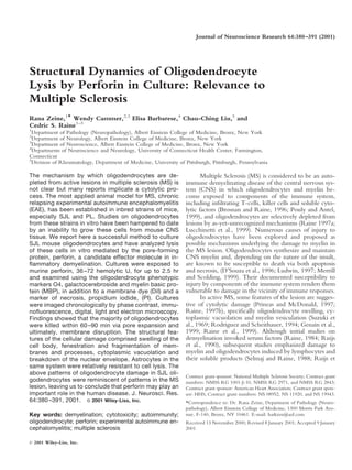
- 7. 386 Zeine et al. Fig. 4. EM of control cells and early perforin-induced cytoplasmic 30,000. C: Thirty minutes PFP. The cytoplasm of this oligodendro- injury. A: Control culture. A normal oligodendrocyte lies to the left of cyte protrudes as villus-like processes into the extracellular space. Note an astrocyte. The cytoplasm of the oligodendrocyte is distinctly denser. the numerous microtubules with the cell body and the villus-like Magnification 12,000. B: Detail of the oligodendrocyte cytoplasm in profiles (arrows). Magnification 75,000. (A). Note the microtubules in cross section (arrows). Magnification
- 8. Oligodendrocyte Lysis by Perforin 387 Fig. 5. Ultrastructural patterns of membrane damage in oligodendro- 30,000. C: Sixty minutes PFP. End-stage lysis is seen in an oligo- cytes exposed to PFP. A: Thirty minutes PFP. This oligodendrocyte dendrocyte that has lost its cell membrane, cytoplasmic integrity and has developed a large vacuole around the perimeter of which myelin- intact nuclear membrane. Magnification 14,000. D: Detail of (C) to like figures are located. Magnification 12,000. B: Detail from A to show beginning disruption of the nuclear membrane. Magnification show the lamellar patterns of the myelin droplets. Magnification 30,000.
- 9. 388 Zeine et al. Fig. 6. Sixty minutes PFP. Two interdigitating, apparently healthy astrocytes display an abundance of intermediate filaments and ro- settes of ribosomes. The nucleus of the cell on the right is normal and a hemidesmosome (typical of subpial astrocytes) is indicated (ar- row). Magnification 19,000. Oligodendrocytes examined after exposure to per- of perforin exposure, cell membranes were lost and nu- forin for 1 hr exhibited complete loss of the cell membrane clear envelopes began to degenerate (Fig. 5C). This is the with only remnants of cytoplasm remaining around the first report demonstrating that SJL mouse oligodendro- nucleus (Fig. 5C). In addition, evidence of degeneration of cytes are susceptible to perforin-induced osmotic lysis, a the nuclear envelope was noted (Fig. 5D). Similar pathol- highly conserved, classical cell-death mechanism (Ojcius ogy, albeit delayed and to a lesser degree, was seen in et al., 1991a; Liu et al., 1995b) and this occurred at a dose astrocytes in the same culture system but on most occa- level similar to that which kills other eukaryotic cells. sions, astrocytes were relatively resistant to damage and Interestingly, oligodendrocyte changes observed maintained a normal ultrastructure well into the experi- here in vitro held much in common with appearances mental period (Fig. 6). described in MS lesions, ultrastructural studies of which DISCUSSION have consistently documented that oligodendrocyte pa- thology exhibits features suggestive of immune osmotic The mechanisms underlying oligodendrocyte deple- lysis or necrosis (Prineas, 1985; Prineas and McDonald, tion from the MS plaque remain to be clarified although cytolytic mechanisms have repeatedly been implicated 1997). In contrast, evidence for oligodendrocyte apoptosis (Raine, 1997b). One candidate molecule in this process is in MS and EAE has been lacking or, at most, rare the pore-forming membranolytic molecule, perforin, and (D’Souza et al., 1996; Bonetti et al., 1997; Bonetti and in this report, we have studied the effects of perforin on Raine, 1997; Waldner et al., 1997). Perforin is a potent oligodendrocytes cultured from a strain of mouse (SJL) mediator of osmotic lysis and perforin-mediated cytotox- most frequently applied to studies on MS and a strain from icity is induced by killer T-cells (Lowin et al., 1994; Kagi which oligodendrocytes have been difficult to grow in the et al., 1994). The presence of killer cells and perforin in past. Our results have shown that during normal develop- demyelinative lesions of EAE and MS is well-known ment in culture, oligodendrocytes extended processes and (Selmaj et al., 1991b; Held et al., 1993; Olive, 1996; Rajan elaborated flattened myelin-like membranes, as described et al., 1996; Gasque et al., 1998; Zeine et al., 1999), and previously for the murine system (Barry et al., 1996). After a dual role has been proposed for these cell types whereby exposure to perforin, the integrity of the membrane veils injury to oligodendrocytes and neurons is detrimental was compromised as microscopic fenestrae appeared and whereas injury to immune cells is protective to the CNS expanded rapidly, transforming the flat membrane sheets (Battistini et al., 1995; Malipiero et al., 1997; Zhang et al., into a delicate lacework (Fig. 2B,C). The cell bodies 1997; Matsumoto et al., 1998; Murray et al., 1998; Zeine swelled and the cytoplasm became vacuolated and dis- et al., 1999). Most likely, the balance between the effector rupted into fragments rich in microtubules. Within 60 min and immunoregulatory arms of the immune response de-
- 10. Oligodendrocyte Lysis by Perforin 389 termines the magnitude of CNS damage inflicted by the Kinase inhibitors block the mobilization and orientation of perforin-based cytotoxicity pathway. In a study on MOG- perforin-containing granules toward the contact zone induced EAE in perforin knockout mice, oligodendrocyte (Wei et al., 1998); and calreticulin blocks perforin lytic pathology was not discussed. activity by stabilizing target cell membranes (Fraser et al., Ultrastructurally, injured oligodendrocytes in MS le- 2000). Thus, the development of strategies to block per- sions appear swollen, with vacuolated cytoplasm and wid- forin activity in vivo holds promise for rescuing oligoden- ened, degenerating or ruptured membranes (Field and drocyte targets during acute episodes of inflammatory de- Raine, 1964; Perier and Gregoire, 1965; Prineas, 1985; ` ` myelination. Rodriguez et al., 1993; Rodriguez and Scheithauer, 1994; The observation that astrocytes were more resistant Raine, 1997b; Raine et al., 1999). Previous studies have than oligodendrocytes to perforin was not surprising con- shown that myelin in acute lesions is transformed into sidering their general resilience to disease. We and others vesicular networks (Kirk, 1979; Lee et al., 1990; Rodri- have shown the expression of perforin within the cyto- guez and Scheithauer, 1994; Genain et al., 1999; Raine et plasm of astrocytes in vivo and in culture (Gasque et al., al., 1999). In this study, some images of oligodendrocytes 1998; Zeine et al., 1999). Astrocytes are the only cells and their membranes undergoing lysis by perforin-induced known to harbor free cytosolic perforin, and they must membrane fragmentation were reminiscent of changes therefore have some protective mechanism to guard their described in acute MS lesions (viz. Suzuki et al., 1969; Fig. own membranes against perforin lysis. Furthermore, as- 11, loc.cit.; Rodriguez and Scheithauer, 1994; Fig. 4A, trocytes may be a source of perforin in the CNS and may loc.cit.; Raine, 1997b; Fig. 5, loc. cit.; Prineas and Mc- play an effector or regulatory role during inflammation. Donald, 1997; Fig. 13.35, loc. cit.; Raine et al., 1999; Figs. Thus, we have shown that oligodendrocytes cultured 6 – 8, loc. cit.). from the CNS of SJL mice are selectively vulnerable to The search for molecules that readily lyse oligoden- cytolytic damage by perforin, and that early-stage cell lysis drocytes has been difficult. Perforin-based cytotoxicity is involves the formation of fenestrae in the cell membranes, most akin to complement toxicity. Although the C9 com- probably as a result of the coalescence of numerous sub- ponent of complement is deposited in MS and EAE le- microscopic perforin-induced pores and the influx of wa- sions, some studies have shown that human oligodendro- ter into the cell. Subsequently, cells either disintegrated cytes are not sensitive to complement damage in culture into cytoplasmic droplets, developed large cytoplasmic (Zajicek et al., 1995). Combining antibodies with com- vacuoles, formed disrupted membranous networks or be- plement results in a reduction in the survival of oligoden- came totally devoid of cell membrane - all these changes drocytes in culture via a toxicity mechanism that can be primary to nuclear disintegration. As stated above, many of inhibited by the addition of normal immunoglobulins these morphologic patterns have been described in MS (Scolding et al., 1989; Scolding et al., 1990; Stangel et al., and EAE previously and are in support of oligodendrocyte 2000). It is most unlikely, however, that immunoglobulin damage in MS being effected by a non-apoptotic process. would be protective in the case of perforin-mediated lysis. In ongoing studies on tissue from EAE and MS (Zeine et We therefore reaffirm a unique role for perforin in irre- al., in preparation), we have documented the upregulation versible oligodendrocyte injury relevant to the pathogen- of perforin by western blotting during acute exacerbations esis of MS and extend previous findings by providing and downregulation during remission. Perforin was present within infiltrating cytotoxic cells and astrocytes by detailed timed images from oligodendrocytes undergoing immunohistochemistry, and the extent of remyelination osmotic lysis. correlated with the absence of perforin. Thus, in the series With regard to previous works on perforin-mediated of molecular events leading to the formation of the MS lysis, our results are in agreement with the findings of lesion, it seems that a role for perforin may very well be Jones et al. (1991) showing that “not all PI-positive cells found at the acute end of the spectrum. proceeded to lysis.” Thus, the possibility of sub-lethal attack by perforin on individual cells remains open and ACKNOWLEDGMENTS raises interest in the mechanism of resistance to perforin We are indebted to Michael Cammer for expert ( Jiang et al., 1990; Ojcius et al., 1990, 1991b). Of ther- technical assistance with digital imaging at the Analytical apeutic relevance would be the conferring of resistance to Imaging Facility of the Albert Einstein College of Medi- selected target cells, e.g., oligodendrocytes, in EAE and cine; to Dr. Hin Hark Gan of New York University and MS. Such a prospect awaits the isolation of ‘L-protectin,’ Howard Hughes Medical Institute for discussion; to Dr. a membrane-bound glycosylated molecule that interferes Jorge Larocca of the Albert Einstein College of Medicine, with perforin function in resistant target cell membranes Department of Neurology, for advice; and to Patricia (Liu et al., 1995b). Currently, some experience has been Cobban-Bond for administrative assistance. This research gained by blocking perforin-mediated lysis via four classes was supported in part by grants from the National Multiple of chemical reagents in vitro. Calcium chelators inhibit Sclerosis Society of the USA (NMSS RG 1001-J-10 exocytosis and perforin binding to membranes (Liu et al., [CSR], NMSS RG 2971 [WC] and NMSS RG 2843 1995b); H -ATPase inhibitors raise the pH within cyto- [EB]), the American Heart Association (CCL), and by toxic granules leading to accelerated degradation of per- HHS Grants NS 08952 (CSR), NS 11920 (CSR), and NS forin (Kataoka et al., 1996; Zeine et al., 1998); MAP- 19943 (EB).
- 11. 390 Zeine et al. REFERENCES Hengartner H, Golstein P. 1994. Fas and perforin pathways as major Antel JP, McCrea E, Ladiwala U, Qin YF, Becher B. 1998. Non-MHC- mechanisms of T cell-mediated cytotoxicity. Science 265:528 –530. restricted cell-mediated lysis of human oligodendrocytes in vitro: relation Kappeler A, Mueller C. 2000. The role of activated cytotoxic T-cells in with CD56 expression. J Immunol 160:1606 –1611. inflammatory bowel disease. Histol Histopathol 15:167–172. Barbarese E. 1991. Spatial distribution of myelin basic protein mRNA and Kataoka T, Shinohara N, Takayama H, Takaku K, Kondo S, Yonehara S, polypeptide in quaking oligodendrocytes in culture. J Neurosci Res Nagai K. 1996. Concanamycin A, a powerful tool for characterization and 29:271–281. estimation of contribution of perforin- and Fas-based lytic pathways in Barry C, Pearson C, Barbarese E. 1996. Morphological organization of cell-mediated cytotoxicity. J Immunol 156:3678 –3686. oligodendrocyte processes during development in culture and in vivo. Kirk J. 1979. Pseudoviral hollow-cored vesicles in multiple sclerosis brain. Dev Neurosci 18:233–242. Acta Neuropathol (Berl) 48:63– 66. Battistini L, Salvetti M, Ristori G, Falcone M, Raine CS, Brosnan CF. Kivisakk P, Stawiarz L, Matusevicius D, Fredrikson S, Soderstrom M, 1995. Gamma delta T cell receptor analysis supports a role for HSP 70 Hindmarsh T, Link H. 1999. High numbers of perforin mRNA express- selection of lymphocytes in multiple sclerosis lesions. Mol Med 1:554 – ing CSF cells in multiple sclerosis patients with gadolinium-enhancing 562. brain MRI lesions. Acta Neurol Scand 100:18 –24. Bonetti B, Pohl J, Gao YL, Raine CS. 1997. Cell death during autoimmune Lee SC, Moore GR, Golenwsky G, Raine CS. 1990. Multiple sclerosis: a demyelination: effector but not target-cells are eliminated by apoptosis. role for astroglia in active demyelination suggested by Class II MHC J Immunol 159:5733–5741. expression and ultrastructural study. J Neuropathol Exp Neurol 49:122– Bonetti B, Raine CS. 1997. Multiple sclerosis: oligodendrocytes display cell 136. death-related molecules in situ but do not undergo apoptosis. Ann Neurol Liu CC, Persechini PM, Young JD. 1995b. Perforin and lymphocyte- 42:74 – 84. mediated cytolysis. Immunol Rev 146:145–175. Brosnan CF, Raine CS. 1996. Mechanisms of immune injury in multiple Liu CC, Walsh CM, Young JD. 1995a. Perforin: structure and function. sclerosis. Brain Pathol 6:243–257. Immunol Today 16:194 –201. Brown A, McFarlin DE, Raine CS. 1982. Chronologic neuropathology of Liu CC, Young LH, Young JD. 1996. Lymphocyte-mediated cytolysis and relapsing experimental allergic encephalomyelitis in the mouse. Lab Invest disease. N Engl J Med 335:1651–1659. 46:171–185. Lowin B, Hahne M, Mattmann C, Tschopp J. 1994. Cytolytic T-cell Cross A, McFarlin DE, McCarron RM, Raine CS. 1987. Adoptively- cytotoxicity is mediated through perforin and Fas lytic pathways. Nature transferred acute and chronic relapsing autoimmune encephalomyelitis in 370:650 – 652. the PL/J mouse with observations on altered pathology by intercurrent Lucchinetti C, Bruck W, Parisi J, Scheithauer B, Rodriguez M, Lassmann infection. Lab Invest 57:499 – 412. H. 1999. A quantitative analysis of oligodendrocytes in multiple sclerosis D’Souza SD, Bonetti B, Balasingam V, Cashman NR, Barker PA, Troutt lesions. A study of 113 cases. Brain 122:2279 –2295. AB, Raine CS, Antel JP. 1996. Multiple sclerosis: Fas signaling in oligo- Ludwin SK. 1997. The pathobiology of the oligodendrocyte. J Neuro- dendrocyte cell death. J Exp Med 184:2361–2370. pathol Exp Neurol 56:111–124. Dal Canto MC, Calenoff MA, Miller SD, Vanderlugt CL. 2000. Lympho- Malipiero U, Frei K, Spanaus KS, Agresti C, Lassmann H, Hahne M, cytes from mice chronically infected with Theiler’s murine encephalo- Tschopp J, Eugster HP, Fontana A. 1997. Myelin oligodendrocyte myelitis virus produce demyelination of organotypic cultures after stim- glycoprotein-induced autoimmune encephalomyelitis is chronic/ ulation with the major encephalitogenic epitope of myelin proteolipid relapsing in perforin knockout mice, but monophasic in Fas- and Fas protein. Epitope spreading in TMEV infection has functional activity. ligand-deficient lpr and gld mice. Eur J Immunol 27:3151–3160. J Neuroimmunol 104:79 – 84. Matsumoto Y, Kohyama K, Aikawa Y, Shin T, Kawazoe Y, Suzuki Y, Field EJ, Raine CS. 1964. Examination of multiple sclerosis biopsy speci- Tanuma N. 1998. Role of natural killer cells and TCR gamma delta men. (Abstract). Third Eur Reg Conf on EM. Prague Czech Acad Sci T-cells in acute autoimmune encephalomyelitis. Eur J Immunol 28:1681– ii:289 –290. 1688. Fraser SA, Karimi R, Michalak M, Hudig D. 2000. Perforin lytic activity is Matusevicius D, Kivisakk P, Navikas V, Soderstrom M, Fredrikson S, Link controlled by calreticulin. J Immunol 164:4150 – 4155. H. 1998. Interleukin-12 and perforin mRNA expression is augmented in Freedman MS, Ruijs TC, Selin LK, Antel JP. 1991. Peripheral blood blood mononuclear cells in multiple sclerosis. Scand J Immunol 47:582– gamma-delta T-cells lyse fresh human brain-derived oligodendrocytes. 590. Ann Neurol 30:794 – 800. Merrill JE, Scolding NJ. 1999. Mechanisms of damage to myelin and Gasque P, Jones J, Singhrao SK, Morgan B. 1998. Identification of an astrocyte oligodendrocytes and their relevance to disease. Neuropathol Appl Neu- cell population from human brain that expresses perforin, a cytotoxic protein robiol 25:435– 458. implicated in immune defense. J Exp Med 187:451– 460. Murray PD, McGavern DB, Lin X, Njenga MK, Leibowitz J, Pease LR, Genain CP, Cannella B, Hauser SL, Raine CS. 1999. Identification of Rodriguez M. 1998. Perforin-dependent neurologic injury in a viral autoantibodies associated with myelin damage in multiple sclerosis. Nat model of multiple sclerosis. J Neurosci 18:7306 –7314. Med 5:170 –175. Norton WT, Farooq M, Fields KL, Raine CS. 1983. The long term culture Held W, Meyermann R, Qin Y, Mueller C. 1993. Perforin and tumor of bulk-isolated bovine oligodendroglia from adult brain. Brain Res necrosis factor alpha in the pathogenesis of experimental allergic enceph- 270:295–310. alomyelitis: comparison of autoantigen induced and transferred disease in Ojcius DM, Jiang SB, Persechini PM, Storch J, Young JD. 1990. Resis- Lewis rats. J Autoimmun 6:311–322. tance to the pore-forming protein of cytotoxic T-cells: comparison of Jiang SB, Ojcius DM, Persechini PM, Young JD. 1990. Resistance of target cell membrane rigidity. Mol Immunol 27:839 – 845. cytolytic lymphocytes to perforin-mediated killing. Inhibition of perforin Ojcius DM, Muller S, Hasselkus-Light CS, Young JD, Jiang S. 1991b. binding activity by surface membrane proteins. J Immunol 144:998 – Plasma membrane-associated proteins with the ability to partially inhibit 1003. perforin-mediated lysis. Immunol Lett 28:101–108. Jones J, Frith S, Piddlesden S, Morgan BP, Compston DA, Campbell AK, Ojcius DM, Persechini PM, Zheng LM, Notaroberto PC, Adeodato SC, Hallett MB. 1991. Imaging Ca2 changes in individual oligodendrocytes Young JD. 1991a. Cytolytic and ion channel-forming properties of the N attacked by T-cell perforin. Immunology 74:572–577. terminus of lymphocyte perforin. Proc Natl Acad Sci USA 88:4621– Kagi D, Vignaux F, Ledermann B, Burki K, Depraetere V, Nagata S, 4625.
- 12. Oligodendrocyte Lysis by Perforin 391 Olive C. 1996. Expression of the T cell receptor delta-chain repertoire in Scolding NJ, Morgan BP, Campbell AK, Compston DA. 1990. Comple- mouse lymph node. Immunol Cell Biol 74:313–317. ment mediated serum cytotoxicity against oligodendrocytes: a comparison Perier O, Gregoire A. 1965. Electron microscopic features of multiple ` ` with other cells of the oligodendrocyte-type 2 astrocyte lineage. J Neurol sclerosis lesions. Brain 88:937–952. Sci 97:155–162. Pouly S, Antel JP. 1999. Multiple sclerosis and central nervous system Selmaj K, Brosnan CF, Raine CS. 1991b. Colocalization of lymphocytes demyelination. J Autoimmun 13:297–306. bearing gamma delta T-cell receptor and heat shock protein hsp65 Prineas JW. 1985. The neuropathology of multiple sclerosis. In: Vinken PJ, oligodendrocytes in multiple sclerosis. Proc Natl Acad Sci USA 88:6452– Bruyn GW, Klawans GW, Koetsier JC, editors. Handbook of clinical 6456. neurology, Vol 47. Demyelinating diseases. Amsterdam: Elsevier. p 213– Selmaj K, Cross AH, Farooq M, Brosnan CF, Raine CS. 1991a. Non- 257. specific oligodendrocyte cytotoxicity mediated by soluble products of Prineas JW, McDonald W. 1997. Demyelinating diseases. In: Graham DI, activated T-cell lines. J Neuroimmunol 35:261–271. Lantos PL, editors. Greenfield’s neuropathology. London: Arnold. p Selmaj KW, Raine CS. 1988. Tumor necrosis factor mediates myelin and 813– 896. oligodendrocyte damage in vitro. Ann Neurol 23:339 –346. Raine CS. 1984. Biology of disease. Analysis of autoimmune demyelina- Stangel M, Compston A, Scolding NJ. 2000. Oligodendroglia are protected tion: its impact upon multiple sclerosis. Lab Invest 50:608 – 635. from antibody-mediated complement injury by normal immunoglobulins Raine CS. 1997a. The lesion in multiple sclerosis and chronic relapsing (IVIg). J Neuroimmunol 103:195–201. allergic encephalomyelitis—a structural comparison. In: Raine CS, Mc- Suzuki K, Andrews JM, Waltz JM, Terry RD. 1969. Ultrastructural studies Farland HF, Tourtellotte WW, editors. Multiple sclerosis: clinical and of multiple sclerosis. Lab Invest 20:444 – 454. pathogenic basis. London: Chapman and Hall. p 243–286. Tschopp J, Schafer S, Masson D, Peitsch MC, Heusser C. 1989. Phospho- Raine CS. 1997b. The Norton lecture: a review of the oligodendrocyte in rylcholine acts as a Ca2 -dependent receptor molecule for lymphocyte the multiple sclerosis lesion. J Neuroimmunol 77:135–152. perforin. Nature 337:272–274. Raine CS, Cannella B, Hauser SL, Genain CP. 1999. Demyelination in Waldner H, Sobel RA, Howard E, Kuchroo VK. 1997. Fas- and FasL- primate autoimmune encephalomyelitis and acute multiple sclerosis le- deficient mice are resistant to induction of autoimmune encephalomyeli- sions: a case for antigen-specific antibody mediation. Ann Neurol 46: tis. J Immunol 159:3100 –3103. 144 –160. Rajan AJ, Gao YL, Raine CS, Brosnan CF. 1996. A pathogenic role for Wei S, Gamero AM, Liu JH, Daulton AA, Valkov NI, Trapani JA, Larner gamma delta T-cells in relapsing-remitting experimental allergic enceph- AC, Weber MJ, Djeu JY. 1998. Control of lytic function by mitogen- alomyelitis in the SJL mouse. J Immunol 157:941–949. activated protein kinase/extracellular regulatory kinase 2 (ERK2) in a Rodriguez M, Scheithauer B. 1994. Ultrastructure of multiple sclerosis. human natural killer cell line: identification of perforin and granzyme B Ultrastruct Pathol 18:3–13. mobilization by functional ERK2. J Exp Med 187:1753–1765. Rodriguez M, Scheithauer BW, Forbes G, Kelly PJ. 1993. Oligodendro- Zajicek J, Wing M, Skepper J, Compston A. 1995. Human oligodendro- cyte injury is an early event in lesions of multiple sclerosis. Mayo Clin cytes are not sensitive to complement. A study of CD59 expression in the Proc 68:627– 636. human central nervous system. Lab Invest 73:128 –138. Ruijs TC, Freedman MS, Grenier YG, Olivier A, Antel JP. 1990. Human Zeine R, Cannella B, Pitt D, Raine CS. 1999. Perforin expression in oligodendrocytes are susceptible to cytolysis by major histocompatibility demyelinative lesions of multiple sclerosis, and a role for perforin in the complex class I-restricted lymphocytes. J Neuroimmunol 27:89 –97. regulation of experimental autoimmune encephalomyelitis. J Neuro- Ruijs TC, Olivier A, Antel JP. 1990. Serum cytotoxicity to human and rat pathol Exp Neurol 58:564. oligodendrocytes in culture. Brain Res 517:99 –104. Zeine R, Pon R, Ladiwala U, Antel JP, Filion LG, Freedman MS. Scolding NJ, Houston WA, Morgan BP, Campbell AK, Compston DA. 1998. Mechanism of gamma delta T cell-induced human oligodendrocyte 1989. Reversible injury of cultured rat oligodendrocytes by complement. cytotoxicity: relevance to multiple sclerosis. J Neuroimmunol 87:49 – 61. Immunology 67:441– 446. Zhang B, Yamamura T, Kondo T, Fujiwara M, Tabira T. 1997. Regulation Scolding NJ, Jones J, Compston DA, Morgan BP. 1990. Oligodendrocyte of experimental autoimmune encephalomyelitis by natural killer (NK) susceptibility to injury by T-cell perforin. Immunology 70:6 –10. cells. J Exp Med 186:1677–1687.