Protein structure
•Descargar como PPTX, PDF•
48 recomendaciones•8,191 vistas

Protein structure and its details

Recomendados
Más contenido relacionado
La actualidad más candente
La actualidad más candente (20)
Destacado
Destacado (6)
Similar a Protein structure
Similar a Protein structure (20)
Último
Último (20)
Protein structure
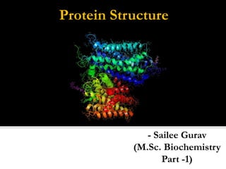
- 1. Protein Structure - Sailee Gurav (M.Sc. Biochemistry Part -1)
- 2. It‟s completely defined & predictable. Each amino acid in one of these giant macromolecules is located at a specific site within the structure , giving the protein the precise shape. It can be described at several levels of organization , each emphasizing a different aspect & each dependent on different types of interactions.
- 5. It‟s simply the sequence of the amino acid polymer. 1 dimensional. By convention, written from amino end to carboxyl end. It is important as it is the foundation in which ultimately the higher structures of the protein are determined, and thus the function of the protein.
- 7. It is a local, regularly occurring structure in proteins and is mainly formed through hydrogen bonds between backbone atoms. Pauling & Corey studied the secondary structures and proposed 2 conformations. The α helix and β sheets.
- 8. Right-handed coiled or spiral conformation (helix) 3.6 residues per turn stabilized by hydrogen bonds Hydrogen bonding between C' = O of residue n and NH of residue n + 4 All C'O and NH groups are joined by H-bonds. Except: Terminal NH and C'O groups
- 9. α-helix is tightly packed Almost no free space within the helix Amino acid side chains are on the outside of the helix Roughly point “downwards” Resembles branches of a Christmas tree Most common location of α helices is along the protein periphery One side facing the solution (exterior) One side facing hydrophobic interior
- 10. α-helix Also called the 4₁₃ π-helix Very loosely coiled Hbonding pattern n + 5 Raerly found in nature. 310-helix Very tightley coiled Hbonding pattern n+3 rarely found in nature
- 12. Beta sheets are another major structural element in globular proteins containing 20–28 % of all residues The basic unit of a beta sheet is a beta strand with approximate backbone dihedral angles phi = -120 and psi = +120 Two types: anti-parallel and parallel strand. Due to the extended nature of the chain, no significant intra-segment hydrogen bonds and van der Waals interactions between atoms of neighboring residues. Sometimes called the beta "pleated" sheet since sequentially neighboring Ca atoms are alternately above and below the plane of the sheet
- 13. Main-chain NH and O atoms within a b sheet are hydrogen bonded to each other. The amino acids in successive strands have alternating directions (anti-parallel).
- 14. Antiparallel beta sheets are considered intrinsically more stable than parallel sheets due to the more optimal orientation of the interstrand hydrogen bonds
- 15. Different types Hairpin loops – often between anti-parallel beta strands Omega loops – beginning and end close (6-16 residues) Extended loops – more than 16 residues Secondary structures are joined together by additional structures called loops. These patterns are called motifs Defining motifs-small, specific combinations of secondary structure elements
- 16. A supersecondary structure is a compact three-dimensional protein structure of several adjacent elements of secondary structure that is smaller than a protein domain or a subunit
- 18. Tertiary structures is defined as the overall arrangement of polypeptide chains in three-dimensional space, describing how secondary structures arrange into supersecondary structures that in turn arrange into domains and domains into tertiary structures. Components: Motif : a recognizable subcomponent of the fold – several motifs usually comprise a domain Fold: used differently in different contexts – most broadly a reproducible and recognizable 3 dimensional arrangement Domain: a compact and self folding component of the protein that usually represents a discreet structural and functional unit
- 19. Tertiary structures can be divided into three main classes: a domain b domains a/b domains The domain is the unit of tertiary structure
- 21. In globular proteins Tertiary interactions are frequently stabilized by sequestration of hydrophobic amino acid residues in the protein core Consequent enrichment of charged or hydrophilic residues on the protein's water-exposed surface. In secreted proteins disulfide bonds between cysteine residue helps to maintain the protein's tertiary structure
- 24. They describes the arrangement of sub-units in a protein consisting of more than one polypeptide chain, where the sub-units may be identical or different. The sub-units in a quaternary structure are held together by non-covalent interactions where the „contact regions‟ between the sub-units resemble the interior of tertiary structure proteins as being hydrophobic. These structures cannot have mirror image superpositioning resulting in symmetrical distribution of the sub-units in the quaternary structure.
- 27. Poly proline -II helix in proteins: Structure & Function. The polyproline type II (PPII) helix in recent years has emerged clearly as a structural class not only of fibrillar proteins but also of the folded and unfolded proteins. The left-handed, extended PPII helix represents the only frequently occurring regular structure . Natively unfolded proteins have a high content of the PPII helices identified by spectroscopic methods. PPII is favorable for protein-protein and protein-nucleic acid interactions and plays a major role in signal transduction and protein complex assembly.
- 28. PPII helices do not necessarily contain proline, but proline has high PPII propensity. PPII helices are involved in transcription, cell motility, self-assembly, elasticity, and bacterial and viral pathogenesis, & has an important structural role in amyloidogenic proteins. PPII helices are not always assigned in experimentally solved structures, & they are rarely used in protein structure modeling.
- 29. Lehninger Principles Of Biochemistry, Fourth Edition. Biochemistry, by Voet & Voet, 3rd edition. Harper’s Illustrated Biochemistry, 26th edition. Biochemistry , by Dr.U.Satyanarayana. www.ncbi.nlm.nih.gov/ biochemistrycourse.blogspot.in/