Muscles of head_&_neck[1]
•Descargar como PPTX, PDF•
3 recomendaciones•1,320 vistas

msc mediacl anatomy

Recomendados
Más contenido relacionado
La actualidad más candente
La actualidad más candente (20)
Similar a Muscles of head_&_neck[1]
Similar a Muscles of head_&_neck[1] (20)
Último
Último (20)
Muscles of head_&_neck[1]
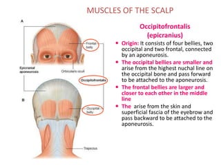
- 1. MUSCLES OF THE SCALP Occipitofrontalis (epicranius) Origin: It consists of four bellies, two occipital and two frontal, connected by an aponeurosis. The occipital bellies are smaller and arise from the highest nuchal line on the occipital bone and pass forward to be attached to the aponeurosis. The frontal bellies are larger and closer to each other in the middle line The arise from the skin and superficial fascia of the eyebrow and pass backward to be attached to the aponeurosis.
- 2. Muscles of facial expression: Associated with (1) the forehead, (2) orbit, (3) mouth, and (4) nose. We will study these muscles in lab, for now think of the facial muscles in groups:
- 3. Buccal fat pat & Buccinator muscle
- 4. 4 Muscles of Face (muscles of facial expression) They are called ms. Of expression because they pull skin of face to produce various expressions. They are arranged in groups around the eye, nose & mouth. They have bony origin. They are inserted into skin of face (no deep fascia in face). They are supplied by branches of facial N., Except levator P.S. by occulomotor N. (striated ms.) + sympathetic N. (smooth ms.).
- 5. 5 Muscles of Face 1- levator palpebrae superioris (the dilator ms. of eyelids, lying in the orbital cavity). 2-Orbicularis oculi (the sphincter ms of eyelids). 3-Corrugator supercilii (deep to orbicularis oculi). 4- Occipitofrontalis (ms. of scalp). A) Muscles of eyelids : B) Muscles of Nose : 1-Procerus. 2-Compressor & dilator naris.
- 6. 6 Muscles of FaceC) Muscles of Lips : Sphincter muscle of the lips : “ Orbicularis Oris”. Dilator muscles of the lips : 1-Levator labii superioris alaeque nasi. 2-Levator labii superioris. 3-Depressor labii inferioris. 4- Zygomaticus minor. 5- Zygomaticus major. 6- Levator anguli Oris (deep to zygomatic ms.). 7- Depressor anguli Oris. 8-Risorius. 9- Mentalis. D) Muscles of Cheek : “Buccinator”
- 7. 7 Muscles of Face (muscles of facial expression) 3 large muscles : 1- Buccinator m. (ms. of cheek). 2- Orbicularis oculi m. 3- Orbicularis oris m. Many small muscles : 1- Dilator ms. of lips (separate lips) : - Levator labii superioris alaeque nasi, levator labii superioris. - Zygomaticus minor & major. - Levator anguli oris, risorius & depressor anguli oris. - Depressor labii inferioris & mentalis. origin : bones & fascia around oral aperature. Insertion : into substance of lips.
- 8. 8 2- Corrugator supercilli : -It lies deep to orbicularis oculi. origin : superciliary arch (bone). Insertion : skin of eyebrow. Action : vertical wrinkles of forehead, as in frowning. 3- Compressor naris & dilator naris : origin : maxilla. Insertion : the fibres are continuous with those of opposite side in front of the bridge of nose to form aponeurosis of bridge of nose. Action : compesses & widens nasal cartilages and aperature. 4- Procerus : - It is continuous with the medial part of occipito-frontalis ms. Origin : nasal bone. Insertion : medial part of skin of eyebrow. Action : wrinkles skin of nose.
- 9. 9 Orbicularis oculi : 1- Orbital part : Origin : medial palpebral ligament + adjoining bone. Insertion :The fibres have no lateral attachment, it loops return to origin. Action : closes eyelids by throwing skin around orbit into folds to protect eyeball. 2- Palpebral part : Origin : medial palpebral ligament. Insertion : lateral palpebral raphe & skin of eyelids. Action : closes palpebral fissure of eyelids gently (sleep) and dilates lacrimal sac.
- 10. 10 Orbicularis oris : Origin : maxilla, mandible & deep skin. Insertion : encircles oral orifice to be inserted to the m.m lining the inner surface of lips. Action : compresses the lips together to close the mouth (sphincter muscle of lips).
- 11. 11 Muscle of Cheek : Buccinator Muscle : Origin : from outer surface of maxilla & mandible opposite the molar teeth + from pterygomandibular ligament. Insertion : 1- upper fibres : into upper lip. 2- lower fibres : into lower lip. 3- middle fibres : decussate at the angle of mouth. N.supply : buccal branch of facial N. Action : 1- it compresses the cheeks & lips against the teeth to prevent accumulation of food in vestibule of mouth. 2- it is used in wistling, when cheeks are distended with air.
- 12. 12 Muscle of Cheek : Buccinator Muscle : It is covered on outside by buccopharyngeal fascia & buccal pad of fat. Its deep surface is lined by buccal mucosa. It is pierced by : 1-parotid duct , opposite upper 2nd molar tooth. 2-Buccal branch of mandibular nerve (sensory) to supply m.m of cheek on the inner surface of buccinator muscle.
- 13. 13 Facial muscle Paralysis • The facial ms. Are innervated by facial N. • Cause : Damage to facial N. (by a tumor in internal acoustic meatus or parotid galnd) /or operation or infection in middle ear / or perineuritis, Bell’s palsy in facial nerve canal. • Results : Lower motor neuron lesion which involves distortion of face+ drooping of lower eyelid + angle of mouth will sag on the affected side. /But Upper motor neuron lesion is due to lesion of pyramidal tract and here the upper face is normal because the neurons supplying this part receive corticobulbar fibres from both cerebral cortices.
- 15. Anterior belly of digastric muscle Posterior belly of digastric Stylohyoid Sternohyoid muscle
- 17. STERNOCLEIDOMASTOID MUSCLE (SCM) Sternal head Clavicular head Mastoid process
- 18. Platysma m. Investing layer of deep cervical fascia
- 19. Splenius capitis m. Levator scapulae m. Post., mid., & ant. scalene mm. Muscles of floor MUSCLES OF FLOOR OF POSTERIOR TRIANGLE
- 20. CAREFREE & CAREFUL ZONES
- 21. OCCIPITAL AND OMOCLAVICULAR TRIANGLES Occipital triangle Omoclavicular triangle (supraclavicular triangle) (subclavian triangle)
- 22. OMOHYOID MUSCLE Omohyoid m. (inferior belly) Omohyoid (superior belly)
- 27. Masseter
- 28. Medial Pterygoid Lateral View Medial View
- 29. Mandibular Sling-Masseter and Medial Pterygoid Lateral View View from Below
- 33. Pharynx • Parts: • Nasopharynx • Oropharynx • Laryngopharynx
- 36. Adductors of the Vocal Folds
- 39. Fascia of the Face Superficial fascia is copious and loose – however, there is no discrete layer of deep fascia of the face except …
- 40. 40 Facial muscle Paralysis • The facial ms. Are innervated by facial N. • Cause : Damage to facial N. (by a tumor in internal acoustic meatus or parotid galnd) /or operation or infection in middle ear / or perineuritis, Bell’s palsy in facial nerve canal. • Results : Lower motor neuron lesion which involves distortion of face+ drooping of lower eyelid + angle of mouth will sag on the affected side. /But Upper motor neuron lesion is due to lesion of pyramidal tract and here the upper face is normal because the neurons supplying this part receive corticobulbar fibres from both cerebral cortices.