

Recommended
More Related Content
What's hot
What's hot (20)
Viewers also liked
Viewers also liked (14)
Similar to Immature teratoma
Similar to Immature teratoma (20)
Recently uploaded
Recently uploaded (20)
Immature teratoma
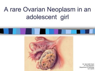
- 1. A rare Ovarian Neoplasm in an adolescent girl Dr. Samriddhi Karki 3rd Year Resident Department of Pathology 13/11/2014
- 2. Clinical history provided 13 year old 06/10/2014 C/O: – Fever X 1month – Abdominal fullness x 20 days – Pain abdomen x 3 days O/E: – P/A: • A firm mass corresponding to ~ 36wks of gestation, apparently arising from the pelvis , extending up to the xiphisternum Clinical Diagnosis: – Right sided Adnexal Mass
- 3. Investigations: Hemogram: • Hb: 10.2gm/dl •Other parameters: within normal limits Tumor Markers: •CA125: 335.8 U/ml (< 35 U/ml) •Alpha-feto protein: 4.99 IU/ml (0.5-0.6) •β-hCG: 0.05mIU/ml (<10.0) USG: • Large heterogenous mass of varied echogenecity mainly cystic, measuring 36 x 20 cm • Impression: Germ cell tumor (Dysgerminoma) CT scan: • Well defined large lobulated, predominantly hypoechoeic cystic lesion in the abdomino-pelvic cavity occupying almost whole abdomen with cystic and solid component with intralesional calcification and foci of fat density. • S/O complex ovarian cyst (Terato-dermoid)
- 4. Provisional Diagnosis: Right sided Ovarian Germ Cell Tumor Exploratory Laparotomy done (12/10/2014)
- 5. Operative findings Ascites 300 ml , straw colored fluid Multiple peritoneal deposits <2cm Deposits in pouch of douglas, utero-vaginal fold Right ovarian mass: – Capsulated mass – Smooth surface – Solid + cystic component Deposits on the contralateral ovary Uterus normal size Deposits in infra colic omentum Undersurface of the diaphragm on either side / Liver: Normal Deposits in the large and small intestine Retroperitoneal + pelvic lymph nodes normal size
- 6. Specimens sent for Histopathological examination 1. Right ovarian mass with fallopian tube 2. Contralateral ovarian biopsy 3. Deposits from Pouch of Douglas 4. Peritoneal tissue from anterior abdominal wall 5. Peritoneal tissue from lateral wall of abdomen 6. Peritoneal tissue from descending colon 7. Deposits from outer surface of small intestine 8. Infra colic omentum
- 8. 1. Right ovarian mass with fallopian tube 2. Contralateral ovarian biopsy 3. Deposits from Pouch of Douglas 4. Peritoneal tissue from anterior abdominal wall 5. Peritoneal tissue from lateral wall of abdomen 6. Peritoneal tissue from descending colon 7. Deposits from small bowel 8. Infra colic omentum … … . . . … .. .. .. . . .
- 13. Heterogenous tumor tissue composed of mixture of immature and mature tissue, predominantly immature embryonal type tissues in the form of neuroectodermal rosettes. Immature mesenchyme in the form of loose, myxoid stroma, with focal differentiation into fat, osteoid, immature cartilage and rhabdomyoblasts. Immature endodermal structure including intestinal type epithelium with basal vacuolation and blood vessels also noted. . . . . . . . . . . ………. ……………… …………………………..
- 14. Mature elements are composed of – Epidermis – Dermal adnexal structures – Fragments of bone and cartilage – Ganglion cells – Glial tissues – Intestinal glands . ……………………………………………………………………………………………………………………………………………………………………….. …………………………………………………………………………………………………………………………………………………………………………………………………………. ….. . …. …. . . ……………………….. ……………….. ………………………………………………..
- 15. TERATOMA • Exclusion of benign solid teratoma. • Identification of other germ cell component. • Determining the grade of the tumor. Grading based on the extent of immature tissue within the teratoma
- 16. Grading of Immature Teratoma (Gonzales-Crussi) Grade I: • Tumors with rare foci of immature neuropeithelial tissue that occupy less than one low power field (40x) in any slide Grade II: • Tumors with similar elements, occupying 1 to 3 low power fields in any slide Grade III: • Tumors with large amount of immature neuroepithelial tissue occupying more than 3 low power fields in any slide. WHO classification of tumors of the breast and female genital organs
- 17. Sections from – Contralateral Ovary, – Pouch of Douglas, – Peritoneal tissue from anterior abdominal wall/ descending colon, – Small intestine, – Infra colic omentum showed tumor deposits. .. . .. . . . . . . ..
- 18. Conclusion: Immature teratoma - Grade III (WHO) pT3bNxMx
- 19. DISCUSSION
- 21. Introduction (Greek- root : Teratos: monster ) The first description of teratoma was made in 1960 by Thürlbeck and Scully. Teratomas are most frequently found in the gonads (ovary and testes). Extragonadal teratomas are rare and arise from midline structures (thyroid, retroperitoneum, mediastinum, pericardium , coccyx and brain). Very rarely, teratomas are found in other solid (e.g. breast, parotid gland, liver) and hollow (e.g. oesophagus, stomach, bladder, uterine cervix) organs. Teratomas may be benign, malignant or a component of a mixed germ cell tumor (GCT).
- 22. Immature Teratoma is a malignant germ cell tumor composed of tissues from three germ layers (ectoderm, mesoderm, and endoderm), with immature embryonal type structures (generally) neuroectodermal tissue. Less than 1% of ovarian tumors. Second most common germ cell tumor. Outwater EK et al. (2001) 10-20% cases in first two decades of life with a peak incidence between 15 and 19 years. Results in 30% of the deaths from ovarian cancer in this age group. Rarely occurs in post –menopausal age group. Netchine I, et al. (2011)
- 23. Etiology Various theories – Most teratomas (65%) are derived from a single germ cell after the first meiotic division with subsequent failure of meiosis II. Over expression of p53 gene (Charoenkwan et al.) Chromosomal abnormalities (Osterhuis et al.) – Grade 1and 2 are diploid (90% ), while most grade 3 are aneuploid (66%) – Lack of 12p amplification
- 24. Clinical features Abdominal pain and distension Abnormal uterine bleeding Urinary and GIT symptoms Anti-NMDA receptor encephalitis [Dabner M et al. (2012)]
- 25. Investigations Tumors markers : – Alpha feto protein (AFP) can be raised in 50% of cases. USG / CT: – Calcification
- 26. GROSS Usually unilateral; rarely bilateral (less than 10%) Large (6-35 cm) Encapsulated with smooth, glistening outer surface. Ruptured in almost half of the cases. Variegated , predominantly solid, fleshy, gray-tan. May be cystic with hemorrhage and necrosis Rosai and Ackerman’s surgical pathology 10th ed. (2011)
- 27. MICROSCOPIC Variable amount of Immature and mature components Immature components: – Immature ectodermal tissue • Mainly neuroectodermal rosettes and tubules – Immature mesenchyme • Loose myxoid stroma with focal differentiation into – Immature cartilage – Immature fat – Osteoid – Rhabdomyoblasts – Immature endodermal tissue • Hepatic tissue • Intestinal type epithelium with basal vacuolization • Embryonic renal tissue with Wilms tumor
- 28. Neuroepithelial mimicker Ameloblastic epithelium (neuroepithelial mimic, which can be a potential pitfall for an erroneous diagnosis of immature teratoma)
- 29. Immunohistochemisty Immature and mature neural tissue : – Glial fibrillary acidic protein (GFAP) – Long chain polysialic acid Immature neuroepithelium of high-grade – Oct 4 Rosai and Ackerman’s surgical pathology 10th ed. (2011); Kwan MY et al. (2004)
- 30. To determine the prognosis To determine the necessity of adjuvant treatment GRADING
- 31. Treatment Preservation of fertility is an important factor. Patients with grade I tumours staging surgery with a unilateral oophorectomy. Patients with grade II or III tumors adjuvant chemotherapy containing bleomycin, etoposide and cisplatin in addition to surgery. The current combination chemotherapy results in overall disease free survival rate of >95%. • Gershenson DM (2007), Pectasides D et al. (2008)
- 32. Prognosis The most important prognostic feature is the grade of the lesion. The 5yr survival rates : – Grade 1 lesions 82% – Grade 2 lesions 63% – Grade 3 lesions 30% Abiko K et al. (2010)
- 33. Risk of recurrence The only predictor of recurrence is the presence of histological foci of yolk sac tumor rather than the grade of the immature component. Most recurrences develop in the first two years. WHO classification of tumors of the breast and female genital organs
- 34. Conclusion Morphological spectrum of immature teratoma of the ovary is varied, complex and offers diagnostic challenges. Histopathological examination is important for accessing the prognosis and recurrence of the tumor. Early diagnosis associated with immediate therapy and close follow-up are essential for long term favourable outcomes.
- 35. References 1. Rosai J. Female reproductive system- ovary. In: Rosai J, editor. Rosai and Ackerman’s surgical pathology. 10 th ed. New Delhi: Elsevier; 2011. p. 1587-88. 2. Tavassoli FA, Mooney A. Sex cord-stromal tumors. In: Fattaneh A, Tavassoli FA, Devilee P, editors. WHO classification of tumors of the breast and female genital organs. Lyon. IARC press; 2003. p. 169- 70. 3. Outwater EK, Siegelman ES, Hunt JL. Ovarian teratomas: tumor types and imaging characteristics. Radiographics. 2001;21(2):475-90. 4. Chabaud-Williamson M, Netchine I, Fasola S, Larroquet M, Lenoir M, Patte C, et al. Ovarian-sparing surgery for ovarian teratoma in children. Pediatr Blood Cancer. 2011;57(3):429-34. 5. Gershenson DM. Management of ovarian germ cell tumors. J Clin Oncol 2007;25:2938-43 6. Pectasides D, Pectasides E, Kassanos D. Anti tumour treatment. Germ cell tumors of the ovary. Cancer Treat Rev 2008;34:427-41 7. Abiko K, Mandai M, Hamanishi J, Matsumura N, Baba T, Horiuchi A, et al. Oct4 expression in immature teratoma of the ovary: relevance to histologic grade and degree of differentiation. Am J Surg Pathol. 2010;34(12):1842-8. 8. Kwan MY, Kalle W, Lau GT, Chan JK:Is gliomatosis peritonei derived from the associated ovarian teratoma? Hum Pathol 2004, 35: 685–688. PubMedAbstract I Publisher Full Text. 9. Thurlbeck WM, Scully RE: Solid teratoma of the ovary. A clinicopathological analysis of 9 cases. Cancer 1960, 13: 804– 811. PubMed I Publisher Full Text.
- 36. S/1238/14
- 37. S/1238/14
- 38. S/1238/14
- 39. . . . . . . . . .. . .
- 40. . . … . . …. . . .
- 41. . . . . . . . . . . . . .. .. .
- 42. H&E x40
- 43. H & E x100 . . . . . . . . . . . . . . . . . . . . .
- 44. H & E X40
- 45. H & E X200
- 46. H & E X100
- 47. H & E X100
- 48. H & E X400
- 49. . . . a . . . . . . . . H & E X100
- 50. Section from Contralateral ovary H & E X200
- 51. Section from POD H & E X40
- 52. Peritoneal tissue from anterior abdominal wall H & E X100
- 53. Peritoneal tissue from descending colon H & E X40
- 54. Deposits form small intestine
- 55. Deposits from infracolic omentum . . . . . . . . . . . . . . . . .. . H & E X40
- 57. GFAP
Editor's Notes
- Contralateral ovary : 2x1x0.2cm ALL 4 less than 2 cm Infra colic omentum: 72 cm in length with tiny whitish deposits
- GCT represent a heterogenous group of tumors reflecting the capacity for multiple lines of differentiation of the main stem cell system. Here also, the tumors are composed of derivatives of different primary germ layers including ectoderm , mesoderm and endoderm. Why we call it immature because this teratoma contains variable amount of immature , embryonal type tissue , generally immature neuroectoderm.
- Enchaphalitis associated with antibodies against N-Methyl-D-aspartate receptor: Potentially lethal but has a high probability of recovery Paraneoplastic syndrome Mediated by autoantibodies that target the NMDA receptors in the brain . These are produced as a cross reactivity with NMDA receptors in the teratoma. psychosis, memory deficits, seizures, and encephalopathy