
Recomendados
Más contenido relacionado
La actualidad más candente
La actualidad más candente (20)
Similar a Nucliec acids and Nucleotide
Similar a Nucliec acids and Nucleotide (20)
Último
Último (20)
Nucliec acids and Nucleotide
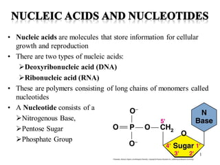
- 1. • Nucleic acids are molecules that store information for cellular growth and reproduction • There are two types of nucleic acids: ➢Deoxyribonucleic acid (DNA) ➢Ribonucleic acid (RNA) • These are polymers consisting of long chains of monomers called nucleotides • A Nucleotide consists of a ➢Nitrogenous Base, ➢Pentose Sugar ➢Phosphate Group 1
- 2. 2
- 3. Nitrogen Bases • The nitrogen bases in nucleotides consist of two general types: - purines: adenine (A) and guanine (G) - pyrimidines: cytosine (C), thymine (T) and Uracil (U) 3
- 4. Pentose Sugars • There are two related pentose sugars: - RNA contains ribose - DNA contains deoxyribose • The sugars have their carbon atoms numbered with primes to distinguish them from the nitrogen bases 4
- 5. Nucleosides and Nucleotides • A nucleoside consists of a nitrogen base linked by a glycosidic bond to C1’ of a ribose or deoxyribose • Nucleosides are named by changing the the nitrogen base ending to -osine for purines and –idine for pyrimidines • A nucleotide is a nucleoside that forms a phosphate ester with the C5’ OH group of ribose or deoxyribose • Nucleotides are named using the name of the nucleoside followed by 5’-monophosphate. 5
- 6. Names of Nucleosides and Nucleotides 6
- 7. 7
- 8. AMP, ADP and ATP • Additional phosphate groups can be added to the nucleoside 5’- monophosphates to form diphosphates and triphosphates • ATP is the major energy source for cellular activity 8
- 9. Primary Structure of Nucleic Acids • The primary structure of a nucleic acid is the nucleotide sequence • The nucleotides in nucleic acids are joined by phosphodiester bonds • The 3’-OH group of the sugar in one nucleotide forms an ester bond to the phosphate group on the 5’-carbon of the sugar of the next nucleotide 9
- 10. Reading Primary Structure • A nucleic acid polymer has a free 5’-phosphate group at one end and a free 3’-OH group at the other end • The sequence is read from the free 5’-end using the letters of the bases • This example reads 5’—A—C—G—T—3’ 10
- 11. Example of RNA Primary Structure • In RNA, A, C, G, and U are linked by 3’-5’ ester bonds between ribose and phosphate 11
- 12. Example of DNA Primary Structure • In DNA, A, C, G, and T are linked by 3’-5’ ester bonds between deoxyribose and phosphate 12
- 13. Secondary Structure: DNA Double Helix • In DNA there are two strands of nucleotides that wind together in a double helix - the strands run in opposite directions (Anti parallel) - the bases are are arranged in step-like pairs - the base pairs are held together by hydrogen bonding • The pairing of the bases from the two strands is very specific • The complimentary base pairs are A-T and G-C - two hydrogen bonds form between A and T - three hydrogen bonds form between G and C • Each pair consists of a purine and a pyrimidine, so they are the same width, keeping the two strands at equal distances from each other 13
- 14. Base Pairing in the DNA Double Helix 14
- 15. 15
- 16. 16
- 17. 17
- 18. 1. The base composition of DNA generally varies from one species to another. 2. DNA specimens isolated from different tissues of the same species have the same base composition. 3. The base composition of DNA in a given species does not change with an organism’s age, nutritional state, or changing environment. 4. In all cellular DNAs, regardless of the species, the number of adenosine residues is equal to the number of thymidine residues (that is, A T), and the number of guanosine residues is equal to the number of cytidine residues (G C). From these relationships it follows that the sum of the purine residues equals the sum of the pyrimidine residues; that is, A G T C. These quantitative relationships, sometimes called Erwin Chargaff “Chargaff’s rules” 18
- 19. Minor groove 3.4 Å Major groove 34 Å Commonly Sequence of DNA is Palindrome. A palindrome is a word, phrase, or sentence that is spelled identically read either forward or backward; for example ROTATOR and NURSES RUN. The term is applied to regions of DNA with inverted repeats. When the inverted repeat occurs within each individual strand of the DNA, the sequence is called a mirror repeat. 19
- 20. Storage of DNA • In eukaryotic cells (animals, plants, fungi) DNA is stored in the nucleus, which is separated from the rest of the cell by a semipermeable membrane • The DNA is only organized into chromosomes during cell replication • Between replications, the DNA is stored in a compact ball called chromatin, and is wrapped around proteins called histones to form nucleosomes 20
- 21. A form Z form B form The Watson and Crick model of DNA is a B form DNA which is most prevalent in nature, However, other forms of DNA are also present in nature. B form is the most stable structure under physiological conditions. A and Z forms are structural variants that have been well characterized in crystal structures and in solutions. Forms of DNA 21
- 22. 22
- 23. Ribonucleic Acid (RNA) • RNA is much more abundant than DNA • There are several important differences between RNA and DNA: - the pentose sugar in RNA is ribose, in DNA it’s deoxyribose - in RNA, uracil replaces the base thymine (U pairs with A) - RNA is single stranded while DNA is double stranded - RNA molecules are much smaller than DNA molecules • There are three main types of RNA: - ribosomal (rRNA), messenger (mRNA) and transfer (tRNA) 23
- 24. Types of RNA 24
- 25. Ribosomal RNA and Messenger RNA • Ribosomes are the sites of protein synthesis - they consist of ribosomal DNA (65%) and proteins (35%) - they have two subunits, a large one and a small one • Messenger RNA carries the genetic code to the ribosomes - they are strands of RNA that are complementary to the DNA of the gene for the protein to be synthesized 25
- 26. Transfer RNA • Transfer RNA translates the genetic code from the messenger RNA and brings specific amino acids to the ribosome for protein synthesis • Each amino acid is recognized by one or more specific tRNA • tRNA has a tertiary structure that is L-shaped - one end attaches to the amino acid and the other binds to the mRNA by a 3-base complimentary sequence 26