Replication Of DNA
•Descargar como PPT, PDF•
2 recomendaciones•421 vistas

DNA,REPLICATION,POLYMERASES,SEMICONSERVATIVE

Recomendados
Más contenido relacionado
La actualidad más candente
La actualidad más candente (20)
Similar a Replication Of DNA
Similar a Replication Of DNA (20)
Más de MSCW Mysore
Más de MSCW Mysore (20)
Último
Último (20)
Replication Of DNA
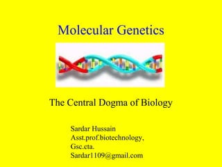
- 1. Molecular Genetics The Central Dogma of Biology Sardar Hussain Asst.prof.biotechnology, Gsc.cta. Sardar1109@gmail.com
- 2. What is a Genetic “Factor”? • From Mendel: – we now accepted that there was genetic transmission of traits. • Traits are transmitted by “factors” – Organisms carry 2 copies of each “factor” • The question now was: what is the factor that carries the genetic information?
- 3. Requirements of Genetic Material • Must be able to replicate, so it is reproduced in each cell of a growing organism. • Must be able to control expression of traits – Traits are determined by the proteins that act within us – Proteins are determined by their sequences • Therefore, the genetic material must be able to encode the sequence of proteins • It must be able to change in a controlled way, to allow variation, adaptation, thus survival in a changing environment.
- 4. Chromosomes – The First Clue • First ability to visualize chromosomes in the nucleus came at the turn of the century – construction of increasingly powerful microscopes – the discovery of dyes that selectively colored various components of the cell • Scientists examined cellular nuclei and observed nuclear structures, which they called chromosomes • Observation of these structures suggested their role in genetic transmission
- 5. Chromosome Observations • Variety of chromosome types found in the nucleus • 2 copies of each type of chromosome in most cells. • All of the cells of an organism, except gametes and rbc’s, have the same number of chromosomes. • All organisms of the same species have the same number of chromosomes. • The number of chromosomes in a cell doubles immediately prior to cell division • Gametes have exactly half of the number of chromosomes as the somatic cells of any organism. – Gametes have just one copy of each chromosome type. – Fertilization produces a diploid cell (a zygote), with the same number of chromosomes as somatic cells of that organism.
- 6. Implications • Chromosomes behaved like Mendel’s “factors” – Mendel's hereditary factors were either located on the chromosomes or were the chromosomes themselves. • Proof chromosomes were hereditary factors – 1905: • The first physical trait was linked to the presence of specific chromosomal material – conversely, the absence of that chromosome meant the absence of the particular physical trait. • Discovery of the sex chromosomes – "X" and "Y." – distinguished from other chromosomes and from each other
- 7. Importance of Sex Chromosomes • Somatic cells taken from female donors always contained 2 copies of the X chromosome • Somatic cells taken from male donors always contained one copy of the X chromosome and one copy of the Y chromosome • All of the other chromosomes in the nucleated cells of both male and female donors appeared identical • Thus, gender was shown to be the direct result of a specific combination of chromosomal material – The first phenotype (physical characteristic) to be assigned a chromosomal location – Specifically the X and Y chromosomes.
- 9. What Carries the Genetic Information? • Chromosomes are about 40% DNA & 60% protein. – Protein is the larger component • Protein molecules are composed of 20 different subunits • DNA molecules are composed of only four • Therefore protein molecules could encode more information, and a greater variety of information – protein had the possibility for much more diversity than in DNA • Therefore, scientists believed that the protein in chromosomes must carry the genetic information
- 10. The Discovery of DNA • First identified in 1868 by Friedrich Miescher, a Swiss biologist, in the nuclei of pus cells obtained from discarded surgical bandages. • He called the substance nuclein • Noted the presence of phosphorous
- 11. The Transforming Principle • Fredrick Griffith - 1928 • Discovered that different strains of the bacterium Strepotococcus pneumonae had different effects on mice – One strain could kill an injected mouse (virulent) – Another strain had no effect (avirulent) – When the virulent strain was heat-killed and injected into mice, there was no effect. – But when a heat-killed virulent strain was co-injected with the avirulent strain, the mice died. • Concluded that some factor in the heat killed bacteria was transforming the living avirulent to virulent? • What was the transforming principle and was this the genetic material?
- 13. The Transforming Principle is DNA • Avery, Macleod, & McCarty – 1943 • Attempted to identify Griffith’s “transforming principle” • Separated the dead virulent cells into fractions – The protein fraction – DNA fraction • Co-injected them with the avirulent strain. – When co-injected with protein fraction, the mice lived – with the DNA fraction, the mice died • Result was IGNORED – Most scientists believed protein was the genetic material. – They discounted this result and said that there must have been some protein in the fraction that conferred virulence.
- 14. The Hershey-Chase Experiment • Hershey & Chase – 1952 • Performed the definitive experiment that showed that DNA was the genetic material. • Bacteriphages = viruses that infect bacteria • Bacteriphage is composed only of protein & DNA • Inject their genetic material into the host
- 16. The Experiment • Prepared 2 cultures of bacteriophages • Radiolabeled sulphur in one culture – there is sulphur in proteins, in the amino acids methionine and cysteine – there is no sulphur in DNA • Radiolabeled phosphorous in the second culture – there is phosphorous in the phosphate backbone of DNA – none in any of the amino acids. • So this one culture in which only the phage protein was labeled, and one culture in which only the phage DNA was labeled.
- 17. Experiment Summary • Performed side by side experiments with separate phage cultures in which either the protein capsule was labeled with radioactive sulfur or the DNA core was labeled with radioactive phosphorus. • The radioactively labeled phages were allowed to infect bacteria. • Agitation in a blender dislodged phage particles from bacterial cells. • Centrifugation pelleted cells, separating them from the phage particles left in the supernatant.
- 18. Results Summary • Radioactive sulfur was found predominantly in the supernatant. • Radioactive phosphorus was found predominantly in the cell fraction, from which a new generation of infective phage was generated. • Thus, it was shown that the genetic material that encoded the growth of a new generation of phage was in the phosphorous-containing DNA.
- 20. Chargaff’s Rule • Once DNA was recognized as the genetic material, scientists began investigating its mechanism and structure. • Erwin Chargaff – 1950 – discovered the % content of the 4 nucleotides was the same in all tissues of the same species – percentages could vary from species to species. • He also found that in all animals (Chargaff’s rule): %G = %C %A = %T • This suggested that the structure of the DNA was specific and conserved in each organism. • The significance of these results was initially overlooked
- 21. Base Pairing in DNA • Not understood ‘til Watson & Crick described double helix • Adenine & guanine are purines – 2 organic rings • Cytosine & guanine are pyrimidines – 1 organic ring • Pairing a purine & a pyrimidine creates the correct 2 nm distance in the double helix • A – T joined by 2 hydrogen bonds • G – C joined by 3 hydrogen bonds
- 22. The Double Helix • James Watson and Francis Crick – 1953 • Presented a model of the structure of DNA. • It was already known from chemical studies that DNA was a polymer of nucleotide (sugar, base and phosphate) units. • X-ray crystallographic data obtained by Rosalind Franklin, combined with the previous results from Chargaff and others, were fitted together by Watson and Crick into the double helix model.
- 23. Watson and Crick shared the 1962 Nobel Prize for Physiology and Medicine with Maurice Wilkins. Rosalind Franklin died before this date.
- 24. DNA Structure • The double helix is formed from two strands of DNA • DNA strands run in opposite directions • They are complementary – attached by hydrogen bonds between complimentary base pairs: – A - T and G - C • This complementary pairing of the bases suggests that, when DNA replicates, an exact duplicate of the parental genetic information is made. – The polymerization of a new complementary strand takes place using each of the old strands as a template.
- 26. Messelson and Stahl • How does DNA replicate? • Matthew Meselson and Franklin Stahl - 1957 • Did an experiment to determine which model best represented DNA replication: • semiconservative replication – the two strands unwind and each acts as a template for new strands – each new strand is half comprised of molecules from the old strand • conservative replication – the strands do not unwind, but somehow generate a new double stranded DNA copy of entirely new molecules
- 29. The Experiment • The original DNA strand was labeled with the heavy isotope of nitrogen, N-15. • This DNA was allowed to go through one round of replication with N-14 (non-labeled) • the mixture was centrifuged so that the heavier DNA would form a band lower in the tube • the intermediate (one N-15 strand and one N- 14 strand) and light DNA (all N-14) would appear as a band higher in the tube
- 30. The expected results for each model were:
- 31. The actual results were as expected for the semiconservative model and thus Watson and Crick's suspicion was borne out.
- 32. Enzymes & Replication • DNA replication is not a passive, spontaneous process. • More than a dozen enzymes & other proteins are required to unwind the double helix & to synthesize & finalize a new strand of DNA. • The molecular mechanism of DNA replication can best be understood from the point of view of the machinery required to accomplish it. • The unwound helix, with each strand being synthesized into a new double helix, is called the replication fork.
- 33. The Enzymes of DNA Replication • Topoisomerase – Responsible for initiation of unwinding of DNA. – The tension holding the helix in its coiled and supercoiled structure is broken by nicking a single strand of DNA. • Helicase – Accomplishes unwinding of the original double strand, once supercoiling is eliminated by topoisomerase. – The two strands want to bind together because of hydrogen bonding affinity for each other, so helicase requires energy (ATP) to break the strands apart.
- 34. Single-stranded Binding Proteins • Important to maintain the stability of the replication fork. • Line up along unpaired DNA strans, holding them apart • Single-stranded DNA is very unstable, so these proteins bind bind to it while it remains single stranded and keep it from being degraded.
- 35. Beginning: Origins of Replication • Replication begins at specific sites called origins of replication • In prokaryotes, the bacterial chromosome has a specific origin • In eukaryotes, replication begins at many sites on the large molecule – 100’s of origins • Proteins that begin replication recognize the origin sequence • These enzymes attach to DNA, separating the strands, and opening a replication bubble • The end of the replication bubble is the “Y” shaped replication fork, where new strands of DNA elongate
- 37. Elongation • Elongation of the new DNA strands is catalyzed by DNA polymerase • Nucleotides align with complimentary bases on the template strand, and are added by the polymerase, one by one, to the growing chain – DNA polymerase proceeds along a single-stranded DNA molecule, recruiting free nucleotides to H-bond with the complementary nucleotides on the single strand • Forms a covalent phosphodiester bond with the previous nucleotide of the same strand – The energy stored in the triphosphate is used to covalently bind each new nucleotide to the growing second strand • Replication proceeds in both directions
- 40. DNA Polymerase • There are different forms of DNA polymerase – DNA polymerase III is responsible for the synthesis of new DNA strands • DNA polymerase is actually an aggregate of several different protein subunits, so it is often called a holoenzyme. • Primary job is adding nucleotides to the growing chain • Also has proofreading activities
- 41. Proofreading & Repair • DNA polymerase proofreads each nucleotide added against its’ template as it is added • Removes incorrectly paired nucleotides & corrects
- 42. DNA Strands are Anti-parallel • The sugar phosphate backbones of the 2 parent strands run in opposite directions – “upside down” to each other • DNA is polar – There is a 3’ end and a 5’ end – At the 3’ end, a OH is attached to the 3’ C of the last deoxyribose – At the other end a phosphate group is attached to the 5’ C of the last nucleotide • DNA polymerase adds nucleotides only to the 3’ end of the growing chain – So new DNA strand elongates only in the 5’ 3” direction
- 44. Why 5’ 3’? • Why can DNA polymerase only add nucleotides to the 3’ end ? • Needs a triphosphate to provide energy for the bond between an added nucleotide & the growing DNA strand. • This triphosphate is very unstable – can easily break into a monophosphate and an inorganic pyrophosphate • At the 5' end, this triphosphate can easily break – It is not be able to attach new nucleotides to the 5' end once the phosphate has broken off • The 3' end only has a hydroxyl group – As long as new nucleotide triphosphates are brought by DNA polymerase, synthesis of a new strand continues, no matter how long the 3' end has remained free.
- 45. Leading & Lagging Strands • The new strand made by adding to the 3’ end = leading strand – Parent strand is 3’ 5’ – New strand is 5’ 3’ • How can DNA polymerase synthesize new copies of the 5' 3' strand, if it can only travel in one direction? • To elongate in the other direction, the process must work away from the replication fork • The new strand formed on the 5’ 3’ parent strand is called the lagging strand
- 46. Building the Lagging Strand • DNA polymerase makes a second copy in an overall 3’ 5’ direction • First, it produces short segments, called Okazaki fragments – These are built in a 5’ –3’ direction • Okazaki fragments are joined together by ligase to produce the new 3’5’ lagging strand
- 47. Ligase • Catalyzes the formation of a phosphodiester bond given an unattached adjacent 3‘ OH and 5‘ phosphate. • This can join Okazaki fragments • This can also fill in the unattached gap left when an RNA primer is removed • DNA polymerase can organize the bond on the 5' end of the primer, but ligase is needed to make the bond on the 3' end.
- 49. Primers • DNA polymerase cannot start synthesizing on a bare single strand. – It only adds to an existing chain • It needs a primer with a 3'OH group on which to attach a nucleotide. • The start of a new chain is not DNA, but a short RNA primer – Only one primer is needed for the leading strand – One primer for each Okazaki fragment on the lagging strand
- 50. Primase • Part of an aggregate of proteins called the primeosome. • Attaches the small RNA primer to the single-stranded DNA which acts as a substitute 3'OH so DNA polymerase can begin synthesis • This RNA primer is eventually removed by RNase H – the gap is filled in by DNA polymerase I.
- 52. Ending the Strand • DNA polymerase only adds to the 3’ end • There is no way to complete the 5’ end of the new strand • A small gap would be left at the 5’ end of each new strand • Repeated replication would then make the strand shorter and shorter, eventually losing genes • Not a problem in prokaryotes, because the DNA is circular – There are no “ends”
- 53. Telomeres • Eukaryotes have a special sequence of repeated nucleotides at the end, called telomeres • Multiple repititions of a short nucleitide sequence – Can be repeated hundreds, or thousands of times – In humans, TTAGGG • Do not contain genes • Protects genes from erosion thru repeated replication • Precvents unfinished ends from activating DNA monitoring & repair mechanisms
- 54. Telomerase • Catalyzes lengthening of telomeres • Includes a short RNA template with the enzyme • Present in immortal cell lines and in the cells that give rise to gametes • Not found in most somatic cells • May account for finite life span of tissues
- 56. Further Proofreading & Repair • Some errors evade initial proofreading or occur after synthesis • DNA can be damaged by reactive chemicals, x-rays, UV, etc • Cells continually monitor DNA for mutations & repair • Contain 100’s of repair enzymes
- 57. Nucleotide Excision & Repair • A segment of DNA containing damage is cut out by a nuclease (a DNA cutting enzyme) • The gap is filled & closed by DNA polymerase and ligase • Thymine dimers • Covalent linking of thymine bases • Causes DNA to “buckle” • One common problem corrected this way
- 59. Proteins of DNA Replication
- 60. Summary View of Replication