Platelet and coagulation disorder
•Descargar como PPTX, PDF•
72 recomendaciones•21,982 vistas


Recomendados
Más contenido relacionado
La actualidad más candente
La actualidad más candente (20)
Destacado
Destacado (14)
Similar a Platelet and coagulation disorder
Similar a Platelet and coagulation disorder (20)
Platelet and coagulation disorder
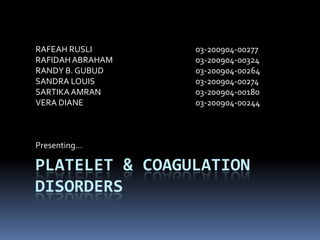
- 1. RAFEAH RUSLI 03-200904-00277 RAFIDAH ABRAHAM 03-200904-00324 RANDY B. GUBUD 03-200904-00264 SANDRA LOUIS 03-200904-00274 SARTIKA AMRAN 03-200904-00180 VERA DIANE 03-200904-00244 Presenting… PLATELET & COAGULATION DISORDERS
- 2. Objective: Describe platelet List the types of platelet & coagulation disorder Describe briefly about the disorders Laboratory diagnosis
- 3. Platelet Platelets are produced in blood cell formation (thrombopoiesis) in bone marrow megakaryoblast > pro-megakaryocyte > immature megakaryocyte > megakaryocyte > Platelet Platelets or thrombocytes are small irregularly shaped non-nucleated 2–3 µm in diameter. lifespan of circulating platelets is 5 to 9 days. platelet production is regulated by thrombopoietin (hormone which produced by the liver and kidneys) Old platelets are destroyed by phagocytosis in the spleen and by Kupffer cells in the liver
- 5. Platelet cont… The platelet structure has 3 zones: Peripheral Structural Organelle Structural zone Consists of the cytoskeleton The cytoskeleton forms the support for the maintenance of the platelet’s discoid shape Regulate contractile system that allows, upon activation, shape change, pseudopod extension, internal contraction, and release of granular constituents.
- 7. Platelet
- 8. Platelet cont… Organelle zone consists of the granules and cellular components These organelles serve in the metabolic processes of the platelet and store enzymes. dense granules contain non-metabolic adenosine triphosphate (ATP) and adenosine diphosphate (ADP), serotonin, and calcium alpha granules contain adhesive proteins such as fibrinogen, fibronectin, von Willebrand factor (VWF), thrombospondin, and vitronectin. alpha granules also contain growth-promoting substances such as platelet- derived growth factor (PDGF), platelet factor 4, and transforming growth factor. Coagulation factors including factor V, high molecular weight kininogen, factor XI, and plasminogen activator inhibitor-1 are also present in the alpha granule.
- 9. Platelet cont… Membrane / peripheral zone Consist of typical phospholipid bilayer membrane Embedded in this structure are different kind of glycoprotein. Glcoprotein Function GP IIb/IIIa Receptor for fibrinogen, VWF, fibronectin, vitronectin and Thrombospondin For platelet aggregation GP Ia/IIa Receptor for Collage GP Ib/IX/V Receptor for insoluble VW For platelet adhesion GP VI Receptor for Collagen
- 10. General function of platelet The function of platelets is the maintenance of hemostasis. Platelets helps in blood clotting. Wound repair Platelets secrete platelet-derived growth factor (PDGF). Granule secretion. Adhesion and aggregation. Pro-coagulation. Cytokine signalling. Phagocytosis. Transport of enzyme and proteins critical to clotting. Formation of a platelet plug to slow blood loss. Contraction of a clot after it has formed, which then reduces the size of the vessel break.
- 11. platelet ultrastructure….. Alpha granules Dense bodies VwF ADP Fibronectin ATP Thrombospondin Serotin
- 12. Types of disorder: Divided into: Coagulation disorder Platelets disorder Coagulation disorder include: Henophilia Von Willebrand disease Platelet disorder include: Deficiency Of Vitamin K. Bernard - Soulier Syndrome. Thrombasthenia Of Glanzmann And Naegeli (Glanzmann Thrombasthenia) Gray Platelet Syndrome. Dense Granule Deficiency Syndrome. Thrombotic Thrombocytopenic Purpura (TTP). Idiopathic Thrombocytopenic Purpura (ITP).
- 13. Hemophilia Definition: disease associated with prolonged bleeding due to the deficiency in clotting factor. Hemophilia is a X-linked disease Types of Hemophilia: Hemophilia A Factor 8 deficiency, x linked disease Hemophilia B Factor 9 deficiency, x-linked disease Hemophilia C Factor 11 deficiency, autosomal genetic disorder Symptoms of hemophilia: Bruising Bleeds easily Bleeding into a joint Bleeding into the muscles Bleeding from injury or bleeding in the brain Other sources of bleeding (eg. Stool & urine)
- 14. Von Willebrand disease The most common hereditary coagulation abnormality Can also be acquired as a result of other medical conditions Due to the deficiency of von Willebrand factor (vWF) Von Willebrand factor - mediates binding of glycoprotein Ib to collagen This binding mediate activation of platelets and formation of primary hemostasis Defect in this factor, resulting glycoprotein IB does not bind to collagen. Thus unable to activate platelets, primary hemostasis does not occur
- 15. Lab findings: comparison between Hemophilia A, Hemophilia B & von Willebrand disease Condition PT APTT Bleeding time Hemophilia A Normal Increased Normal Hemophilia B Normal Increased normal Von Willebrand disease Normal Increased increased Symptoms of von Willebrand disease: Abnormal menstrual bleeding Bleeding of the gums Bruising Nosebleeds Skin rash
- 16. Bernard-Soulier syndrome Also known as hemorrhagiparous thrombocytic dystrophy It is due to deficiency of glycoprotein Ib (GPIb), the receptor for von willebrand factor Lacks of GPIb cause vWF unable to bind to the glycoprotein finally lead to decrease in primary clot formation / primary hemostasis Characterized by Characterized by abnormally large platelets / giant platelets Characterized by prolonged bleeding time, thrombocytopenia, increased megakaryocytes, and decreased platelet survival Some of the symptoms: Purpura. Epistaxis. Menorrhagia. Gingival and gastrointestinal bleeding.
- 17. Deficiency of Vitamin K Role of Vitamin K in blood coagulation: Important in maturation of clotting factor. modification of certain proteins required for blood coagulation If the clotting factor does not mature, it is useless in the hemostasis process. Factor which causes the deficiency of vitamin K Disturbed intestinal uptake. By therapeutic or accidental intake of vitamin k-antagonists or very rarely. By nutritional vitamin k deficiency Some of the possible symptoms of vitamin K deficiency: Risk of massive uncontrolled bleeding Hematomas
- 18. THROMBASTHENIA OF GLANZMANN AND NAEGELI extremely rare coagulopathy can be inherited in an autosomal recessive manner or acquired as an autoimmune disorder due to deficiency in glycoprotein IIb/IIIa (GpIIb/IIIa) glycoprotein IIb/IIIa (GpIIb/IIIa) is receptor for fibrinogen. When glycoprotein IIb/IIIa (GpIIb/IIIa) receptor is dysfunction, fibrinogen cannot bind to the platelets. As a result, no fibrinogen bridging of platelets to other platelets occur In other words, primary hemostasis inhibited and prevent platelets aggregation Bleeding time is significantly prolonged
- 19. THROMBASTHENIA OF GLANZMANN AND NAEGELI Symptoms includes: Increased mucosal bleeding. Epistaxis. Menorrhagia. Increased bleeding post-operatively. The bleeding tendency is variable but may be severe. Platelet numbers and morphology are normal. Platelet aggregation is normal with ristocetin, but impaired with other agonists such as ADP, thrombin, collagen or epinephrine.
- 20. GRAY PLATELET SYNDROME Gray platelet syndrome (GPS) is a rare inherited bleeding disorder. The abnormal alpha-granules appear grey on blood films stained by the May-Grünwald-Giesma stain - hence, the syndrome's name. It caused by the inability of platelets to store alpha-granule proteins. The platelets' haemostatic proteins are not released at the site of vascular injury. Thus slows aggregation and vessel repair And contribute to the bleeding tendency. Symptoms may include: platelets that have a gray appearance severe thrombocytopenia myelofibrosis splenomegaly
- 21. DENSE GRANULE DEFICIENCY SYNDROME it is a bleeding disorder caused by a deficiency in dense granules in the platelets Dense granules release chemicals in the clotting process and help platelets stick together to form clots. Lacks of dense granules in platelets means lacks of storage sites for substances for clotting. Therefore no chemicals which facilitates in the clotting process will be released. Finally leads to slow platelet activation, and prolonged bleding.
- 22. THROMBOTIC THROMBOCYTOPENIC PURPURA (TTP) A blood disorder that causes blood clots to form in small blood vessels around the body, and leads to a low platelet count. The two main types of TTP are inherited and acquired. In inherited TTP, the ADAMTS13 gene is faulty and doesn't prompt the body to make a normal ADAMTS13 enzyme. As a result, enzyme activity is lacking or changed. Acquired TTP is the more common type of the disorder. The ADAMTS13 gene isn't faulty. Instead, the body makes antibodies (proteins) that block the activity of the ADAMTS13 enzyme. A lack of activity in the ADAMTS13 enzyme causes TTP. The ADAMTS13 gene controls the enzyme, which is involved in blood clotting. The enzyme breaks up a large protein called von Willebrand factor that clumps together with platelets to form blood clots.
- 23. IDIOPATHIC THROMBOCYTOPENIC PURPURA (ITP) Also known as immune thrombocytopenic purpura, is classified as an autoimmune disease. The term “idiopathic” indicates that the disease is of an unknown cause or origin: in other words, modern medicine has not yet figured out what it is. And the word “purpura” comes from a description of the bruise- colored skin of someone afflicted with the disease: the purple color caused by blood that leaked under the skin.
- 24. IDIOPATHIC THROMBOCYTOPENIC PURPURA (ITP) Idiopathic thrombocytopenic purpura is a bleeding disorder in which the immune system destroys platelets Persons with the disease have too few platelets in the blood The two types of ITP are acute (temporary or short-term) and chronic (long-lasting). Acute ITP generally lasts less than 6 months. It mainly occurs in children—both boys and girls—and is the most common type of ITP. Acute ITP often occurs after a viral infection. Chronic ITP lasts 6 months or longer and mostly affects adults. However, some teenagers and children do get this type of ITP. Chronic ITP affects women two to three times more often than men. Symptoms: Abnormally heavy menstruation. Bleeding into the skin causes a characteristic skin rash that looks like pinpoint red spots. (petechial rash) Easy bruising. Nosebleed or bleeding in the mouth.
- 25. Lab diagnosis Status of platelet & coagulation process can be screened by using the following simple lab test: Bleeding Time Clotting time Activated partial thromboplastin time (APTT) Prothrombin time (PT) Full Blood Count Peripheral Blood Film May-Grünwald-Giesma stain
- 26. Bleeding time Function: Basic screening test of platelet function Procedure: Place a blood pressure cuff on the arm and inflare it to 40mm Hg Clean the area of the fore arm below the anticubital fossa with 70% alcohol. There should be no superficial veins in the area selected Make 2 skin punctures in rapid succession, each 3mm deep, by using disposable lancets Start the stop-watch as soon as bleeding starts. Wipe off the blood at 15 seconds interval by touching lightly with a blotting paper Record the time taken for the blood to stop flowing in the both punctures and determine the mean time. Remove the blood pressure cuff and cover the puncture site with plaster Result: Normal range : 2-6 minutes Borderline : 6-10 minutes (Test should be repeated)
- 27. Bleeding time:
- 28. Clotting time: Function: To measure he time required for a sample of blood to coagulate in vitro under standard conditions Procedure: Make a clean venipuncture with minimum trauma to the connective tissue Start stop-watch as soon as the blood enters the glass syringe. Draw 4ml of blood and deliver 1ml each into 4 glass tubes previously warmed to 37ºC Place the tube immediately at 37ºC water bath At the end of each minutes, gently tilt one tube to see if the blood is clotted. Continue tilting the tube one after the other at one minute interval till one of them can be tilted at 90º without the blood flowing out. Note the time Continue with the remaining tubes and take the average of the clotting time in all the 4 tubes Reference values: Normal values : 5-10 minutes Prolonged clotting time is a marked deficiency in one of the coagulation factors of intrinsic pathway. Replaced by APTT
- 29. Activated partial thromboplastin time Function: Evaluate intrinsic pathway Measure all coagulating factor except factor VII and XIII Monitor heparin therapy Procedure: Pre-warmed APTT reagent at 37ºC for at least 10 minutes. Collect blood specimen in blue top tube Centrifuge and separate the plasma from blood Add 100µl of APTT reagent into a test tube, then add 100µl of plasma into the test tube and incubate for 2-3 minutes Then add 100µl calcium chloride reagent to the plasma Start stop-watch until clot formed Record the time at which clot form Reference value: Normal range : 35-45 seconds
- 30. Prothrombin time Function: Used to evaluate the extrinsic pathway (factor 1,2,5,7, and 10) Also used to monitor warfarin therapy Procedure: Collect blood in blue top tube. Centrifuge blood and separate plasma from blood Make sure to pre-warmed PT reagent 37ºC for at least 10 minutes before use. Place 100µl plasma into test tube. Pre-warm plasma for 2-3 minutes at 37ºC Then add 200µl of PT reagent to the tube, simultaneously start the stop-watch and record the time required for clot formation. Reference value: Normal range : 11-14 seconds
- 32. Other lab test: Full blood count: Normal range : 150,000 – 400,000 /ul Those with coagulation / platelet disorder : thrombocytopenia PBF: Giant platelet may present thrombocytopenia Special staining: MGG stain for Gray platelet syndrome
- 33. Giant Platelet Giant Platelet Thrombocytosis Thrombocytopenia