How high frequency ultrasound imaging is supporting preclinical research applications
•
1 recomendación•396 vistas

This free webinar hosted by Scintica Instrumentation introduced participants to some of the basics of high frequency ultrasound imaging and reviewed the most common preclinical research applications for this exciting technology. Our presenter, Ms. Tonya Coulthard, discussed the basic principles of ultrasound imaging, presented sample images from various organs and tissues as a way of surveying common research applications, and introduced a few novel techniques that will be of interest to many researchers. Throughout the webinar, technical information, product specifications and sample images where shown from the Prospect T1 compact, high-frequency, preclinical ultrasound imaging system.

Recomendados
Más contenido relacionado
La actualidad más candente
La actualidad más candente (19)
Similar a How high frequency ultrasound imaging is supporting preclinical research applications
Similar a How high frequency ultrasound imaging is supporting preclinical research applications (20)
Más de Scintica Instrumentation
Más de Scintica Instrumentation (20)
Último
Último (20)
How high frequency ultrasound imaging is supporting preclinical research applications
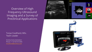
- 1. Tonya Coulthard, MSc. Team Leader Scintica Instrumentation Phone: +1 (519) 914 5495 tcoulthard@scintica.com Overview of High Frequency Ultrasound Imaging and a Survey of Preclinical Applications
- 2. • Basics of Ultrasound Imaging • Prospect T1 System Overview • Key Research Applications Topics of Discussion
- 3. Basics of Ultrasound Imaging • Understanding how ultrasound works • Review of acquisition modes
- 4. • Ultrasound is a non-invasive imaging technique which does not required the use of ionizing radiation, instead it uses sound waves • The transducer both sends and receives the ultrasound waves and the computer interprets the returned signal into an image Ultrasound Imaging Note – these images were not taken with the Prospect T1 system
- 5. • High-frequency ultrasound waves are necessary to resolve the small anatomical targets in preclinical research • Compromise is shorter penetration depth Ultrasound Imaging
- 6. • B-Mode images are a 2D grey scale representation of anatomical structures – still or cine loop images • The sound waves cannot penetrate through air or bone • Ultrasound gel, or some other liquid, is used to couple the transducer to the surface Acquisition Modes – Brightness (B) Mode
- 7. • M-Mode displays moving structures along a single line • As data is only taken along a single line, the sampling frequency is very high (1000 frames per second) • The user defines the sample volume on a B-mode image Acquisition Modes – Motion (M) Mode
- 8. • Doppler modes detect moving structures, i.e. blood • Velocity and direction of the blood can be determined by the “Doppler Shift” • Color/Power Doppler are displayed as an overlay on a B-Mode image, while Pulsed Wave Doppler is displayed as a spectrogram Acquisition Modes – Doppler Modes
- 9. • Microbubble contrast agents may be used to image the vasculature or targeted cellular markers; using non- targeted or targeted microbubbles respectively • Reference subtracted or harmonic imaging may be used to visualize the contrast agents Acquisition Modes – Contrast Mode
- 10. Prospect T1 System Overview • System components and standard configuration • Add-on hardware and software components
- 11. Prospect T1 System Components
- 12. Prospect T1 System Components • The Prospect T1 is the first tablet based high-frequency ultrasound system specifically designed for preclinical imaging of small animals • System components: • Tablet • Probe • Scanning Platform
- 13. Prospect T1 System Components: Tablet • The powerful tablet reduces the footprint of the system, taking up less lab space, making it easy to move when necessary • Multiple data formats exist for either still, cine loop, or RAW data storage • Offline software analysis is possible to preserve time on the system for imaging • The intuitive workflow and touch screen allow researchers to start acquire images and generating data quickly
- 14. Prospect T1 System Components: Probes • Two single element probes are available: • 20 MHz (user selectable between 15-30 MHz) • Primarily used for rat imaging, as well as harmonic contrast imaging • 40 MHz (user selectable between 30-50 MHz) • Primarily used for mouse imaging, and superficial anatomical targets in larger species like rats
- 15. Prospect T1 System Components: Scanning Platforms • The platform is compact in design, again to limit the footprint of the system • The scanning platform has been designed for ergonomical positioning of the probe • Animal beds have integrated heating, and ECG and respiratory monitoring • Interchangeable beds are available for mice or rats • Animal beds can be precisely adjusted in the X, Y, and Z axis
- 16. Standard System Configuration • Standard system configuration for mouse • B-Mode • M-Mode • Pulsed Wave / Color / Power / Tissue Doppler Mode • Contrast Mode • Comprehensive Measurement and Analysis Tools • Scanning Platform – with mouse bed • 40 MHz probe • Standard system configuration for rat • B-Mode • M-Mode • Pulsed Wave / Color / Power / Tissue Doppler Mode • Contrast Mode • Comprehensive Measurement and Analysis Tools • Scanning Platform – with rat bed • 20 MHz probe
- 17. Prospect T1 System Add-Ons
- 18. Add-Ons: 3D Motor • The 3D motor expands the capabilities of the Prospect T1 to acquire 3D B-mode images • Add-on includes the software analysis package to view the 3D images and perform volume calculations
- 19. Add-Ons: Image Guided Needle Injection Mount • The image guided needle injection mount integrates with probe • Injections may be performed with a regular syringe and steel needle, or pulled glass capillary needle • Injections may be made into developing embryos, adult myocardium, or abdominal/muscle targets E15.5 mouse embryo Adult mouse myocardium
- 20. Add-Ons: Shear Wave Elastography • Shear wave elastography is used to quantify mechanical and elastic properties of tissues • The acoustic radiation force is generated by a push probe mounted on the side of the imaging probe • The software analysis generates a colored elastogram which is overlaid on a B-mode image
- 21. Add-Ons: Integrated Sonoporation • Sonoporation is the controlled cavitation or bursting of microbubbles with the intention of increasing the permeability of the cell membrane or to open to blood brain barrier • Sonoporation is performed by a secondary, non-imaging, probe directed at the anatomical target • Software integration and control of the sonoporation probe is included with this add-on
- 22. Key Research Applications • Cardiovascular Research • Cancer Biology • Abdominal & Anatomical Imaging • Developmental Biology • Ophthalmology • Other Animal Models – Zebrafish, Chick Embryos
- 24. Cardiovascular Research: Mouse Systolic Function – B-Mode Long Axis View IVS LV LVPW AO LA PM Mitral valve Short Axis View IVS LV LVPW LVAW PM LV : left ventricle LVAW: left ventricular anterior wall LVPW : left ventricular posterior wall PM: papillary muscle IVS : interventricular septum AO : aortic orifice LA : left atrium
- 25. Cardiovascular Research: Mouse Systolic Function – B-Mode; Area Length Measurement (ALM) • End diastolic volume; End systolic volume • Stroke volume • Ejection fraction • Fractional area change (from short axis) • Fractional shortening • Left ventricular mass • Left ventricular mass index
- 26. Cardiovascular Research: Mouse Systolic Function – M-Mode
- 27. Cardiovascular Research: Mouse Systolic Function – M-Mode • Can be done on either the long or short axis M-mode image • LV mass • LV mass index • Fractional shortening • End diastolic volume; end systolic volume • Stroke volume • Ejection fraction • Cardiac output
- 28. Cardiovascular Research: Mouse Systolic Function – PW Doppler & B-mode • Stroke Volume is calculated as a function of the Velocity Time Interval (VTI) and vessel diameter • Cardiac Output is simply stroke volume x heart rate • VTI can be manually or automatically traced on the PW Doppler: • Peak velocity • Peak pressure gradient • Mean velocity • Mean pressure gradient • Acceleration & Deceleration
- 29. Cardiovascular Research: Mouse Diastolic Function – Color & PW Doppler LV R V L A R A MV T V LV : left ventricle RV: right ventricle LA: left atrium RA: right atrium MV: mitral valve TV: tricuspid valve Mitral Valve Tricuspid Valve
- 30. Cardiovascular Research: Mouse Diastolic Function – Color & PW Doppler • Acceleration rate of E wave • Peak velocity of E & A waves • Deceleration time of E wave • E:A ratio • Isovolumic relaxation/contraction time (IVRT & IVCT) • Ejection time • Myocardial performance index (Tei index)
- 31. Cardiovascular Research: Mouse Diastolic Function – Tissue Doppler • Peak velocity of E & A waves • Isovolumic relaxation/contraction time (IVRT & IVCT) • Ejection time • Filling time
- 32. Cardiovascular Research: Mouse Aortic Arch RPA IA LCCA LSCA AAr AAr : Aortic Arch RPA : Right Pulmonary Artery IA: Innominate Artery LCCA : Left Common Carotid Artery LSCA : Left Subclavian Artery AAr AAo IA LCCA LSCA
- 33. Cardiovascular Research: Rat Carotid Artery Intima-Media Thickness
- 34. Cardiovascular Research: Mouse Carotid Artery ICA ECA RCCA Right Common Carotid Artery (RCCA) Internal Carotid Artery (ICA) External Carotid Artery (ECA)
- 35. Cardiovascular Research: Mouse Peripheral Vasculature – Color & PW Doppler • Automatic peak analysis is used to help with peripheral vascular measurements • Systolic:Diastolic ratio • Peak systolic velocity • End diastolic velocity • Resistive index • Pulsatility index
- 36. Cardiovascular Research: Image Guided Needle Injection; Adult Mouse Myocardium • Image guided injection may be done into the myocardium or other anatomical target • Stem cells or other therapy may injected into the myocardium to study the effect on myocardial infarction lesion size, for example
- 37. Cancer Biology
- 38. Cancer Biology: Early detection of tumors Primary liver tumors (mouse)
- 39. Cancer Biology: Surrounding Structures Mouse Axillary Lymph Node • Visualizing the surrounding structures helps visualize additional changes which may occur • This lymph node is normal, however they may be enlarged and have an altered appearance with advanced disease
- 40. Cancer Biology: Tumor measurement • Linear and area measurement tools allow tumor sizing on 2D B-mode images Orthotopic Liver Tumor - Mouse Orthotopic Breast Tumor - Mouse
- 41. Cancer Biology: Tumor measurement • The 3D motor add-on can be used to acquire 3D B- mode images • Orthotopic breast tumor in a mouse • Volume = 211mm3
- 42. Cancer Biology: Tumor Perfusion • Microbubble contrast agents are inject intravenously to study perfusion • Two types of contrast imaging are possible • Reference subtracted – with green color overlay applied on cineloop image (20 or 40MHz probe) • Harmonic imaging – microbubble specific harmonic signal is detected (20MHz probe)
- 44. Abdominal & Anatomical: Mouse Liver and Gallbladder Liver Liver vessels Gallbladder
- 45. Abdominal & Anatomical: Mouse Spleen and Pancreas Spleen Splenic vein Pancreas Spleen Kidney
- 46. Abdominal & Anatomical: Mouse Kidney Kidney
- 47. Abdominal & Anatomical: Mouse Abdominal Aorta
- 48. Abdominal & Anatomical: Mouse Knee and Lower Leg Tibiofemoral tendon Synovial membrane Cartilage Patella Muscle Lower Leg
- 49. Abdominal & Anatomical: Mouse Ovary Ovary Ovary Cyst
- 51. Developmental Biology: Mouse Embryos Embryonic heart and neural tube _E9.5 Embryos_E7.5 Embryonic brain_E12.5 Embryonic spinal cord_E12.5 Embryonic head and forelimb_E14.5
- 52. Developmental Biology: Mouse Embryos – Color and PW Doppler Mitral Valve Umbilical cord Dorsal Aorta
- 53. Developmental Biology: Mouse Embryos – M-Mode Heart Ventricles
- 54. Developmental Biology: Image Guided Needle Injection; Mouse Embryo • Image guided injection may be done into a variety of anatomical targets within the embryo • The uterine horn is exposed from the dame and injections done into the exposed embryos
- 55. Opthamology
- 56. Ophthalmology: Anterior Structures and Retinal Vasculature; mouse eyeEyelid CorneaLens Cornea Ciliary body
- 57. Ophthalmology: 3D Imaging; rat eye Volume = 57.6mm3
- 58. Ophthalmology: Anterior Structures; rabbit eye Normal Thickened Cornea
- 60. Other Animal Models: Zebrafish Gills Fin Eye Spinal Cord Ventricular Inflow E A
- 61. Other Animal Models: Chick Embryo 5 Day 7 Day 7 Day
- 62. • Basics of Ultrasound Imaging • Prospect T1 System Overview • Key Research Applications Topics of Discussion
- 63. Tonya Coulthard, MSc. Team Leader Scintica Instrumentation Phone: +1 (519) 914 5495 tcoulthard@scintica.com Q&A SESSION: To ask a question, click the Q&A Button, type your question and click send. Any questions that are not addressed during the live webinar will be answered following the event. Thank you for participating!