SlideShare una empresa de Scribd logo
1 de 63
Endocrine System
Pituitary Gland
- The main endocrine gland.
- Called the “master” gland.
- Acts as the control center
for the endocrine system which
controls all the hormones
produced by other glands in the
body.
PITUITARY FOSSA
Some of the neurons within the
hypothalamus - neurosecretory
neurons - secrete hormones that
strictly control secretion of hormones
from the anterior pituitary.
The hypothalamic hormones are
referred to as releasing hormones
and inhibiting hormones, reflecting
their influence on anterior pituitary
hormones.
Hypothalamic releasing
and inhibiting hormones
are carried directly to the
anterior pituitary gland
via hypothalamic-
hypophyseal portal veins
.
Specific hypothalamic
hormones bind to
receptors on specific
anterior pituitary cells,
modulating the release
of the hormone they
produce.
Anterior
Pituitary
Posterior
pituitary Stimulates milk
ejection and uterine
contractions
Ovary and testisOxytocin
Conservation of
body water
KidneyAntidiuretic hormone
Control of reproductive
function
Ovary and testisFollicle stimulating
hormone
Control of reproductive
function
Ovary and testisLuteinizing hormone
Milk productionMammary glandProlactin
Stimulates secretion of
glucocorticoids
Adrenal gland cortexAdrenocorticotrophic
hormone
Stimulates secretion of
thyroid hormones
Thyroid glandThyroid stimulating
hormone
Promotes growth (indirectly),
control of protein, lipid and
carbohydrate metabolism
Liver, adipose tissueGrowth hormone
Major physiologic
effects
Major target organsHormone
• The portion of the adenohypophysis known as the pars tuberalis
contains cords of epithelial cells and is filled with hypophyseal portal
vessels. It reportedly contains gonadotropes and thyrotropes, plus
other secretory cells of unknown function.
• The pars intermedia is closely associated with pars nervosa and
separated from the pars distalis by the hypophyseal cleft.
Melanocyte-stimulating hormone is the predominant hormone
secreted by the pars intermedia.
The pituitary gland has two distinct
parts, the anterior and the posterior
lobes, each of which releases
different hormones.
• Histology of the Adenohypophysis
• The bulk of the adenohypophysis is
pars distalis. That tissue is composed
of winding cords of epithelial cells
flanked by vascular sinusoids.
• In sections stained with dyes such as
hematoxylin and eosin, three distinct
cell types are seen among epithelial
cells:
• Acidophils have cytoplasm that stains
red or orange
• Basophils have cytoplasm that stains
a bluish color
• Chromophobes have cytoplasm that
stains very poorly
Acidophils: contain the polypeptide hormones:
– Somatotropes which produce growth hormone
– Lactotropes which produce prolactin
Basophils: contain the glycoprotein hormones:
– Thyrotropes which produce thyroid stimulating hormone
– Gonadotropes which produce luteinizing hormone or
follicle-stimulating hormone
– Corticotropes which produce adrenocorticotrophic hormone
Chromophobes
• These are cells that have minimal or no hormonal content.
– may be acidophils or basophils that have degranulated and thereby
are depleted of hormone
– may also represent stem cells that have not yet differentiated into
hormone-producing cells.
• Although classification of cells as acidophils or
basophils is useful in some situations, specific
identification of anterior pituitary cells requires
immunostaining for the hormone in question.
• In addition to differential staining characteristics, the
size of secretory granules varies among different
types of cells in the anterior pituitary.
Somatotropes and lactotropes tend to have the
largest size granules.
Hyperpituitarism and
Pituitary Adenomas
Pituitary tumor causes symptoms by any of
three mechanisms:
1. By producing too much of one or more
hormones.
2. By compressing the pituitary gland, and thus
making it produce too little of one or more
hormones.
3. By compressing the optic nerves or (less
commonly) the nerves controlling eye
movements, and thus causing either loss of part
or all of the visual field, or double vision.
ENLARGING PITUITARY MASS
Type of Adenoma Secretion Staining Pathology
Corticotrophic
adenomas
Secrete adrenocotrophic
hormone (ACTH) and
Proopiomelanocortin
(POCM)
Basophilic Cushing’s disease
Somatotrophic
adenomas
Secrete growth hormone
(GH)
Acidophilic Acromegaly
(Gigantism)
Thyrotrophic
adenomas (rare)
Secret thyroid stimulating
hormone (TSH)
Basophilic Occasionally
hyperthyroidism
usually does not cause
symptoms
Gonadotrophic
adenomas
Secrete luteinizing
hormone (LH), follicle
stimulating hormone (FSH)
Basophilic Usually does not cause
symptoms
Prolactinomas Secret prolactin Acidophilic Galactorrhea,
hypogonadism,
amenorrhea, infertility
and impotence
Null cells adenomas Do not secrete hormones May stain positive for
synaptophysin
By producing too much of one or more hormones
• Growth hormone: causes ACROMEGALY
– a syndrome that includes:
• excessive growth of soft tissues and bones
• high blood sugar
• high blood pressure
• heart disease
• sleep apnea
• excess snoring
• carpal tunnel syndrome
• pain symptoms (including headache).
By producing too much of one or more hormones
• Thyroid stimulating hormone:
– causes high production of thyroid hormone
– Thyroid hormone:
• leads to nervousness and irritability
• fast heart rate and high blood pressure
• heart disease
• excess sweating and thin skin
• and weight loss.
By producing too much of one or more hormones
• Prolactin:
– causes inappropriate secretion of breast milk (even in
men)
– osteoporosis (bone weakening)
– loss of sex drive
– Infertility
– irregular menstrual cycles
– and impotence
By producing too much of one or more hormones
• Adrenocorticotropic hormone:
– causes weight gain (particularly in the body’s trunk,
not the legs or arms)
– high blood pressure
– high blood sugar
– brittle bones
– emotional changes
– stretch marks on the skin
– easy bruising.
By producing too much of one or more hormones
• Gonadotropins (FSH and LH):
– usually not elevated enough to produce direct
symptoms
– but in extreme cases can cause infertility (inability to
have a child)
– irregular menstrual cycles in women.
Cushing’s Syndrome
• Definition
– a condition that occurs when your body is exposed
to high levels of the hormone cortisol for a long time.
– Referred to as hypercortisolism
– use of oral corticosteroid medication (most common
cause)
– The condition can also occur when your body
makes too much cortisol.
Hallmark signs of Cushing's syndrome:
- fatty hump between shoulders
- rounded face
- pink or purple stretch marks on your skin
- can also result in high blood pressure, bone loss and, on
occasion, diabetes.
By compressing the pituitary gland, and thus making it produce
too little of one or more hormones.
• Growth hormone: causes poor muscle strength, irritability,
weakening of bone strength, and overall feeling of malaise (feeling
unwell). DWARFISM in children.
Thyroid stimulating hormone: causes fatigue, low energy, and
weight gain.
• Prolactin: causes inability to breastfeed after a woman gives birth to
a baby.
• Adrenocorticotropic hormone: causes fatigue and low energy, low
blood pressure, low blood sugar, and upset stomach.
• Gonadotropins (FSH and LH): cause infertility, decrease in sex
drive, impotence, and irregular menstrual cycles.
Histology of the Neurohypophysis
• The neurohypophysis is known also as the pars
nervosa.
• Three areas of this organ, starting closest to the
hypothalamus:
– the median eminence
– infundibular stalk
– infundibular process
• The infundibular - bulk of the neurohypophysis (usually
referred to as the posterior pituitary)
• Composed on largely unmyelinated axons from
hypothalamic neurosecretory neurons.
• These neurons secrete oxytocin and antidiuretic
hormone.
• The neurohypophysis shown here resembles neural
tissue, with glial cells, nerve fibers, nerve endings, and
intra-axonal neurosecretory granules.
• The hormones vasopressin (antidiuretic hormone, or
ADH) and oxytocin made in the hypothalamus
(supraoptic and paraventricular nuclei) are transported
into the intra-axonal neurosecretory granules where they
are released.
Anterior pituitary Posterior pituitary
• Oxytocin increases uterine contractions and
stimulates milk secretion.
• Underproduction of ADH results in a disorder called
diabetes insipidus characterized by inability to
concentrate the urine. The consequence is excess
urination leading potentially to dehydration.
• Antidiuretic hormone (ADH) increases reabsorption
of water by the tubules of the kidney.
Syndrome of Inappropriate ADH Secretion
(SIADH)
= Excess resorption of water -> hyponatremia
= CAUSES:
Secretion of ectopic ADH (small cell ca of the lung)
Injury to hypothalamus or pituitary or both.
Parathyroid Gland
They are hard to differentiate from the thyroid or fat.
The parathyroid glands are four or more small glands, about the size of a
grain of rice, located on the posterior surface (back side) of the thyroid
gland.
The parathyroid glands are named for their proximity to the thyroid but serve
a completely different role than the thyroid gland.
They are quite easily recognizable from the thyroid as they have densely
packed cells, in contrast with the follicle structure of the thyroid.
Thyroid Gland: colloid-filled follicles Parathyroid Gland
The sole purpose of the parathyroid glands is to control calcium
within the blood in a very tight range between 8.5 and 10.5.
Parathyroid hormone (PTH)
- mobilizes calcium release from bone when there is a decrease the
blood level of calcium.
- enhances intestinal absorption of calcium.
PARATHYROID HORMONE
Hyperparthyroidism
• The most common cause of excess hormone
production (hyperparathyroidism) is the
development of a benign tumor in one of the
parathyroid glands.
• Over-production of parathyroid hormone.
• Occur in 94 percent of all patients with primary
hyperparathyroidism.
Parathyroid adenoma
Hyperprathyroidism
Symptoms of Parathyroid Disease
• Loss of energy.
• Don't feel like doing much. Tired all the time.
• Just don't feel well; don't quite feel normal. Hard to explain but just feel kind of bad.
• Feel old. Don't have the interest in things that you used to.
• Can't concentrate, or can't keep your concentration like in the past.
• Depression.
• Osteoporosis and Osteopenia. Bones hurt; typically it's bones in the legs and arms but
can be most bones.
• Don't sleep like you used to. Wake up in middle of night. Trouble getting to sleep.
• Tired during the day and frequently feel like you want a nap.
• Spouse claims you are more irritable and harder to get along with (cranky, bitchy).
• Forget simple things that you used to remember very easily.
• Gastric acid reflux; heartburn; GERD.
• Decrease in sex drive.
• Thinning hair (predominately in older females).
• Kidney Stones.
• High Blood Pressure (sometimes mild, sometimes quite severe; up and down a lot).
• Recurrent Headaches (usually patients under the age of 40).
• Heart Palpitations (arrhythmias). Typically atrial arrhythmias.
Adrenal Glands
• Located immediately anterior to the
kidneys, encased in a connective tissue
capsule and usually partially buried in an
island of fat.
• Retroperitoneal.
Adrenal Gland
Adrenal cortex
- secretes several classes of steroid hormones (glucocorticoids and
mineralocorticoids)
- with three concentric zones of cells that differ in the major steroid
hormones they secrete.
Adrenal Gland
Adrenal medulla
- source of the catecholamines epinephrine and norepinephrine.
- chromaffin cell is the principle cell type.
- The medulla is richly innervated by preganglionic sympathetic fibers and
is, in essence, an extension of the sympathetic nervous system.
HORMONE SYNTHESIS:
– Corticosteroid.
– Androgen such as testoterone.
– Aldosterone.
– Function is regulated by the neuroendocrine hormones from the
pituitary, hypothalamus and renin-angiotensin system.
– Adrenal medulla is regulated by direct innervation.
ADRENAL CORTEX
The adrenal cortex comprises three zones each produces and secretes
distinct hormones.
• Zona glomerulosa (outer)
– for production of mineralocorticoids, mainly aldosterone, which is largely
responsible for the long-term regulation of blood pressure.
• Zona fasciculata
– responsible for producing glucocorticoids, chiefly cortisol in humans.
• Zona reticularis (innermost)
– produces androgens, mainly dehydroepiandrosterone (DHEA) and
DHEA sulfate (DHEA-S) in humans.
ADRENAL CORTEX
Adrenal Medullary Hormones
– Cells in the adrenal medulla synthesize and secrete
epinephrine and norepinephrine.
Adrenergic Receptors and Mechanism of Action
– The physiologic effects of epinephrine and
norepinephrine are initiated by their binding to
adrenergic receptors on the surface of target cells.
Receptor Effectively Binds Effect of Ligand
Binding
Alpha1
Epinephrine,
Norepinephrine
Increased free calcium
Alpha2 Epinephrine,
Norepinephrine
Decreased cyclic AMP
Beta1 Epinephrine,
Norepinephrine Increased cyclic AMP
Beta2 Epinephrine Increased cyclic AMP
Adrenal cortical hyperplasia.
causes:
- due to a pituitary
adenoma secreting ACTH
(Cushing's disease)
- Cushing's syndrome from
ectopic ACTH production
- idiopathic adrenal
hyperplasia.
Adrenal atrophy (with either
Addison's disease or long-
term corticosteroid therapy).
Normal adrenal glands
Adrenal adenoma with Cushing's syndrome
- remaining atrophic adrenal is seen at the right.
- composed of yellow firm tissue just like adrenal cortex.
- well-circumscribed.
Histologically
- composed of well-differentiated cells resembling
cortical fasciculata zone. It is benign.
Hallmark signs of Cushing's syndrome:
- fatty hump between shoulders
- rounded face
- pink or purple stretch marks on your skin
- can also result in high blood pressure, bone loss and, on occasion,
diabetes.
PRIMARY HYPERALDOSTERONISM
• Generic term for a closely related, uncommon
syndromes characterized by chronic excess
aldosterone secretion independent of the RA system.
• Characterized by suppression of plasma renin activity.
• Secondary increased aldostrerone due to renal
ischemia (2ndary hyperladosteronism)
Causes of Primary Hyperaldosteronism
1. Conn’s Syndrome:
-solitary aldosterone-secreting adenoma
2. Bilateral Idiopathic Hyperplasia of adrenals.
3. Glucocorticoid-suppressible hyperaldosteronism
4. Adrenal Cortical Carcinoma
Solitary aldosterone secreting adenoma - Conn’s Syndrome
- patient had hypokalemia.
- with high serum aldosterone and a low serum renin,
- This lesion accounts for about two-thirds of cases of primary
hyperaldosteronism (PHA)
- Bilateral adrenal hyperplasia accounts for about 30% of PHA.
• Microscopically, the adrenal cortical adenoma at
the right resembles normal adrenal fasciculata.
The capsule is at the left. There may be some
cellular pleomorphism.
• This is a large adrenal
cortical carcinoma which
is displacing the left
kidney downward.
• Such neoplasms are
usually functional
(secreting corticosteroids
or sex steroids).
• They have a poor
prognosis.
• High power microscopic appearance of an adrenal cortical carcinoma
• loosely resembles normal adrenal cortex
• It is difficult to determine malignancy in endocrine neoplasms based upon
cytology alone.
• Thus, invasion (as seen here in a vein) and metastases are the most
reliable indicators.
• Luckily, most endocrine neoplasms are benign adenomas.
BV
TC
• Here is an adrenal cortical carcinoma seen microscopically at high
power to demonstrate cellular pleomorphism with nuclear
hyperchromatism.
• Both benign and malignant endocrine neoplasms demonstrate some
degree of cellular pleomorphism, so it is not easy to tell benign from
malignant on histologic grounds alone. The larger the neoplasm, the
more likely it is malignant, but the best indicators are invasion and
metastasis.
ADRENAL MEDULLA
A. PHEOCHROMOCYTOMA:
- associated with catecholamine-induced
hypertension.
- occassionally, this tumor produces other
biogenic steroids or peptides asociated with
Cushing’s Syndrome.
-morphology: - ave . weight of 100 gms
- Zellballen appearance
• This large adrenal neoplasm has been sectioned in half.
Note the grey-tan color of the tumor compared to the
yellow cortex stretched around it and a small remnant of
remaining adrenal at the lower right. This patient had
episodic hypertension. This is a tumor arising in the
adrenal medulla--a pheochromocytoma.
T
N
• There is some residual adrenal cortical tissue at
the lower center right, with the darker cells of
pheochromocytoma seen above and to the left.
HYPOADRENALISM
• Caused by any anatomic or metabolic lesion of the
adrenal cortex that impairs output of the cortical steroids.
• Primary Acute Adrenal Insufficiency
- Waterhouse Friderichsen Syndrome
due to overwhelming septicemic
infection caused by meningococci
but occasionally other virulent
organism such as gonococci,pneumococi
and staphylococci.
morphology: massive bilateral adrenal hemorrhage
HYPOADRENALISM
• Primary Chronic Adrenal Insufficiency:
- Addison’s Disease
- caused by any destructive process in the
adrenal cortex.
a. Autoimmune Adrenalitis
b. Infection
c. Metastatic Ca to the adrenal cortex

Más contenido relacionado

La actualidad más candente (20)

Disorders of pituitary gland
Disorders of pituitary glandDisorders of pituitary gland
Disorders of pituitary gland
 
Hypopituitarism & Hyperpituitarism
Hypopituitarism & HyperpituitarismHypopituitarism & Hyperpituitarism
Hypopituitarism & Hyperpituitarism
 
Thyroid gland
Thyroid glandThyroid gland
Thyroid gland
 
Endocrine System
Endocrine SystemEndocrine System
Endocrine System
 
HYPERTHYROIDISM GRAVE'S DISEASE
HYPERTHYROIDISM GRAVE'S DISEASEHYPERTHYROIDISM GRAVE'S DISEASE
HYPERTHYROIDISM GRAVE'S DISEASE
 
Thyroid gland
Thyroid  glandThyroid  gland
Thyroid gland
 
Hypothalamus pituitary axis
Hypothalamus pituitary axisHypothalamus pituitary axis
Hypothalamus pituitary axis
 
Acromegaly & gigantism
Acromegaly & gigantismAcromegaly & gigantism
Acromegaly & gigantism
 
Common Endocrine Disorders
Common Endocrine DisordersCommon Endocrine Disorders
Common Endocrine Disorders
 
Adrenal gland physiology
Adrenal gland physiologyAdrenal gland physiology
Adrenal gland physiology
 
Endocrine disorder
Endocrine disorderEndocrine disorder
Endocrine disorder
 
Anti Diuretic Hormone
Anti Diuretic HormoneAnti Diuretic Hormone
Anti Diuretic Hormone
 
Endocrinology: anatomy and physiology
Endocrinology: anatomy and physiologyEndocrinology: anatomy and physiology
Endocrinology: anatomy and physiology
 
Gigantism
GigantismGigantism
Gigantism
 
Pituitary and hypothalamus
Pituitary and hypothalamusPituitary and hypothalamus
Pituitary and hypothalamus
 
pathology of Adrenal gland ppt
pathology of Adrenal gland pptpathology of Adrenal gland ppt
pathology of Adrenal gland ppt
 
Hyperthyroidism & hypothyroidism
Hyperthyroidism & hypothyroidismHyperthyroidism & hypothyroidism
Hyperthyroidism & hypothyroidism
 
Endocrinology
EndocrinologyEndocrinology
Endocrinology
 
02. thyroid physiology
02. thyroid physiology02. thyroid physiology
02. thyroid physiology
 
Cushing's syndrome
Cushing's syndromeCushing's syndrome
Cushing's syndrome
 

Destacado

Endocrine system & disorders, gland by gland
Endocrine system & disorders, gland by glandEndocrine system & disorders, gland by gland
Endocrine system & disorders, gland by glandjugafoce
 
Chap17 powerpoint l
Chap17 powerpoint lChap17 powerpoint l
Chap17 powerpoint lkevperrino
 
Endocrine disorders and therapeutic management
Endocrine disorders and therapeutic managementEndocrine disorders and therapeutic management
Endocrine disorders and therapeutic managementPhoebe Morandarte
 
Endocrine disorders
Endocrine disordersEndocrine disorders
Endocrine disordersVibhuti Kaul
 
Pituitary Gland - Melissa Santos
Pituitary Gland - Melissa SantosPituitary Gland - Melissa Santos
Pituitary Gland - Melissa Santosfrancesbautista
 
The Endocrine System
The Endocrine SystemThe Endocrine System
The Endocrine Systemshabeel pn
 
Endocrine System 11f
Endocrine System 11fEndocrine System 11f
Endocrine System 11fcshaffar
 
Endocrine system
Endocrine systemEndocrine system
Endocrine systemkslaughter6
 
Barr Body Staining
Barr Body StainingBarr Body Staining
Barr Body StainingAmna Jalil
 

Destacado (14)

Pituitary Gland
Pituitary GlandPituitary Gland
Pituitary Gland
 
Endocrine system & disorders, gland by gland
Endocrine system & disorders, gland by glandEndocrine system & disorders, gland by gland
Endocrine system & disorders, gland by gland
 
Chap17 powerpoint l
Chap17 powerpoint lChap17 powerpoint l
Chap17 powerpoint l
 
Endocrine disorders and therapeutic management
Endocrine disorders and therapeutic managementEndocrine disorders and therapeutic management
Endocrine disorders and therapeutic management
 
Endocrine disorders
Endocrine disordersEndocrine disorders
Endocrine disorders
 
Endocrine
EndocrineEndocrine
Endocrine
 
Pituitary Gland - Melissa Santos
Pituitary Gland - Melissa SantosPituitary Gland - Melissa Santos
Pituitary Gland - Melissa Santos
 
The Endocrine System
The Endocrine SystemThe Endocrine System
The Endocrine System
 
Endocrine System 11f
Endocrine System 11fEndocrine System 11f
Endocrine System 11f
 
Endocrine system
Endocrine systemEndocrine system
Endocrine system
 
ppt on Digestive system akki
ppt on Digestive system akkippt on Digestive system akki
ppt on Digestive system akki
 
Human endocrine system
 Human endocrine system Human endocrine system
Human endocrine system
 
Endocrine
EndocrineEndocrine
Endocrine
 
Barr Body Staining
Barr Body StainingBarr Body Staining
Barr Body Staining
 

Similar a Endocrine disorder

Disorders of endocrine system - Pituitary gland
Disorders of endocrine system - Pituitary glandDisorders of endocrine system - Pituitary gland
Disorders of endocrine system - Pituitary glandGuvera Vasireddy
 
Lect 1-pituitary insufficiency
Lect 1-pituitary insufficiencyLect 1-pituitary insufficiency
Lect 1-pituitary insufficiencyMohanad Aljashamy
 
ENDOCRINE SYSTEM.pptx
ENDOCRINE SYSTEM.pptxENDOCRINE SYSTEM.pptx
ENDOCRINE SYSTEM.pptxAditibarman2
 
Comparative anatomy endocrine system
Comparative anatomy  endocrine systemComparative anatomy  endocrine system
Comparative anatomy endocrine systemOmer Rasool
 
Endocrine System
Endocrine SystemEndocrine System
Endocrine Systempooja singh
 
Hormones of pituitary gland
Hormones of pituitary gland Hormones of pituitary gland
Hormones of pituitary gland zainab akram
 
The Endocrine System Pathology
The Endocrine System PathologyThe Endocrine System Pathology
The Endocrine System PathologyDr. Roopam Jain
 
PITUITARY GLAND PATHOLOGY
PITUITARY GLAND PATHOLOGYPITUITARY GLAND PATHOLOGY
PITUITARY GLAND PATHOLOGYDr. Roopam Jain
 
The Endocrine System
The Endocrine SystemThe Endocrine System
The Endocrine Systembunnyheart28
 
Endocrine basic and advanced by dr ashvini jakhar
Endocrine basic and advanced by dr ashvini jakharEndocrine basic and advanced by dr ashvini jakhar
Endocrine basic and advanced by dr ashvini jakharLt Cdr (Dr) Ashvini Jakhar
 
The endocrine system
The endocrine systemThe endocrine system
The endocrine systemIndia™
 
Chemical Coordination and Integration_NEET_XI_NCERT-1.pptx
Chemical Coordination and Integration_NEET_XI_NCERT-1.pptxChemical Coordination and Integration_NEET_XI_NCERT-1.pptx
Chemical Coordination and Integration_NEET_XI_NCERT-1.pptxsaabitkhan280
 
Coordination & Response Part 2 - The Endocrine System
Coordination & Response Part 2 - The Endocrine SystemCoordination & Response Part 2 - The Endocrine System
Coordination & Response Part 2 - The Endocrine SystemNirmala Josephine
 
Pituitary Gland, its Hormones and Functions
Pituitary Gland, its Hormones and FunctionsPituitary Gland, its Hormones and Functions
Pituitary Gland, its Hormones and FunctionsMuhammad Yousaf
 

Similar a Endocrine disorder (20)

Disorders of endocrine system - Pituitary gland
Disorders of endocrine system - Pituitary glandDisorders of endocrine system - Pituitary gland
Disorders of endocrine system - Pituitary gland
 
TheEndocrine System
TheEndocrine SystemTheEndocrine System
TheEndocrine System
 
Endocrine System
Endocrine SystemEndocrine System
Endocrine System
 
Lect 1-pituitary insufficiency
Lect 1-pituitary insufficiencyLect 1-pituitary insufficiency
Lect 1-pituitary insufficiency
 
Endocrine system
Endocrine systemEndocrine system
Endocrine system
 
ENDOCRINE SYSTEM.pptx
ENDOCRINE SYSTEM.pptxENDOCRINE SYSTEM.pptx
ENDOCRINE SYSTEM.pptx
 
Comparative anatomy endocrine system
Comparative anatomy  endocrine systemComparative anatomy  endocrine system
Comparative anatomy endocrine system
 
Endocrine System
Endocrine SystemEndocrine System
Endocrine System
 
Hormones of pituitary gland
Hormones of pituitary gland Hormones of pituitary gland
Hormones of pituitary gland
 
The Endocrine System Pathology
The Endocrine System PathologyThe Endocrine System Pathology
The Endocrine System Pathology
 
The Endocrine Pathology
The Endocrine PathologyThe Endocrine Pathology
The Endocrine Pathology
 
PITUITARY GLAND PATHOLOGY
PITUITARY GLAND PATHOLOGYPITUITARY GLAND PATHOLOGY
PITUITARY GLAND PATHOLOGY
 
Endocrine system
Endocrine systemEndocrine system
Endocrine system
 
pitutiary gland.pptx
pitutiary gland.pptxpitutiary gland.pptx
pitutiary gland.pptx
 
The Endocrine System
The Endocrine SystemThe Endocrine System
The Endocrine System
 
Endocrine basic and advanced by dr ashvini jakhar
Endocrine basic and advanced by dr ashvini jakharEndocrine basic and advanced by dr ashvini jakhar
Endocrine basic and advanced by dr ashvini jakhar
 
The endocrine system
The endocrine systemThe endocrine system
The endocrine system
 
Chemical Coordination and Integration_NEET_XI_NCERT-1.pptx
Chemical Coordination and Integration_NEET_XI_NCERT-1.pptxChemical Coordination and Integration_NEET_XI_NCERT-1.pptx
Chemical Coordination and Integration_NEET_XI_NCERT-1.pptx
 
Coordination & Response Part 2 - The Endocrine System
Coordination & Response Part 2 - The Endocrine SystemCoordination & Response Part 2 - The Endocrine System
Coordination & Response Part 2 - The Endocrine System
 
Pituitary Gland, its Hormones and Functions
Pituitary Gland, its Hormones and FunctionsPituitary Gland, its Hormones and Functions
Pituitary Gland, its Hormones and Functions
 

Más de specialclass

Global burden of hbv
Global burden of hbvGlobal burden of hbv
Global burden of hbvspecialclass
 
Alcoholic liver 2005 ust
Alcoholic liver  2005  ustAlcoholic liver  2005  ust
Alcoholic liver 2005 ustspecialclass
 
Acute pancreatitis
Acute pancreatitisAcute pancreatitis
Acute pancreatitisspecialclass
 
Respiratory disorders in children
Respiratory disorders in childrenRespiratory disorders in children
Respiratory disorders in childrenspecialclass
 
Dermpath surgical pathology
Dermpath   surgical pathologyDermpath   surgical pathology
Dermpath surgical pathologyspecialclass
 
Liver surgical pathology
Liver surgical pathologyLiver surgical pathology
Liver surgical pathologyspecialclass
 
Pancreatic disorder
Pancreatic disorderPancreatic disorder
Pancreatic disorderspecialclass
 
Perioperative fluid therapy
Perioperative fluid therapyPerioperative fluid therapy
Perioperative fluid therapyspecialclass
 
Glaucoma dr.gosiengfiao dr.pangalanan
Glaucoma dr.gosiengfiao dr.pangalananGlaucoma dr.gosiengfiao dr.pangalanan
Glaucoma dr.gosiengfiao dr.pangalananspecialclass
 
Part 3 hodgkins lymphoma
Part 3 hodgkins  lymphomaPart 3 hodgkins  lymphoma
Part 3 hodgkins lymphomaspecialclass
 
Part i neoplastic proliferation of wbc
Part i neoplastic proliferation of wbcPart i neoplastic proliferation of wbc
Part i neoplastic proliferation of wbcspecialclass
 
Surg path thyroid.special
Surg path thyroid.specialSurg path thyroid.special
Surg path thyroid.specialspecialclass
 
Original tcvs lecture fatima 3rd year
Original  tcvs lecture  fatima  3rd yearOriginal  tcvs lecture  fatima  3rd year
Original tcvs lecture fatima 3rd yearspecialclass
 

Más de specialclass (20)
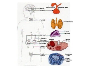
Global burden of hbv
Global burden of hbvGlobal burden of hbv
Global burden of hbv
 
Fibrosis[1]
Fibrosis[1]Fibrosis[1]
Fibrosis[1]
 
Chapter31.liver
Chapter31.liverChapter31.liver
Chapter31.liver
 
Biliary tract
Biliary tractBiliary tract
Biliary tract
 
Alcoholic liver 2005 ust
Alcoholic liver  2005  ustAlcoholic liver  2005  ust
Alcoholic liver 2005 ust
 
Acute pancreatitis
Acute pancreatitisAcute pancreatitis
Acute pancreatitis
 
Respiratory disorders in children
Respiratory disorders in childrenRespiratory disorders in children
Respiratory disorders in children
 
Dermpath surgical pathology
Dermpath   surgical pathologyDermpath   surgical pathology
Dermpath surgical pathology
 
Liver surgical pathology
Liver surgical pathologyLiver surgical pathology
Liver surgical pathology
 
Fmc postop period
Fmc postop periodFmc postop period
Fmc postop period
 
Pancreatic disorder
Pancreatic disorderPancreatic disorder
Pancreatic disorder
 
Perioperative fluid therapy
Perioperative fluid therapyPerioperative fluid therapy
Perioperative fluid therapy
 
Glaucoma dr.gosiengfiao dr.pangalanan
Glaucoma dr.gosiengfiao dr.pangalananGlaucoma dr.gosiengfiao dr.pangalanan
Glaucoma dr.gosiengfiao dr.pangalanan
 
Part 3 hodgkins lymphoma
Part 3 hodgkins  lymphomaPart 3 hodgkins  lymphoma
Part 3 hodgkins lymphoma
 
Part 2 nhl
Part 2 nhlPart 2 nhl
Part 2 nhl
 
Part i neoplastic proliferation of wbc
Part i neoplastic proliferation of wbcPart i neoplastic proliferation of wbc
Part i neoplastic proliferation of wbc
 
Surg path thyroid.special
Surg path thyroid.specialSurg path thyroid.special
Surg path thyroid.special
 
Appendix
AppendixAppendix
Appendix
 
Original tcvs lecture fatima 3rd year
Original  tcvs lecture  fatima  3rd yearOriginal  tcvs lecture  fatima  3rd year
Original tcvs lecture fatima 3rd year
 
Eye emergencies2
Eye emergencies2Eye emergencies2
Eye emergencies2
 

Endocrine disorder

  • 1.
  • 2. Endocrine System Pituitary Gland - The main endocrine gland. - Called the “master” gland. - Acts as the control center for the endocrine system which controls all the hormones produced by other glands in the body.
  • 4.
  • 5. Some of the neurons within the hypothalamus - neurosecretory neurons - secrete hormones that strictly control secretion of hormones from the anterior pituitary. The hypothalamic hormones are referred to as releasing hormones and inhibiting hormones, reflecting their influence on anterior pituitary hormones.
  • 6. Hypothalamic releasing and inhibiting hormones are carried directly to the anterior pituitary gland via hypothalamic- hypophyseal portal veins . Specific hypothalamic hormones bind to receptors on specific anterior pituitary cells, modulating the release of the hormone they produce.
  • 7.
  • 8. Anterior Pituitary Posterior pituitary Stimulates milk ejection and uterine contractions Ovary and testisOxytocin Conservation of body water KidneyAntidiuretic hormone Control of reproductive function Ovary and testisFollicle stimulating hormone Control of reproductive function Ovary and testisLuteinizing hormone Milk productionMammary glandProlactin Stimulates secretion of glucocorticoids Adrenal gland cortexAdrenocorticotrophic hormone Stimulates secretion of thyroid hormones Thyroid glandThyroid stimulating hormone Promotes growth (indirectly), control of protein, lipid and carbohydrate metabolism Liver, adipose tissueGrowth hormone Major physiologic effects Major target organsHormone
  • 9. • The portion of the adenohypophysis known as the pars tuberalis contains cords of epithelial cells and is filled with hypophyseal portal vessels. It reportedly contains gonadotropes and thyrotropes, plus other secretory cells of unknown function. • The pars intermedia is closely associated with pars nervosa and separated from the pars distalis by the hypophyseal cleft. Melanocyte-stimulating hormone is the predominant hormone secreted by the pars intermedia. The pituitary gland has two distinct parts, the anterior and the posterior lobes, each of which releases different hormones.
  • 10. • Histology of the Adenohypophysis • The bulk of the adenohypophysis is pars distalis. That tissue is composed of winding cords of epithelial cells flanked by vascular sinusoids. • In sections stained with dyes such as hematoxylin and eosin, three distinct cell types are seen among epithelial cells: • Acidophils have cytoplasm that stains red or orange • Basophils have cytoplasm that stains a bluish color • Chromophobes have cytoplasm that stains very poorly
  • 11. • Histology of the Adenohypophysis • The bulk of the adenohypophysis is pars distalis. That tissue is composed of winding cords of epithelial cells flanked by vascular sinusoids. • In sections stained with dyes such as hematoxylin and eosin, three distinct cell types are seen among epithelial cells: • Acidophils have cytoplasm that stains red or orange • Basophils have cytoplasm that stains a bluish color • Chromophobes have cytoplasm that stains very poorly
  • 12. Acidophils: contain the polypeptide hormones: – Somatotropes which produce growth hormone – Lactotropes which produce prolactin Basophils: contain the glycoprotein hormones: – Thyrotropes which produce thyroid stimulating hormone – Gonadotropes which produce luteinizing hormone or follicle-stimulating hormone – Corticotropes which produce adrenocorticotrophic hormone Chromophobes • These are cells that have minimal or no hormonal content. – may be acidophils or basophils that have degranulated and thereby are depleted of hormone – may also represent stem cells that have not yet differentiated into hormone-producing cells.
  • 13. • Although classification of cells as acidophils or basophils is useful in some situations, specific identification of anterior pituitary cells requires immunostaining for the hormone in question. • In addition to differential staining characteristics, the size of secretory granules varies among different types of cells in the anterior pituitary. Somatotropes and lactotropes tend to have the largest size granules.
  • 15. Pituitary tumor causes symptoms by any of three mechanisms: 1. By producing too much of one or more hormones. 2. By compressing the pituitary gland, and thus making it produce too little of one or more hormones. 3. By compressing the optic nerves or (less commonly) the nerves controlling eye movements, and thus causing either loss of part or all of the visual field, or double vision.
  • 16.
  • 18.
  • 19. Type of Adenoma Secretion Staining Pathology Corticotrophic adenomas Secrete adrenocotrophic hormone (ACTH) and Proopiomelanocortin (POCM) Basophilic Cushing’s disease Somatotrophic adenomas Secrete growth hormone (GH) Acidophilic Acromegaly (Gigantism) Thyrotrophic adenomas (rare) Secret thyroid stimulating hormone (TSH) Basophilic Occasionally hyperthyroidism usually does not cause symptoms Gonadotrophic adenomas Secrete luteinizing hormone (LH), follicle stimulating hormone (FSH) Basophilic Usually does not cause symptoms Prolactinomas Secret prolactin Acidophilic Galactorrhea, hypogonadism, amenorrhea, infertility and impotence Null cells adenomas Do not secrete hormones May stain positive for synaptophysin
  • 20. By producing too much of one or more hormones • Growth hormone: causes ACROMEGALY – a syndrome that includes: • excessive growth of soft tissues and bones • high blood sugar • high blood pressure • heart disease • sleep apnea • excess snoring • carpal tunnel syndrome • pain symptoms (including headache).
  • 21. By producing too much of one or more hormones • Thyroid stimulating hormone: – causes high production of thyroid hormone – Thyroid hormone: • leads to nervousness and irritability • fast heart rate and high blood pressure • heart disease • excess sweating and thin skin • and weight loss.
  • 22. By producing too much of one or more hormones • Prolactin: – causes inappropriate secretion of breast milk (even in men) – osteoporosis (bone weakening) – loss of sex drive – Infertility – irregular menstrual cycles – and impotence
  • 23. By producing too much of one or more hormones • Adrenocorticotropic hormone: – causes weight gain (particularly in the body’s trunk, not the legs or arms) – high blood pressure – high blood sugar – brittle bones – emotional changes – stretch marks on the skin – easy bruising.
  • 24. By producing too much of one or more hormones • Gonadotropins (FSH and LH): – usually not elevated enough to produce direct symptoms – but in extreme cases can cause infertility (inability to have a child) – irregular menstrual cycles in women.
  • 25. Cushing’s Syndrome • Definition – a condition that occurs when your body is exposed to high levels of the hormone cortisol for a long time. – Referred to as hypercortisolism – use of oral corticosteroid medication (most common cause) – The condition can also occur when your body makes too much cortisol.
  • 26. Hallmark signs of Cushing's syndrome: - fatty hump between shoulders - rounded face - pink or purple stretch marks on your skin - can also result in high blood pressure, bone loss and, on occasion, diabetes.
  • 27. By compressing the pituitary gland, and thus making it produce too little of one or more hormones. • Growth hormone: causes poor muscle strength, irritability, weakening of bone strength, and overall feeling of malaise (feeling unwell). DWARFISM in children. Thyroid stimulating hormone: causes fatigue, low energy, and weight gain. • Prolactin: causes inability to breastfeed after a woman gives birth to a baby. • Adrenocorticotropic hormone: causes fatigue and low energy, low blood pressure, low blood sugar, and upset stomach. • Gonadotropins (FSH and LH): cause infertility, decrease in sex drive, impotence, and irregular menstrual cycles.
  • 28. Histology of the Neurohypophysis • The neurohypophysis is known also as the pars nervosa. • Three areas of this organ, starting closest to the hypothalamus: – the median eminence – infundibular stalk – infundibular process • The infundibular - bulk of the neurohypophysis (usually referred to as the posterior pituitary) • Composed on largely unmyelinated axons from hypothalamic neurosecretory neurons. • These neurons secrete oxytocin and antidiuretic hormone.
  • 29. • The neurohypophysis shown here resembles neural tissue, with glial cells, nerve fibers, nerve endings, and intra-axonal neurosecretory granules. • The hormones vasopressin (antidiuretic hormone, or ADH) and oxytocin made in the hypothalamus (supraoptic and paraventricular nuclei) are transported into the intra-axonal neurosecretory granules where they are released.
  • 31. • Oxytocin increases uterine contractions and stimulates milk secretion. • Underproduction of ADH results in a disorder called diabetes insipidus characterized by inability to concentrate the urine. The consequence is excess urination leading potentially to dehydration. • Antidiuretic hormone (ADH) increases reabsorption of water by the tubules of the kidney.
  • 32. Syndrome of Inappropriate ADH Secretion (SIADH) = Excess resorption of water -> hyponatremia = CAUSES: Secretion of ectopic ADH (small cell ca of the lung) Injury to hypothalamus or pituitary or both.
  • 33. Parathyroid Gland They are hard to differentiate from the thyroid or fat. The parathyroid glands are four or more small glands, about the size of a grain of rice, located on the posterior surface (back side) of the thyroid gland. The parathyroid glands are named for their proximity to the thyroid but serve a completely different role than the thyroid gland. They are quite easily recognizable from the thyroid as they have densely packed cells, in contrast with the follicle structure of the thyroid.
  • 34. Thyroid Gland: colloid-filled follicles Parathyroid Gland
  • 35. The sole purpose of the parathyroid glands is to control calcium within the blood in a very tight range between 8.5 and 10.5. Parathyroid hormone (PTH) - mobilizes calcium release from bone when there is a decrease the blood level of calcium. - enhances intestinal absorption of calcium. PARATHYROID HORMONE
  • 36. Hyperparthyroidism • The most common cause of excess hormone production (hyperparathyroidism) is the development of a benign tumor in one of the parathyroid glands. • Over-production of parathyroid hormone. • Occur in 94 percent of all patients with primary hyperparathyroidism.
  • 38. Hyperprathyroidism Symptoms of Parathyroid Disease • Loss of energy. • Don't feel like doing much. Tired all the time. • Just don't feel well; don't quite feel normal. Hard to explain but just feel kind of bad. • Feel old. Don't have the interest in things that you used to. • Can't concentrate, or can't keep your concentration like in the past. • Depression. • Osteoporosis and Osteopenia. Bones hurt; typically it's bones in the legs and arms but can be most bones. • Don't sleep like you used to. Wake up in middle of night. Trouble getting to sleep. • Tired during the day and frequently feel like you want a nap. • Spouse claims you are more irritable and harder to get along with (cranky, bitchy). • Forget simple things that you used to remember very easily. • Gastric acid reflux; heartburn; GERD. • Decrease in sex drive. • Thinning hair (predominately in older females). • Kidney Stones. • High Blood Pressure (sometimes mild, sometimes quite severe; up and down a lot). • Recurrent Headaches (usually patients under the age of 40). • Heart Palpitations (arrhythmias). Typically atrial arrhythmias.
  • 39.
  • 41. • Located immediately anterior to the kidneys, encased in a connective tissue capsule and usually partially buried in an island of fat. • Retroperitoneal.
  • 42.
  • 43. Adrenal Gland Adrenal cortex - secretes several classes of steroid hormones (glucocorticoids and mineralocorticoids) - with three concentric zones of cells that differ in the major steroid hormones they secrete.
  • 44. Adrenal Gland Adrenal medulla - source of the catecholamines epinephrine and norepinephrine. - chromaffin cell is the principle cell type. - The medulla is richly innervated by preganglionic sympathetic fibers and is, in essence, an extension of the sympathetic nervous system.
  • 45. HORMONE SYNTHESIS: – Corticosteroid. – Androgen such as testoterone. – Aldosterone. – Function is regulated by the neuroendocrine hormones from the pituitary, hypothalamus and renin-angiotensin system. – Adrenal medulla is regulated by direct innervation. ADRENAL CORTEX
  • 46. The adrenal cortex comprises three zones each produces and secretes distinct hormones. • Zona glomerulosa (outer) – for production of mineralocorticoids, mainly aldosterone, which is largely responsible for the long-term regulation of blood pressure. • Zona fasciculata – responsible for producing glucocorticoids, chiefly cortisol in humans. • Zona reticularis (innermost) – produces androgens, mainly dehydroepiandrosterone (DHEA) and DHEA sulfate (DHEA-S) in humans. ADRENAL CORTEX
  • 47. Adrenal Medullary Hormones – Cells in the adrenal medulla synthesize and secrete epinephrine and norepinephrine. Adrenergic Receptors and Mechanism of Action – The physiologic effects of epinephrine and norepinephrine are initiated by their binding to adrenergic receptors on the surface of target cells.
  • 48. Receptor Effectively Binds Effect of Ligand Binding Alpha1 Epinephrine, Norepinephrine Increased free calcium Alpha2 Epinephrine, Norepinephrine Decreased cyclic AMP Beta1 Epinephrine, Norepinephrine Increased cyclic AMP Beta2 Epinephrine Increased cyclic AMP
  • 49. Adrenal cortical hyperplasia. causes: - due to a pituitary adenoma secreting ACTH (Cushing's disease) - Cushing's syndrome from ectopic ACTH production - idiopathic adrenal hyperplasia. Adrenal atrophy (with either Addison's disease or long- term corticosteroid therapy). Normal adrenal glands
  • 50. Adrenal adenoma with Cushing's syndrome - remaining atrophic adrenal is seen at the right. - composed of yellow firm tissue just like adrenal cortex. - well-circumscribed. Histologically - composed of well-differentiated cells resembling cortical fasciculata zone. It is benign.
  • 51. Hallmark signs of Cushing's syndrome: - fatty hump between shoulders - rounded face - pink or purple stretch marks on your skin - can also result in high blood pressure, bone loss and, on occasion, diabetes.
  • 52. PRIMARY HYPERALDOSTERONISM • Generic term for a closely related, uncommon syndromes characterized by chronic excess aldosterone secretion independent of the RA system. • Characterized by suppression of plasma renin activity. • Secondary increased aldostrerone due to renal ischemia (2ndary hyperladosteronism)
  • 53. Causes of Primary Hyperaldosteronism 1. Conn’s Syndrome: -solitary aldosterone-secreting adenoma 2. Bilateral Idiopathic Hyperplasia of adrenals. 3. Glucocorticoid-suppressible hyperaldosteronism 4. Adrenal Cortical Carcinoma
  • 54. Solitary aldosterone secreting adenoma - Conn’s Syndrome - patient had hypokalemia. - with high serum aldosterone and a low serum renin, - This lesion accounts for about two-thirds of cases of primary hyperaldosteronism (PHA) - Bilateral adrenal hyperplasia accounts for about 30% of PHA.
  • 55. • Microscopically, the adrenal cortical adenoma at the right resembles normal adrenal fasciculata. The capsule is at the left. There may be some cellular pleomorphism.
  • 56. • This is a large adrenal cortical carcinoma which is displacing the left kidney downward. • Such neoplasms are usually functional (secreting corticosteroids or sex steroids). • They have a poor prognosis.
  • 57. • High power microscopic appearance of an adrenal cortical carcinoma • loosely resembles normal adrenal cortex • It is difficult to determine malignancy in endocrine neoplasms based upon cytology alone. • Thus, invasion (as seen here in a vein) and metastases are the most reliable indicators. • Luckily, most endocrine neoplasms are benign adenomas. BV TC
  • 58. • Here is an adrenal cortical carcinoma seen microscopically at high power to demonstrate cellular pleomorphism with nuclear hyperchromatism. • Both benign and malignant endocrine neoplasms demonstrate some degree of cellular pleomorphism, so it is not easy to tell benign from malignant on histologic grounds alone. The larger the neoplasm, the more likely it is malignant, but the best indicators are invasion and metastasis.
  • 59. ADRENAL MEDULLA A. PHEOCHROMOCYTOMA: - associated with catecholamine-induced hypertension. - occassionally, this tumor produces other biogenic steroids or peptides asociated with Cushing’s Syndrome. -morphology: - ave . weight of 100 gms - Zellballen appearance
  • 60. • This large adrenal neoplasm has been sectioned in half. Note the grey-tan color of the tumor compared to the yellow cortex stretched around it and a small remnant of remaining adrenal at the lower right. This patient had episodic hypertension. This is a tumor arising in the adrenal medulla--a pheochromocytoma. T N
  • 61. • There is some residual adrenal cortical tissue at the lower center right, with the darker cells of pheochromocytoma seen above and to the left.
  • 62. HYPOADRENALISM • Caused by any anatomic or metabolic lesion of the adrenal cortex that impairs output of the cortical steroids. • Primary Acute Adrenal Insufficiency - Waterhouse Friderichsen Syndrome due to overwhelming septicemic infection caused by meningococci but occasionally other virulent organism such as gonococci,pneumococi and staphylococci. morphology: massive bilateral adrenal hemorrhage
  • 63. HYPOADRENALISM • Primary Chronic Adrenal Insufficiency: - Addison’s Disease - caused by any destructive process in the adrenal cortex. a. Autoimmune Adrenalitis b. Infection c. Metastatic Ca to the adrenal cortex