Cell membrane physiology and pharmacology
•Descargar como PPTX, PDF•
44 recomendaciones•11,863 vistas

The cell membrane is a thin semi-permeable membrane that surrounds the cytoplasm. It is composed of lipids, proteins, and carbohydrates arranged in a fluid mosaic structure. The membrane regulates what enters and exits the cell through diffusion, facilitated diffusion, active transport, endocytosis, and exocytosis. Membrane proteins carry out important functions like transport, signaling, enzyme activity, and attaching to cytoskeleton. Dysfunctions of ion channels and receptors in the membrane can cause various disorders. Many drugs like local anesthetics, antimicrobials, and antifungals act by disrupting the structure and function of the cell membrane.

Recomendados
Más contenido relacionado
La actualidad más candente
La actualidad más candente (20)
Destacado
Destacado (17)
Similar a Cell membrane physiology and pharmacology
Similar a Cell membrane physiology and pharmacology (20)
Más de Dr Shahid Saache
Último
Último (20)
Cell membrane physiology and pharmacology
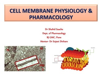
- 1. CELL MEMBRANE PHYSIOLOGY & PHARMACOLOGY Dr Shahid Saache Dept. of Pharmacology BJ GMC, Pune Mentor- Dr Sujeet Divhare
- 2. Cell Membrane The cell membrane is a thin semi-permeable membrane that surrounds the cytoplasm of a cell, enclosing its contents.
- 7. Why cell membrane is important to study??
- 9. History • 1895-Charles Ernest Overton- layers surrounding cells are ”lipoids” made of lipids and cholesterol • 1925-Gorter and Grendel proposed lipid bilayer model of cell membrane • 1935-Danielli and Davson earliest molecular model of biomembranes including proteins with lipids. • 1958-Robertsons says two protein layers are adsorbed to lipid bilayer. All membrane have same composition. • 1972- The Fluid Mosaic Model of Singer and Nicolson. • 1984-The Mattress Model by Mouritsen and Bloom.
- 10. COMPOSITION OF CELL MEMBRANE 1. Lipids • Phosopholipids • Sterols 2. Proteins • Integral • Peripheral 3. Carbohydrates • Glycolipids • Glycoproteins
- 11. Phospholipids Fatty acid Phosphate Fatty acid tails hydrophobic Phosphate group head hydrophilic Arranged as a bilayer
- 13. Lipid composition varies across the two leaflets of the same membrane Changes in distribution have biological consequences Platelet is able to play its role in clot formation only when phosphatidylserine moves to outer leaflet. Phosphatidylserine exposure also act as marker for programmed cell death
- 14. 14
- 16. • Important for exocytosis and endocytosis • For membrane biogenesis Factors altering fluidity • Temperature ↑….. Fluidity • Cholesterol content ↑….. Fluidity 16 Role of Fluidity of membrane
- 17. 17
- 18. 18 More than lipids… In 1972, S.J. Singer & G. Nicolson proposed that membrane proteins are inserted into the phospholipid bilayer It’s like a fluid… It’s like a mosaic… It’s the Fluid Mosaic Model!
- 19. Membrane is a collage of proteins & other molecules embedded in the fluid matrix of the lipid bilayer Extracellular fluid Cholesterol Cytoplasm Glycolipid Transmembrane proteins Filaments of cytoskeleton Peripheral protein Glycoprotein Phospholipids
- 20. Membrane proteins • Classified depending on type of interaction with bilayer. i. Integral membrane proteins- pass through bilayer ii. Peripheral membrane proteins- associate with bilayer by non-covalent interactions iii. Lipid-anchored proteins.
- 21. Lipid-anchored membrane proteins • Covalently linked to membrane by short oligosachharide linked to a molecule of GPI embedded on outer leaflet. • Example –Scrapie protein PrPc • Some linked to inner leaflet like Ras and Src proteins have been implicated in transformation of normal cell to malignant cell.
- 22. Lipid-anchored membrane proteins • One of protein is responsible for sleeping sickness. • Protozoan parasite carried by tsetse flies survives in blood by virtue of dense cell surface coat made of a GPI anchored glycoprotein.(Eg- Transamidase complex) • Several hundreds of glycoprotein variants to invade host immune system. Trypanosome brucie
- 23. Six major functions of membrane proteins Transport Enzymatic activity Signal transduction Cell-cell recognition Intercellular joining Attachment to the cytoskeleton ECM
- 24. • Mechanical structure – maintain the physical integrity of cell and hold the cytoskeleton in place. • Selective permeability – Gases, hydrophobic and small non polar molecules can easily pass through it. • Transport – certain molecules pass through passively other need various transporters. • Markers and signalling – some surface proteins act as cell marker and helps in cell signalling. functions of cell membrane
- 25. Movement across the Cell Membrane
- 26. Diffusion 2nd Law of Thermodynamics governs biological systems universe tends towards disorder (entropy) Diffusion movement from high low concentration
- 27. Diffusion Move from HIGH to LOW concentration “passive transport” no energy needed diffusion osmosis movement of water
- 28. Diffusion across cell membrane Cell membrane is the boundary between inside & outside… separates cell from its environment IN food carbohydrates sugars, proteins amino acids lipids salts, O2, H2O OUT waste ammonia salts CO2 H2O products IN OUT
- 29. Diffusion through phospholipid bilayer What molecules can get through directly? fats & other lipids inside cell outside cell lipid salt aa H2Osugar NH3 What molecules can NOT get through directly? polar molecules H2O ions salts, ammonia large molecules starches, proteins
- 30. Factors affecting rate of diffusion •Temperature- Higher temperature → diffuse faster •Surface area- Larger surface area → diffuse faster •Concentration gradient- Higher gradient→ diffuse faster •Size of particles- smaller particles → diffuse faster •Diffusion medium- • Solid → slowest • Liquid → faster • Gas → fastest
- 31. Channels through cell membrane Membrane becomes semi-permeable with protein channels specific channels allow specific material across cell membrane inside cell outside cell sugaraaH2O saltNH3
- 32. Facilitated Diffusion Diffusion through protein channels channels move specific molecules across cell membrane no energy needed open channel = fast transport facilitated = with help high low
- 33. Active Transport conformational change Cells may need to move molecules against concentration gradient shape change transports solute from one side of membrane to other protein “pump” “costs” energy = ATP ATP low high
- 34. symportantiport Active transport Many models & mechanisms ATP ATP
- 35. How about large molecules? Moving large molecules into & out of cell through vesicles & vacuoles endocytosis phagocytosis = “cellular eating” pinocytosis = “cellular drinking” exocytosis exocytosis
- 36. Endocytosis phagocytosis pinocytosis receptor-mediated endocytosis fuse with lysosome for digestion non-specific process triggered by molecular signal
- 37. 2007-2008 The Special Case of Water Movement of water across the cell membrane
- 38. Osmosis is diffusion of water Water is very important to life, Diffusion of water from high concentration of water to low concentration of water across a semi-permeable membrane
- 39. Concentration of water Direction of osmosis is determined by comparing total solute concentrations Hypertonic - more solute, less water Hypotonic - less solute, more water Isotonic - equal solute, equal water hypotonic hypertonic water net movement of water
- 40. freshwater balanced saltwater Managing water balance Cell survival depends on balancing water uptake & loss
- 41. RECEPTORS
- 42. Cell Surface Receptors Can be divided into several categories g b a a b N C
- 43. Ligand gated Ion channels Pentamers – 5 subunits Cell membrane
- 44. Ligand gated Ion channels are molecules that act like keys that fit certain binding pockets or locks on the receptor. Activated or turned on by Cell membrane
- 45. GABA Glycine Nicotinic acetylcholine Serotonin Glutamate Examples of these:
- 47. G protein Effector pathway Substrates Gs Adenylyl cyclase Beta-receptors, H2, D1,serotonin Gi Adenylyl cyclase Muscarinic M2 D2, alpha-2,opioid Gq Phospholipase C Alph-1, AT1, M1, M3 Go Ca++ channel K+ channel in heart, SM G-protein coupled receptors
- 48. g b a G-protein coupled receptors Cell membrane • G-protein composed of one alpha, beta, and gamma subunit • 2 primary signaling cascades: cAMP or phosphatidylinositol pathways • Pathway activated depends on alpha subunit type • (Gαs, Gαi/o, Gαq/11, Gα12/13) • GDP bound to a when g b a
- 49. g b a G-protein coupled receptors Cell membrane • When a ligand binds, the receptor changes conformation, allowing G-protein to be activated (GDP is exchanged for GTP) • G-protein dissociates from receptor then subunits from each other. GTP a GTP
- 50. g b a cAMP pathway Cell membrane GTP a GTP • Gαs binds to Adenylate Cyclase (AC) and stimulates cAMP synthesis from ATP • Gαi/o binds to AC and inhibits cAMP synthesis
- 52. g b a Phosphatidylinositol pathway GTP a GTP • Gαq/11 binds to Phospholipase C (PLC) and catalyzes the cleavage of phosphatidylinositol 4,5-biphosphate (PIP2) into the second messengers inositol (1,4,5) trisphosphate (IP3) and diacylglycerol (DAG). P P P To sarcoplasmic
- 53. EXAMPLES • Muscarinic cholinergic receptors • M1, M3, M5- Gq • M2, M4- Gi •Adrenergic receptors • α1 - Gq • α2 - Gi • β1 - Gs • β2 - Gs • β3 - Gs •Dopamine receptors • D1 - Gs • D2 - Gi
- 54. • GABA receptors – GABA- B – Gi • TSH receptors- Gs • LH receptors • ACTH receptors • Rhodopsin receptors • Oxytocin • Neuropeptides Vasopressin – V1- Vascular receptor (Vasocinstriction) – V2- Collecting duct – V3- Anterior pituitary • AT II • VIP
- 55. Receptor tyrosine kinase Tyr Tyr Tyr Tyr Tyr Tyr • Two inactive monomers contain tyrosine (Tyr) residues • Ligand binding to the monomers leads to dimer formation Tyr Tyr Tyr Tyr Tyr Tyr Cell membrane
- 56. Receptor tyrosine kinase Tyr Tyr Tyr Tyr Tyr Tyr ATP ATP ATP ATP ATP ATP P P P P P P ATP molecules donate a phosphate (P) to each of the tyrosines. Inactive relay proteins bind to the phosphorylated tyrosine residues and trigger cellular responses downstream Cell membrane
- 58. Insulin Vascular Endothelial Growth Factor (VEGF) Platelet-derived Growth Factor (PDGF) Epidermal Growth Factor (EGF) Fibroblast Growth Factor (FGF) Nerve Growth Factor (NGF) Examples of these:
- 59. JAK STAT Signaling Ligand Binding JAK Activation Cell membrane JAKJAK PP
- 60. JAK STAT Signaling Phosphorylation of Tyrosine Residues Tyr Tyr JAKJAK PP Cell membrane STAT
- 61. JAK STAT Signaling Phosphorylation of STAT Tyr Tyr JAKJAK PP Cell membrane STATSTAT PP
- 62. STAT Cytokine Responsive Gene Gene Transcription Nucleu s STAT P P JAK STAT Signaling STAT Translocation to Nucleus
- 63. Growth Hormone Prolactin IL-2 Cytokines T cell receptors B cell receptors Examples of these:
- 65. Serine Threonine kinase pathway/ B Raf
- 66. Examples: •TGF-Beta •BMP B-Raf Inhibitors •Sorafenib- Approved for Liver and Kidney cancers •vemurafenib and dabrafenib are approved by FDA for treatment of late-stage melanoma. •Trametinib- FDA-approved to treat BRAF-mutated melanoma.
- 67. ANP receptors
- 68. Integrins a b • Join the cytoskeleton on the inside of the cell to the extracellular matrix on the outside. • Heterodimers of alpha and beta subunits Cell membrane
- 69. Toll-like receptors nucleus Cell membrane DNA • Involved in the immune response • Signals between downstream proteins result in enhanced transcription of inflammatory Immune response
- 70. Disorders Some disease resulting from or attributed to abnormalities of Membranes
- 71. Disorder due to defect in ion channels
- 72. Some drugs acting on cell membrane
- 76. Polymyxin B & Colistin • They are active against gram-negative bacteria only. • Colistin is more potent on Pseudomonas, Salmonella and Shigella. • Rapidly acting bactericidal agents • They have high affinity for phospholipids: the peptide molecules (or their aggregates) orient between the phospholipid and protein films in gram-negative bacterial cell membrane causing membrane distortion or pseudopore formation →ions, amino acids, etc. leak out.
- 77. • Given orally, side effects are limited to the g.i.t. • Systemic toxicity of these drugs (when injected) is high: flushing and paresthesias (due to liberation of histamine from mast cells), marked kidney damage, neurological disturbances, neuromuscular blockade. • Uses: skin infections, burns, otitis externa, conjunctivitis, corneal ulcer—caused by gram- negative bacteria including Pseudomonas. • Gram-negative bacillary (E. coli, Salmonella, Shigella) diarrhoeas, especially in infants and children
- 78. Daptomycin • Lipopeptide antibiotic used in the treatment of systemic and life-threatening infections caused by Gram-positive organisms • Inserts into the cell membrane →aggregates → alters the curvature of the membrane → creates holes that leak ions → rapid depolarization → loss of membrane potential → inhibition of protein, DNA, and RNA synthesis → bacterial cell death. • Use: Skin and skin structure infections, MRSA • Dose: 4 mg/kg IV
- 79. Cell membrane active Antifungals
Notas del editor
- -Cell membrane or plasma membrane or plasmalemma or cytoplasmic membrane 10nm thick
- Plants have cell walls made up of cellulose which function in the support and protection of the cell. Animals have only cell membranes made up of phospholipid bilayer and protein which protects and hold together the cell and its parts, it does not need to provide the support that plant cell was provide because animals have other forms of support ( i.e. exoskeletons and endoskeletons) and the lack of the cell wall allows for increased flexibility and advanced cell/tissue specialization, which plants cannot achieve. Bacterial cell walls are made up of peptidoglycan and provide support for the cells much like plant cell walls.
- Fungal cell membrane
- Memb lipids are amphipathic
- Cell membrane is Asymmetric because- Phospholipids in outer and inner leaflets are different Proteins are also slightly different in outer & inner leaflet Carbohydrate mainly on outer leaflets
- Cholesterol molecules- Mobile Polar end towards cell membrane Speed of Lateral movement 107 /second Flip flop – once in a month
- There is no hole in the cell membrane
- GPI- Glycosylphosphatidylinositol Anchor Src family kinase is a family of non-receptor tyrosine kinases
- cell needs materials in & products or waste out
- Stereospecific
- e.g transport of glucose by insulin by glucose transport
- Hormone release from glands, neurotransmitters release
- Ligands- substances which binds to receptors.. Hormone, NT, Drugs, toxins Macromolecule to which a particular ligand binds and receptor undergo some transformational changes show biological actions.
- Epinephrine- 1st messenger C AMP 2nd messenger PK A- phosphorylation of enzymes, transcription factors, ion channels Gi binds to K Channels Clinical significance: Pertusis, Cholera
- PDE inhibitors and caffeine
- Antagonist: Atropine, scopolamine, pirenzepine Non selective alpha blocker- phenoxybenzamine, phentolamine Aplfa1 selective- prazosin, terazosin, doxazosin Alfa2 selective- Yohimbine D Receptors- tell about Parkinsons disease
- GABA B Agonist- Baclofen GABA B antagonist – Sclofen TSH mutant rece- follicular adenoma LH R dysfunction- precocious puberty
- We will see signaling in one pass receptors
- The first drug licensed to act on this pathway is sorafenib — a Raf kinase inhibitor.
- Grb2- Growth factor receptor-bound protein2 SOS- Sons of seven protein Ras- H-ras and K-ras Harvey and Kirsten sarcoma virus first identified in rats
- STAT= Signal transducer and activator of transcription
- TGF-Beta BMP
- Ligands in ECM – collagen, fibronectin, and laminin Important for cellular processes including: cell adhesion, cell migration, signal transduction, and cell growth/death
- Ligands include: Lipopolysaccharide, double-stranded RNA, flagellin, heat shock proteins Important for the innate immune response to bacterial and viral stimuli.
- Prostaglandins (PGs) and Leukotrienes (LTs) and PAF are biologically active derivatives of 20 carbon atom polyunsaturated essential fatty acids that are released from cell membrane phospholipids. They are the major lipid derived autacoids.
- The dibucaine number (DN) is the percent of pseudocholinesterase (PChE) enzyme activity that is inhibited by dibucaine. Together, the DN and the PChE enzyme activity results can help to identify individuals at risk for prolonged paralysis following the administration of succinylcholine.
- They are useful for tachycardias and re entrants arrhythmias
- They exhibit synergism with many other AMAs by improving their penetration into the bacterial cell.
- -occasional nausea, vomiting, diarrhoea. -Pseudomonas superinfection enteritis.
- Azole- inhibit cyt p450 lanosterole 14 demethylase Allyl amine- Terbinafine- inhibit squalene epoxide