
Recomendados
Más contenido relacionado
La actualidad más candente
La actualidad más candente (20)
Similar a Rh incompatibility
Similar a Rh incompatibility (20)
Último
Último (20)
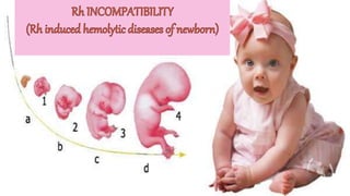
Rh incompatibility
- 2. INTRODUCTION Hemolytic diseases of the fetus and newborn occurs as a result of paternally derved RBC antigens presenting on fetal redcells which is absent in the mother.Rhesus iso immunisation seen in Rh negative pregnant mothers remains the most common cause of haemolytic diseases of the fetus and newborn. When an Rh negative mother causes an Rh positive fetus, transplacental haemorrhage from the fetus to the mother. Fetal RBC enter the mothers blood stream stay in the maternal circulation for their normal lifespan, facilitating stimulation of maternal immune response and production of antibodies , the fetus if Rh positive is affected resulting erythroblastosis fetalis.
- 4. CAUSES Factors that influence an Rh negative pregnant female chance of developing Rh incompatibility include the following Ectopic pregnancy Placenta Previa Placental abruption Abdominal trauma In uterofetal death Spontaneous abortion It is a condition that develops when a pregnant women has Rh negative blood and the baby in her womb has Rh positive blood. DEFINITION
- 9. ETIO PATHOLOGY Iso immunization can occur either following in compatible blood transfusion.The normal serum in the human body contains small amount of natural antibodies of IgM,IgG,Ig A isotypes that can react with foreign or self antigens and these complexes may have the potential to elicit an immune response. The immune complexes between antigens and antibodies may form at the onset and during the course of an immune response. The initial response in case of a women exposed to the RhD antigen is week.It is primarily composed of IgM , a large molecule that cannot cross the placenta.
- 10. However this immune response changes to the production of IgG within a period of 6 weeks to 6 months, which can cross the placenta resulting in various complications in the fetus and the mother.Rh iso immunization is generally observed n case of 2nd pregnancies . As the maternal antibodies cross the placenta and destroy the fetal Rh positive red cell, haemolytic anemia ensures along with increased levels of bilirubin. Severe anemia may be followed by erythroblastosis fetalis that is characterized by extramedullary hematopoiesis,Heart failure,edema and Percardial effusion.
- 12. PATHOGENESIS Hematopoisis in the fetus begin as early as 8th week of gestation up to 40% of pregnancy.These cells pass through the placenta into maternal circulation. If the fetus is Rh positive and mother Rh negative, the mother forms antibodies against fetal blood cells. First IgM antibodies that are too large to pass through the placenta and later IgG antibodies that can cross the placenta. This process of antibody formation is called Maternal sensitization. During subsequent pregnancies antibodies form in response to repeated contact with antigen from the fetal blood resulting in lysis. Severe Rh incompatibility results in marked fetal haemolytic anemia because the fetal erythrocytes are destroyed by maternal Rh positive antibodies.
- 13. Placenta usually clear the bilirubin generated by RBC breakdown.In extreme cases fetal bilirubin level increases.This results in fetal jaundice known as Icterus gravis. The fetus compensates for the anemia by producing large number of immature erythrocytes to replace those hremolyzed called as Erythroblastosis fetalis. Severe form ---Hydrops fetalis. The fetus has marked anemia cardiomegaly, hepatosplenomegaly, cardiac decompression. Hypoxia results from the severe anemia because decreased intravascular oncotic pressure involved.
- 14. CLINICAL MANIFESTATION OF HEMOLYTIC DISEASES OF NEWBORN ICTERUS GRAVIS NEONATORUM HYDROPS FETALIS CONGENITAL ANAEMIA OF NEWBORN
- 15. ICTERUS GRAVIS NEONATORUM The baby is born without evidences of jaundice but soon develops within 24 hours. While fetus is in utero, there is destruction of fetal red cells with liberation of un conjucated bilirubin which is mostly excreted the placenta in to the maternal system. As the umbilical cord is clamped with continuing haemolysis, the bilirubin concentration is increased. Sooner or later baby develops jaundice. If the bilirubin rises to the critical level of 20 mg per 100 ml (340micromol/l) {normal—30 micromol/l} The bilirubin crosses the blood brain barrier to damage the basal nuclei of the brain permanently producing the clinical manifestation of kernicterus
- 17. CONGENITAL ANEMIA OF THE NEWBORN The destruction of the red cells continues up to 6 weeks after which the antibodies are not available for Haemolysis. The liver and spleen are enlarged. CAUSES: Rh incompatibility Risk factors: Pregnant woman with Rh negative blood who had a prior pregnancy with fetus that was Rh positive. Pregnant woman who had a prior blood transfusion. Pregnant woman with Rh negative blood who did not receive Rh immunization prophylaxis during a prior pregnancy with Rh positive fetus.
- 18. SYMPTOMS • High bilirubin level(greater than 18mg/cc) • Extreme jaundice, absent startle reflex,Poor sucking reflex, Lethargy EARLY • High pitch cry, Arched back with neck Hyperextended backwards • Bulging fontanel, Seizure INTER MEDIATE • High pitched hearing loss, intellectual disability, muscle rigidity, speech difficulties, seizures, movement disorders LATE
- 19. DIAGNOSIS: Blood test for Rh factor TREATMENT: IMMUNE GLOBULIN INJECTION: Give an injection of Rho immune globulin week 28th of the pregnancy. This desensitizes mothers blood to Rh positive blood. Injection of immune globulin within 72 hours after delivery .This is help ful for future pregnancy.
- 22. HYDROPS FETALIS Excessive destruction of the fetal red cells lead to severe anemia , tissue anoxaemia and metabolic acidosis. Hyperplasia of the placental tissue occurs in an effort to increase the transfer of oxygen but the available fetal red cells are progressively diminished due to hemolysis. As a result of fetal anoxaemia, there is damage to the liver leading to hupoproteinaemia which responsible for generalised edema, ascities and hydrothorax. Fetal death occurs sooner due to cardiac failure. The baby is either still born or macerated and even if born alive, dies soon after.
- 24. TREATMENT Amniocentesis to determine severity Intrauterine fetal transfusion Early induction of labour A direct transfusion of packed red cells and also exchange transfusion of the newborn. This is done to rid the infants blood of the maternal antibodies that are destroying the red blood cells. Control the congestive failure and fluid retention.
- 25. FACTORS INFLUENCING Rh IMMUNIZATION DURATION OF PREGNANCY AMOUNT OF TRANSPLACENTAL HEMORRHAGE ABO INCOMPATIBILITY ANTI-D PROPHYLAXIS POST PARTUM PROPHYLAXIS
- 26. DURATION OF PREGNANCY Entry of fetal cell (trans placental hemorrhage) in to the maternal circulation mostly occurs at the time of expulsion of the fetus. It can occur during pregnancy as well. Both the frequency and magnitude of bleeding increases as pregnancy advances. Even spontaneous abortion is associated with transplacental haemorrhage , but the incidence is less than 1% Abortion in first trimester—3% Abortion in second trimester--- 12% Maximum risk—46% in the time of delivery. As fetal Rh antigen can be detected early, when the fetus is only 38 days old, sensitisation can occur as early as 7 week of gestation.
- 27. AMOUNT OF TRANS PLACENTAL HEMORRHAGE The volume of fetal blood cells entering the maternal circulation is not uniform in all pregnancies. It may vary from 0.1 ml to more on average the blood may not be more than 15 ml in term pregnancy. The risk of immunisation is 3% with 0.1ml of fetal red cells, 25% with 0.25-1 ml and increases to 65% with more than 5 ml.
- 29. IDENTIFICATION OF Rh INCOMPATIBILITY If the pregnant mother is Rh negative, then the husband’s blood group is checked. In case husband Rh negative no further investigation is required and pregnancy is managed as a normal does not need any additional care. If the pregnant mother’s blood group is Rh negative, the husband is Rh positive then at the first visit, Indirect coomb’s test is done to detect maternal serum anti –D antibodies which unbound, If antibodies are not detected, the test is repeated in the second and third trimester. Anti partum prophylaxis with a dose of 300micro gram of anti-D has been recommended.
- 30. DOPPLER STUDIES DOPPLER USG of the middle cerebral artery of the fetus to determine the Middle Cerebral Artery Peak Systolic Velocity (MCA-PSV )is being studied fetal anemia. The wave forms of the MCA are observed using colour Doppler and highest point of the Doppler waveform is measured as MCA-PSV. It has been propose that absolute MCA-PSV may be helpful in the management od red cell iso immunization and rate of change in MCA-PSV over time help predict fetal anemia. It is advised periodically for evaluation of fetal heart size, edema, pericardial effusion, ascites and AFI ULTRA SONOGRAPHY
- 31. MANAGEMENT OF UNSENSITIZED PREGNANCY-STANDARD PROTOCOL Repeat Rh antibody titre. USG examination of fetus. IM Rh immunoglobulin 300 micro gram. Analysis of the husband’s blood type. To prevent this from occurring , an injection of Rh –D immunoglobulin is offered to all unsensitised Rh negative women during the 28th week if a repeat Rh antibody titre is still negative. No need to perform an USG examination of the fetus. The antibody screening must be repeated at 35th week gestation and if negative , the women kept on observation .If positive, she should be managed on the lines of a sensitised patient.
- 32. VARIOUS DIAGNOSTIC MODALITIES Rh ANTIBODY ASSAY USG EVALUATION OF THE FETUS MIDDLE CEREBRAL ARTERY DOPPLER AMNIOCENTESIS AND EXAMINATION OF AMNIOTIC FLUID
- 33. Rh ANTIBODY ASSAY Maternal titre Human antiglobulin titre(Indirect coomb’s test)-to determine the degree of allo immunisation. Critical titre-the titre associated with high risk for fetal Hydrops. If the coomb’s test is positive in dilution of further evaluation of the fetus is required1:16. If the titre is less than it is repeated every trimester. Titre are repeated every 2 weeks thereafter to determine whether there is trend.
- 34. METHOD OF TERMINATION Indication: Previous H/O still birth with father being homozygous. Sudden rise in maternal antibody titre. Sonographic features of fetal affection. Mild affection-Pregnancy may be continue up to 38 weeks. Severe affection-terminate the pregnancy before 34 weeks.
- 35. METHOD OF TERMINATION AMNIOTOMY(LOW RUPTURE OF THE MEMBRANE) Termination is done near term –vaginal PG E2 to make cervix ripe. Oxytocin drip may be added. If it is fail-caesarean section is preferred. CAESAREAN SECTION: When termination has to be done prematurely (34-37 weeks) if it is unfavourable. CARE DURING VAGINAL DELIVERY: Careful fetal monitoring. Prophylactic ergometrine during second stage. Gentle handling of the uterus in third stage To take care of PPH.
- 36. INTRA UTERINE FETAL TRANSFUSION INTRA PERIOTONEAL TRANSFUSION INTRA VASCULAR TRANSFUSION
- 37. INTRA PERITONEAL TRANSFUSION Blood is transfused to fetal peritoneal cavity under USG guidance. Correct the fetal anemia when transfuse the erythrocytes are taken up by sub- diaphragmatic lymphatics. It can be started at 18 weeks and repeated at intervals of 1-3 weeks to 32-34 weeks. Type and amount of blood –blood group “o”. Rh negative packed cell cross matched with the mother are to be transfused. The quantity of blood is to be calculate as number of weeks of gestation. The blood is to be infused slowly(5-10ml) through a polythene tube that has been threaded through an introducing needle inserted in to the fetal abdomen under guidance.
- 38. INTRA VASCULAR TRANSFUSION Transfusion is made through umbilical cord vessel near its insertion in to the placenta under guidance of USG. Blood group “o” Rh negative packed cells compatible with mothers blood are transfused. Check the Hematocrit value at intervals during the procedure to determine the volume. Goal: Achieve haematocrit of 50% ;Repeat transfusion is given 2 weeks. Volume overload –fetal injury, preterm labour, fetal maternal hemorrhage,. Fetal surveillance with USG and continuous electronic fetal monitoring. Betamethasone (24 mg in divided dose) should be administered to the mother 24 hours before transfusion from 26 weeks onwards to enhance pulmonary maturity.
- 39. PREVENTION Anti-D immunoglobulin 300 micro gram of D Antibody is given to D-negative , non sensitized mothers to prevent the hazards f sensitization. Anti-D globulin is provided to D-negative mothers after miscarriage , evacuation of molar pregnancy or ectopic pregnancy. It is also administered when there is heavy feto maternal bleeding such as placental abruption, intrauterine manipulation. Dose of 300 micro gram D-antibody will neutralize about 15ml of red cells. Rosette test is done to all such mothers to know the accurate amount of Anti-D required.