
Recomendados
Más contenido relacionado
La actualidad más candente
Destacado
Destacado (13)
Similar a Chapter 6
Similar a Chapter 6 (20)
Chapter 6
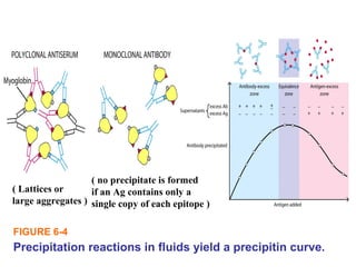
- 1. Precipitation reactions in fluids yield a precipitin curve. FIGURE 6-4 ( Lattices or large aggregates ) ( no precipitate is formed if an Ag contains only a single copy of each epitope )
- 2. FIGURE 6-5 Diagrammatic representation of radial & double immunodiffusion. : precipitation reactions in gels yield visible precipitin lines; no visible precipitate forms in regions of Ab or Ag excess. in the Ab-containing semisolid medium The region of equivalence -> The area is proportional to the conc. of Ag.
- 3. FIGURE 6-6 (a) Immunoelectrophoresis. - an antigen mixture is first electrophoresed to separate its components by charge - diffusion & producing lines of precipitation.
- 4. FIGURE 6-7 Demonstration of humaglutination using Ab against sheep red blood cells (SRBCs): a constant # of SRBCs plus serial two-fold dilutions of anti-SRBC serum + + + (control) -visible clumping by interaction between Ab & a particulate antigen suchas RBC, latex beads. -depend on the crosslinking of polyvalent antigens, similar to precipitation rxns (lgM is a good agglutinin) -provide a way to type bacteria with a panel of typing antisera. -routinely performed to type RBCs for blood transfusion.
- 5. FIGURE 6-8 -The original home pregnancy test kit employed hapten inhibition (agglutination inhibition) to determine the presence or absence of human chorionic gonadotropin (HCG) >>> The kits currently on the market use ELISA-based assays. -Also used to determine the use of illegal drugs, & immunity (Ab) to virus (rubella). 6-
- 6. Sensitivity of various immunoassays
- 7. FIGURE 6-9 A solid-phase radioimmunoassay (RIA) to detect hepatitis B virus in blood samples & A standard curve to determine the conc. of HBsAg in unknown serum. Radioimmuno AssayRadioimmuno Assay - One of the most sensitive technique for measuring hormones, drugs, & vitamins at conc. Of<0.001 ㎍ / ㎖ first discovered by Dr. Berson & Yalow in 1960 (1977 Novel prize to Yalow) - The principle involves competitive binding of radiolabeled Ag and unlabeled Ag to the limited supply of a high affinity Ab.
- 8. FIGURE 6-10 Variations in the enzyme-linked immunosorbent assay (ELISA) technique, similar to RIA except using an Enzyme (alkaline , horseradish peroxidase, & β-galactosidase)ⓟ : safer & less costly. to detect Ab (HIV, HCV) to detect Ag to detect Ag ELISAELISA
- 9. FIGURE 6-11 The ELISPOT assay, a modification of the ELISA assay to determine qucontitatively the # of cells in a population that are producing specific Ab or cytokine. ELISAELISA -> precipitates & forms a spot only on the areas of the well where cytokine-secreting cells had been deposited.
- 10. FIGURE 6-12 Western blotting : separates the components according to their molecular weight. : the proteins in the gel are transferred to the sheet of nitrocellulose or nylon by the passage of an electric current. : probed with Ab & then radiolabeled or enzyme-linked 2nd Ab. : a position is visualized by means of an ELISA reaction.
- 11. FIGURE 6-13 Immunoprecipitates can be collected using magnetic beads coupled to a secondary antibody. Immunoprecipitation - Ag-Ab attached to a synthetic bead complex >>> 0 - labeling Ag with radiolabeled leucine, cysteine, or methionine → A radiolabeled Ag-Ab complex 0 SDS→ → •PAGE autoradiography→ - Ag-Ab complex + 2nd Ab attatched to a synthetic bead or magnetic beads >>> 0 or magnet EM showing a cell with magnetic beads attached to its surface via antibodies.
- 12. Immunofluorescence mIgM-producing B cells indirectly stained with rhodamine-conjurated secondary Ab under a fluorescence microscope. FIGURE 6-14 Fluorochromes -Fluorescein (490 517nm)→ -Rhodamine (515 546nm)→ -Phycoerythrin : absorb light of one wavelength & emit fluorescence at a longer wavelength than fluorescein.
- 13. FIGURE 6-15 Separation of fluorochrome-labeled cells with the flow cytometer which uses a laser beam & light detector. : different Ag in different cells / different levels of Ag in the same type of cell → fluorescence intensity / the size of cells. labeled with fluorescein (green) labeled with rhodamine (red) each dot represents a cell (small electrical change) (exciting the fluorochrome) ↓ each droplet (cell) emits the fluorescence Flow cytometry & Fluorescence
- 14. Alternalives to Ag-Ab Reactions Ag-Ab-Ab* Ag-IgG-A/G* or Ag-Ab-biotin-(a)vidin*→ ① Protein A (from staphylococcus) & protein G (from streptococcus) - bind to rhe Fc region of lgG molecules (ka ~ 108 ) - used to detect lgG molecules in the Ag-Ab complexes - used to isolate lgG molecules in the affinity columns ② Avidin (from egg whites) & streptavidin (from streptomyces avidinii) conjugated with an enzyme, fluorechrome, radioactive label) - bind to biotin (a vitamin) with higher affinity (ka ~ 1015 ) - Ab can be labeled with (ka ~ 1018 )
- 15. FIGURE 6-16 An immunoelectronmicrograph of the surface of a B-cell lymphoma was stained with two antibodies (Ab against class II MHC labeled sith 30nm gold particles, & another Ab against class I MHC w/ 15nm gold particles. (The density of class I exceeds that of class II) - Electron-dense label (ferritin or colloidal gold) is conjugated to the Fc portion. Immuno EM. electron-denselabels Absorbs electrons.
- 16. CLINICAL FOCUS Distribution of selected markers on some leukemic cell types (leukemia can wise at any maturational stage of any one of the hematopoietic lineages) → Immuno phenotyping: the determination of the profile of selected cell- surface markers displayed by the leukemic cell, using “flow cytometry & mAb” [:acute lymphocytic leukemia] [:chronic lymphocytic leukemia]