
Recomendados
Más contenido relacionado
Último
Último (6)
Destacado
Destacado (20)
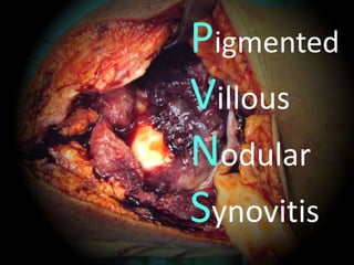
Pigment villos nodular Synovitis PVNS
- 2. • 基本資料:32 歲 男性,勞力工作 • 兩個月前,左腳 腫痛,走路走不 遠,後來疼痛加 劇 • 一個月前,左後 膝窩處出現一個 軟軟的腫瘤
- 3. • 腫瘤變的更大 病患至醫院就診 • Doctor:為一個 貝克式囊腫 • 建議手術! • 病人接受手術 …………但是 B
- 4. • 切除術開了 七個小時 • 醫生後來說不是 囊腫… • 那切下來的東西 是什麼?? 病人:醫生說再看看 ?
- 5. 膝蓋週圍常見的囊腫
- 6. Synovial cysts • herniation of synovial tissue into the surrounding soft tissue • 有時連接的通道 消失了和其他的 Ganglion/Bursa 不易區分 S
- 7. Popliteal (Baker‘s) cyst • 膝蓋最常見的腫瘤 • Communication posterior joint capsule /gastrocnemius- semimembranosus recess. • "ball-valve” • knee flexion/open extension/close S
- 9. 有時會有骨頭 漂至囊腫內
- 10. • 最近……膝後的腫 瘤更大,整個膝 蓋腫無法彎曲 • PE: ROM 0~30 Severe swelling and tenderness , soft tissue mass over operation scar . • PVNSMRI ??
- 13. MRI 報 告:PVNS Excision biopsy Posterior 順原傷口 Anterior ` Arthroscope ? Open arthrotomyOpen arthrotomy
- 17. • 病理報告: • Anterior: PMN, lymphoid follicles, Hemosiderin PVNS • Posterior: Bursitis with inflammation
- 18. PVNS Recurrent hemoarthrosis Benign disease with malignant behavior Easily local recurrence • High recurrence rate • Long recurrence period
- 19. • 正常關節/肌腱的滑 膜會不斷的增生 • 關節腔會發炎,增厚 • 組織內含Hemosiderin 外觀看起來暗紅色 • 形成絨毛(Villous) 或結節狀(Nodular) Pigmented Villous Nodular Synovitis
- 22. • Etiology --- unknown • Location – Monarticular: Knee (75%) > Hip > Wrist > Ankle > Shoulder – Polyarticular 少見 • 臨床症狀 :痛,活動受限,腫脹
- 23. • Diffuse type Large joint Localized type Small joint • 診斷 關節抽吸:Sero-sanguineou “Without” trauma history • MRI • T2 image: hemosiderin depositionLow signal
- 24. • 好發於20~50歲年輕人 • Non-surgical Treatment – Intra-articular injection • Steroid • Yttrium-90 silicate (RA, knee) – Radiation therapy • Post-op. (35 Gy / 15 fractions) • 術後行放療證實可以減少 復發
Notas del editor
- 這種複雜的表格千萬不要去讀他,萃取重要的意義放旁邊即可。 Calibri 的數字、標點跟英文,整整齊齊,在科學界比較適合。表達一種嚴謹跟仔細的感覺。 Calibri 的圓邊,又帶一點圓滑與謙虛的意涵。 如果您想給人更精準、更 sharp 的感覺,可以考慮 Verdana。
- 這種複雜的表格千萬不要去讀他,萃取重要的意義放旁邊即可。 Calibri 的數字、標點跟英文,整整齊齊,在科學界比較適合。表達一種嚴謹跟仔細的感覺。 Calibri 的圓邊,又帶一點圓滑與謙虛的意涵。 如果您想給人更精準、更 sharp 的感覺,可以考慮 Verdana。
- Popliteal cyst. (A) radiograph demonstrates coarse calcifications (arrow) in the popliteal fossa. (B) USG image at the level of popliteal fossa demonstrates a cystic lesion containing echogenic calcifications (arrow) with posterior acoustic shadowing. (C) Axial T2W image through right knee demonstrates the hyperintense popliteal cyst fluid arising between semimembranosus (arrowhead) tendon and medial head of gastrocnemius (asterisk), with hypointense loose bodies (arrow) layering dependently
- 如果你真的要打字的話,請打關鍵字就好,不要把整段文章 copy 上去。 強調請用黃色,單字大寫、放大、粗體都很好用,不要用紅色。盡量行數控制在六行以內,越少越好。
- 另一個選擇是淡藍色,一樣,大寫、粗體、加大,都是好用的強調方法。 不要用深藍,很難閱讀與辨識。
- 這種複雜的表格千萬不要去讀他,萃取重要的意義放旁邊即可。 Calibri 的數字、標點跟英文,整整齊齊,在科學界比較適合。表達一種嚴謹跟仔細的感覺。 Calibri 的圓邊,又帶一點圓滑與謙虛的意涵。 如果您想給人更精準、更 sharp 的感覺,可以考慮 Verdana。