
Recomendados
Más contenido relacionado
Similar a Apoptosis and CANCER biology 2020.pdf
Similar a Apoptosis and CANCER biology 2020.pdf (20)
Último
Último (20)
Apoptosis and CANCER biology 2020.pdf
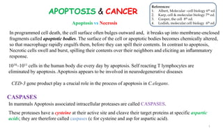
- 1. APOPTOSIS & CANCER 1010–1011 cells in the human body die every day by apoptosis. Self reacting T lymphocytes are eliminated by apoptosis. Apoptosis appears to be involved in neurodegenerative diseases CED-3 gene product play a crucial role in the process of apoptosis in C.elegans. In mammals Apoptosis associated intracellular proteases are called CASPASES. These proteases have a cysteine at their active site and cleave their target proteins at specific aspartic acids; they are therefore called caspases (c for cysteine and asp for aspartic acid). In programmed cell death, the cell surface often bulges outward and, it breaks up into membrane-enclosed fragments called apoptotic bodies. The surface of the cell or apoptotic bodies becomes chemically altered, so that macrophage rapidly engulfs them, before they can spill their contents. In contrast to apoptosis, Necrotic cells swell and burst, spilling their contents over their neighbors and eliciting an inflammatory response. Apoptosis vs Necrosis CASPASES 1 References: 1. Albert, Molecular –cell biology 6th ed. 2. Karp, cell & molecular biology 7th ed. 3. Cooper, the cell 8th ed. 4. Lodish, molecular cell biology 6th ed.
- 2. Target Proteins of Caspases: FAK, PKB, PKC, and Raf1. Inactivation of FAK, for example, is presumed to disrupt cell adhesion. Lamins - Cleavage leads to the disassembly of the nuclear lamina and shrinkage of the nucleus. Proteins of the cytoskeleton- Cleavage of these proteins lead to changes in cell shape. Caspase activated DNase (CAD), an endonuclease, attacks DNA, severing it into fragments. During apoptosis, a phospholipid “scramblase” moves phosphatidylserine molecules to the outer leaflet of the plasma membrane where they are recognized as an “eat me” signal by specialized macrophages. This entire apoptotic program can be executed in less than an hour. The apoptotic bodies are recognized by the presence of phosphatidylserine on their surface. Phosphatidylserine is a phospholipid that is normally present only on the inner leaflet of the plasma membrane. 2
- 3. Caspase activation during Apoptosis Caspases are synthesized in the cell as inactive precursors and are activated only during apoptosis. There are two major classes of apoptotic caspases: initiator caspases and executioner caspases. The major function of the initiator caspases is to activate the executioner caspases. Once activated, executioner caspases catalyze the widespread protein cleavage events that kill the cell. Executioner caspases are initially formed as inactive dimers. Presence of initiator caspase, the executioner caspase dimer undergoes an activating conformational change. The executioner caspases then cleave a variety of key proteins, leading to the controlled death of the cell. An initiator caspase contains a protease domain in its C-terminal region and a small protein interaction domain near its N- terminus. It is initially made in an inactive, monomeric form, sometimes called procaspase. Apoptotic signals trigger the binding to the adaptor proteins, initiator caspases dimerize and activated, leading to cleavage of a specifc site in their protease domains. 3
- 4. The Extrinsic Pathway of Apoptosis How is the initiator caspase frist activated in response to an apoptotic signal? The two best-understood activation mechanisms in mammalian cells are called the extrinsic pathway and the intrinsic, or mitochondrial, pathway . Extracellular signal proteins binding to cell-surface death receptors trigger the extrinsic pathway of apoptosis. Death receptors contain an extracellular ligand-binding domain, a single transmembrane domain, and an intracellular death domain.The receptors are homotrimers and belong to the tumor necrosis factor (TNF) receptor family, which includes a receptor for TNF itself and the Fas death receptor. When TNF binds to a TNF receptor (TNFR1), the activated receptor binds two different cytoplasmic adaptor proteins (TRADD and FADD-Fas associated death domain) and procaspase-8 to form a multiprotein complex at the inner surface of the plasma membrane . 4
- 5. The Intrinsic Pathway of Apoptosis Activation of the intrinsic pathway is regulated by members of the Bcl-2 family of proteins, which are characterized by the presence of one or more BH domains. Bcl-2 family members subdivided into 3 groups: (1) Proapoptotic members ... promote apoptosis (e.g., Bax and Bak), (2) Anti-apoptotic members (e.g., Bcl-xL, Bcl-w, and Bcl-2) (3) BH3-only proteins.... promote apoptosis (e.g., Bid, Bad, Puma and Bim) (So-named because they share only one small domain—the BH3 domain—with other Bcl-2 family members) Internal stimuli, such as irreparable genetic damage, lack of oxygen (hypoxia), extremely high concentrations of cytosolic Ca2+, viral infection, or severe oxidative stress trigger apoptosis by the intrinsic pathway . 5
- 6. The intrinsic (mitochondria-mediated) pathway of apoptosis. In mammalian cells, internal cell death signals induce apoptosis as a result of damage to mitochondria. When active, the proapoptotic effector proteins Bax and Bak form oligomers in the outer membrane of mitochondria, resulting in the release of cytochrome c from the intermembrane space. Release of cytochrome c leads to the formation of apoptosomes containing Apaf-1 and caspase-9 in which caspase-9 is activated initiator. Caspase-9 then activates downstream caspases, such as caspase-3, by proteolytic cleavage. Under normal conditions of cell survival, cytochrome c is localized to the mitochondrial intermembrane space while Apaf-1 and caspase-9 are found in the cytosol, so caspase-9 remains inactive. 6
- 7. CANCER THE CAUSES OF CANCER DNA tumor viruses and RNA tumor viruses: DNA viruses are... polyoma virus, simian virus 40 (SV40), adenovirus, and herpes-like viruses. Human papilloma virus (HPV) causes cervical cancer.An effective vaccine against this virus is now available. Epstein Barr virus– casuses Burkitt’s lymphoma in areas where malaria is common. Herpes virus (HHV-8), Kaposi’s sarcoma. Helicobacter pylori is responsible for ulcers and gastric lymphomas 7
- 8. THE GENETICS OF CANCER The most common solid tumors—such as those of the breast, colon, prostate, and lung—arise in epithelial tissue. Leukaemia develops in rapidly dividing blood-forming tissues Expression of telomerase have a tremendous growth advantage of cancer cells. The Pap smear is a test for detecting precancerous cells in the epithelial lining of the cervix. 8
- 9. Tumor-Suppressor Genes and Oncogenes The oncogene is not a viral gene, but a cellular gene that had become incorporated into the viral genome during a previous infection (e.g. Src). Normal cells possess a variety of proto-oncogenes, functions in a cell’s normal activities Proto-oncogene to Oncogene Conversion 1. By gene mutation 2. By gene Duplication one or more times 3. By Chromosome rearrangement Genes class, in which a gain-of-function mutation can drive a cell toward cancer, are called protooncogenes; their mutant, overactive or overexpressed forms are called oncogenes. 9
- 10. 10
- 11. Cancer-critical mutations –Dominant & Recessive (A)Oncogenes act in a dominant manner: a gain-of-function mutation in a single copy of the cancer-critical gene can drive a cell toward cancer. (B) Mutations in tumor suppressor genes, on the other hand, generally act in a recessive manner: the function of both alleles of the cancer critical gene must be lost to drive a cell toward cancer. 11
- 13. The transformation of a normal cell to a cancer cell is accompanied by the loss of function of one or more tumor-suppressor genes. Protein encoded by the RB gene, pRB, helps regulate the passage of cells from the G1 stage of the cell cycle into S phase. E2F family of transcription factors are required for G1 to S phage transition. E2F proteins are normally bound to pRB and prevents them from activating a number of genes encoding proteins required for S-phase activities (e.g., cyclin E and DNA polymerase α). The first tumor-suppressor gene to be studied and eventually cloned—and one of the most important—is associated with a rare childhood cancer of the retina of the eye, called retinoblastoma. 13
- 14. The role of pRB in controlling transcription of genes During most of G1, the unphosphorylated pRB is bound to the E2F protein. The E2F–pRB complex binds to the promoter regions of numerous genes involved in cell cycle progression, acting as a transcriptional repressor. Activation of the cyclin-dependent kinase (Cdk) leads to the phosphorylation of pRB, which can no longer bind the E2F protein (step 2). Loss of the bound pRB converts the DNA- bound E2F into a transcriptional activator, leading to expression of the genes being regulated (step 3). The mRNA is translated into proteins (step 4) that are required for the progression of cells from G1 into S phase of the cell cycle (step 5). 14
- 15. The Role of p53: Guardian of the Genome p53 is a transcription factor that activates the expression of a large number of genes involved in cell cycle regulation and apoptosis. p53 increases the expression of the protein p21, that inhibits the cyclin-dependent kinase that normally drives a cell through the G1 checkpoint. p53 rises in the damaged G1 cell and progression through the cell cycle is arrested. p53 can direct a genetically damaged cell to death by apoptosis. p53 can bind to Bax proteins at the outer mitochondrial membrane and triggers apoptosis. 15
- 16. p53 degradation is facilitated by a protein called MDM2 ( a ubiquitin ligase). DNA damage trigger the phosphorylation of p53 (by ATM related pathway), that is no longer able to interact with MDM2, which stabilizes existing p53 molecules in the nucleus. DNA damage lead to stabilization of p53? A model for the function of p53. Action of p53 p53 is required for both cell cycle arrest and apoptosis induced by DNA damage. Cell cycle arrest is mediated by induction of the Cdk inhibitor p21 and apoptosis by induction of the proapoptotic Bcl-2 family members PUMA and Noxa. The MDM2 oncogene protein is a ubiquitin ligase that targets p53. 16
- 17. Another tumor-suppressor gene, APC associated with colon cancer. APC suppresses the Wnt pathway, which activates the transcription of genes (e.g., MYC and CCND1) that promote cell proliferation. Two genes, BRCA1 and BRCA2 are responsible for the majority of the inherited cases of breast cancer. The BRCA proteins respond to DNA damage and activate DNA repair by means of homologous recombination. DNA damage initiates activity of both tumor- suppressor genes and proto-oncogenes DNA damage is seen to cause double-strand breaks in the DNA (step 1) that are repaired by a proposed multiprotein complex that includes BRCA1 and BRCA2 (step 2a). Mutations in either of the genes that encode these proteins can block the repair process (step 2b). If DNA damage is not repaired, a checkpoint is activated that leads to a rise in the level of p53 activity (step 3a). 17
- 18. Mutations in the PI3K/Akt/mTOR Pathway Drive Cancer Cells to Grow The phosphoinositide 3-kinase (PI 3 kinase)/ Akt/mTOR intracellular signaling pathway is critical for cell growth control. Extracellular signal proteins, including insulin and insulin-like growth factors, activate this pathway. The abnormal activation of the protein kinases Akt and mTOR not only stimulates protein synthesis, but also greatly increases both glucose uptake and glycolysis (Warburg effect) in tumor cells. Tumor-suppressor gene, PTEN (phosphatase) suppresses the PI 3-kinase/Akt/mTOR pathway by dephosphorylating the PI (3,4,5) P3 molecules that the PI 3-kinase in normal cells. In obese or type 2 diabetes, insulin levels are abnormally high, driving cancer cell growth without need of mutation in the PI 3-kinase/Akt/mTOR pathway. 18
- 19. The oncogene mutated most frequently in human tumors is RAS, a GTPase. Oncogenes That Encode Growth Factors or Their Receptors simian sarcoma virus oncogene (sis) derived from the cellular gene for platelet-derived growth factor (PDGF). Avian erythroblastosis virus oncogene (erbB) encodes an EGF receptor. Oncogenes That Encode Cytoplasmic Protein Kinases Raf is a serine-threonine protein kinase. Oncogenes That Encode Nuclear Transcription Factors Transcription factor MYC is a oncogen Myc regulates the expression of a huge number of proteins and miRNAs involved in cell growth and proliferation. Oncogenes 19
- 20. These include .. growth factors (1), receptors for growth factors (2), protein kinases and the proteins that activate them (3), proteins that regulate the cell cycle (4), transcription factors (5), proteins that modify chromatin (6), Metabolic enzymes (7), and proteins that inhibit apoptosis (8). Proteins involved in mitosis, tissue invasion, and metastasis are not included. Proteins encoded by proto-oncogenes 20
- 21. Tumor suppressors and tumor suppression are shown in red, whereas oncogenes and tumor stimulation are shown in blue. Arrows indicate activation, perpendicular lines indicate inhibition. Among the proteins depicted in this figure are transcription factors (p53, MYC, and E2F), a transcriptional coactivator or corepressor (pRB), a lipid kinase (PI3K) and lipid phosphatase (PTEN), a cytoplasmic tyrosine kinase (RAF) and its activator (RAS), a GTPase activating protein for RAS (NF1), a protein kinase that promotes cell survival (PKB/AKT), a protein that senses DNA breaks (BRCA), subunits of a cyclin-dependent kinase (CYCLIN D1 and CDK4), a Cdk inhibitor (p21), an antiapoptotic protein (BCL-2), a ubiquitin ligase (MDM2), an enzyme that elongates DNA (telomerase), and a protein that binds growth factors (e.g., EGFR). The arrows and lines do not necessarily represent direct activation or inhibition. For example, PTEN inhibits PKB through removal of a phosphate from PIP3 and EGFR activates RAS via GRB2 and SOS. The dashed line indicates indirect action by activation of expression of the MYC gene. An overview of several of the signaling pathways involved in tumorigenesis 21
- 22. Pathways affected by human oncogenes and tumor suppressor gene The oncogenes and tumor suppressor genes mutated in human cancers can be organized into 12 pathways that confer a selective growth advantage by regulating cell survival, genome maintenance, and cell fate 22
- 23. Molecular Approaches to Cancer Treatment Oncogene-targeted drugs Fifty percent of cancers could be prevented by changes in lifestyle. 23
- 24. Many cancers may be treatable by enhancing the immune response against the specifc tumor. Immunotherapy (A) Tumor cells will produce many mutant proteins which are displayed on MHC complexes on the tumor-cell surface and would normally activate a T cell response that destroys the tumor but the cancer cells have evolved immunosuppressive mechanisms that protect them from such killing. (B) Several monoclonal antibodies against PD1 (programmed cell death protein 1) and a hypothetical protein X have been developed that prevent the downregulation of T cells, and these antibodies (such as pembrolizumab or Keytruda) have been found to be effective against some types of cancer, including melanoma and some kinds of lung cancer. 24