11.3 kidney
•Descargar como PPTX, PDF•
3 recomendaciones•4,021 vistas

2015 IB Biology Curriculum

Recomendados
Recomendados
Más contenido relacionado
La actualidad más candente
La actualidad más candente (20)
Similar a 11.3 kidney
Similar a 11.3 kidney (20)
Más de Bob Smullen
Más de Bob Smullen (20)
Último
Último (20)
11.3 kidney
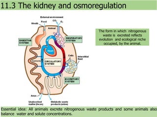
- 1. Essential idea: All animals excrete nitrogenous waste products and some animals also balance water and solute concentrations. 11.3 The kidney and osmoregulation The form in which nitrogenous waste is excreted reflects evolution and ecological niche occupied, by the animal.
- 2. Understandings Statement Guidance 11.3 U.1 Animals are either osmoregulators or osmoconformers. 11.3 U.2 The Malpighian tubule system in insectsand the kidney carry out osmoregulation and removal of nitrogenous wastes. 11.3 U.3 The composition of blood in the renal arteryis different from that in the renalvein. 11.3 U.4 The ultrastructure of the glomerulus and Bowman’s capsule facilitate ultrafiltration. 11.3 U.5 The proximal convoluted tubule selectively reabsorbs useful substances by active transport. 11.3 U.6 The loop of Henle maintains hypertonic conditions in the medulla. 11.3 U.7 ADH controls reabsorption of water in the ADH will be used in preference to vasopressin. collecting duct. 11.3 U.8 The length of the loop of Henle is positively correlated with the need for water conservation in animals. 11.3 U.9 The type of nitrogenous waste in animals is correlated with evolutionary history and habitat.
- 3. Applications and Skills Statement Guidance 11.3 A.1 Consequences of dehydration and overhydration. 11.3 A.2 Treatment of kidney failure by hemodialysis or kidney transplant. 11.3 A.3 Blood cells, glucose, proteins and drugsare detected in urinary tests. 11.3 S.1 Drawing and labelling a diagram of the human kidney. 11.3 S.2 Skill: Annotation of diagrams of the nephron. The diagram of the nephron shouldinclude glomerulus, Bowman’s capsule, proximal convoluted tubule, loop of Henle, distal convoluted tubule; the relationship betweenthe nephron and the collecting duct should be included.
- 4. Types of metabolic waste produced by living systems 1. Digestive waste 2. Respiratory waste 3. Excess water and salts (through osmoregulation) 4. Nitrogenous waste (through excretion)
- 5. ANIMAL PHYSIOLOGY OSMOREGULATORS: • All terrestrial animals, freshwater animals and some marine organisms are osmoregulators because they maintain constant internal solute concentration, even when living in marine environments with very different osmolarities • Typically these organisms maintain their solute concentration at about one third of the concentration of seawater and about 10 times that of fresh water • (Terrestrial Animals/ Freshwater Animals/ Boney Fish) OSMOCONFORMERS: • Animals that have similar internal solute concentration in comparison to the solute concentration to their surrounding environment • (Marine Invertebrates/ Cartilaginous Fish) 11.3 U.1 Animals are either osmoregulators or osmoconformers.
- 6. Osmoregulation Example •Maintaining osmotic homeostasis • Balancing water and solute concentrations (Salts/nitrogen) • Maintains cell integrity • Maintains enzyme function • etc. Osmoregulators When they live in fresh water, their bodies tend to take up water because the environment is relatively hypotonic. In such hypotonic environments, these fish do not drink much water. Instead, they pass a lot of very dilute urine, and they achieve electrolyte balance by active transport of salts through the gills. 11.3 U.1 Animals are either osmoregulators or osmoconformers.
- 7. Osmoconformers Example maintain an internal conditions that are equal to osmolarity of their environment. Minimizing the osmotic gradient minimizes the water movement in and out of cells. A disadvantage is that internal conditions may be sub- optimal. When they move to a hypertonic marine environment, these fish start drinking sea water; they excrete the excess salts through their gills and their urine. • Marine fish lose water by osmosis Actively excrete salt to maintain homeostasis • Freshwater fish lose water by osmosis Excrete excess water 11.3 U.1 Animals are either osmoregulators or osmoconformers.
- 8. Two forms of excretory systems Malpighian tubules – Insects, arthropods Kidneys - Vertebrates 1. Malpighian tubes: remove Nitrogen waste from hemolymph, located near digestive tract. Secretes dry waste with feces. 2. Kidneys: compact organs containing tubules surrounded by capillaries. Responsible for water and blood filtration, excretion of Nitrogen waste and salt 11.3 U.9 The type of nitrogenous waste in animals is correlated with evolutionary history and habitat.
- 9. Types of Nitrogenous Wastes: 1. Ammonia – water soluble, very toxic; aquatic animals 2. Urea – produced by liver; less toxic, conserves water; most vertebrates 3. Uric acid – in birds & reptiles ammonia is convert as uric acid. Uric acid does not require water and is highly concentrated. This is beneficial to these organisms as they do not have to carry the extra water around excreted as paste or crystals. Important in reducing weight for flight 11.3 U.9 The type of nitrogenous waste in animals is correlated with evolutionary history and habitat. When animals breakdown amino and nucleic acids, nitrogenous waste is formed in the form of ammonia. Ammonia is highly basic, toxic and can be very reactive. What a Ammonia becomes after this step is determined by the organisms evolutionary history and habitat. As an example: Marine and freshwater organisms can release the ammonia directly into the surrounding water where it becomes dilute.
- 10. 11.3 U.2 The Malpighian tubule system in insects and the kidney carry out osmoregulation and removal of nitrogenous wastes. The removal of nitrogenous waste and osmoregulation in insects by the Malpighian tubule • Nitrogenous wastes are broken down into URIC ACID in the insects. • Malpighian tubules branch off from their intestinal tract. • Uric acid, Na+, and K+ are actively transported from the hemolymph into the lumen of the tubules. • This draws water into the tubules by osmosis. • The water, ions, and uric acid move into the hindgut. • In the rectum, most of the water (osmosis) and salts (pumped) are selectively REABSORBED while the dehydrated uric acid is eliminated as a semisolid paste with the feces.
- 11. 11.3 U.2 The Malpighian tubule system in insects and the kidney carry out osmoregulation and removal of nitrogenous wastes. Malpighian tubules are longer and more convoluted than shown in this simplified illustration, they extend into the body cavity, where they are surrounded by hemolymph. Hemolymph is a fluid (analogous to the blood) that circulates in the interior of the insect’s body remaining in contact with the tissues. The removal of nitrogenous waste and osmoregulation in insects by the Malpighian tubule
- 12. Osmoregulation: control solute concentrations and balance water gain/loss Excretion: removal of nitrogenous wastes from body Diffusion is a form of passive transport, a net movement of particles from an area of high concentration to an area of low concentration. This is often through a partially permeable membrane. PASSIVE: DOES NOT REQUIRE ENERGY Concentration gradient: the difference in concentration of substances between two locations
- 13. Osmosis Most cell are partially permeable membrane, water flows with the concentration gradiant. When a cell is submerged in water, the water molecules pass through the cell membrane from an area of low solute concentration (outside the cell) to one of high solute concentration (inside the cell)
- 14. Osmolarity is the measure of the concentration of solute inside of a fluid or a cell. Cells can be in three types of Osmotic Environments:
- 15. How to make urine: Water and solutes enter filtrate; blood cells and proteins (nitrogen waste) remain in body fluid. Reclaim glucose, vitamins, hormones Add toxins and excess ions Filtrate leaves body as urine
- 16. The urine produced by each kidney is transported via a URETER to be stored in the BLADDER. The bladder empties through the URETHRA. 11.3 S.1 Drawing and labelling a diagram of the human kidney
- 17. •The RENAL CORTEX is the outer layer of tissue under the capsule where the blood is filtered. •The RENAL MEDULLA is found as a “middle” layer of tissue. Water and salt balance take place here. •Urine that has been produced by the filtration/reabsorption processes of the kidney is collected in the RENAL PELVIS. Structure of the Kidney 11.3 S.1 Drawing and labelling a diagram of the human kidney
- 18. 11.3 S.1 Drawing and labelling a diagram of the human kidney
- 19. The kidney causes changes in the composition of blood renal vein (filtered blood) renal artery (unfiltered blood) ureter (urine) blood in the renal vein compared and contrasted with the renal artery has … • no change in proteins – not filtered • less urea and toxins# • less oxygen* • more carbon dioxide* • less salts and ions$ (if in excess) • less water$ (if in excess) • less glucose* *Oxygen and glucose are used for cell respiration in the kidney and carbon dioxide is produced. urea toxins water salts ions # Undesired waste is removed from the blood. $ The blood water and salt concentration needs to be balanced (osmoregulation). The kidney helps by removing these molecules if in excess. 11.3 U.3 The composition of blood in the renal artery is different from that in the renal vein.
- 20. 11.3 S.1 Drawing and labelling a diagram of the human kidney
- 21. • Each kidney is made up of 1.25 million filtering units called nephrons. • 1,100 to 2000 L of blood flow through the kidneys each day. • The nephrons and collecting ducts create 180 L of initial filtrate. • Nearly all of the sugar, vitamins, and organic nutrients and 99% of water are reabsorbed into the blood. • Only about 1.5 L of urine are produced. Nephron- the functional units of the kidney. 11.3 S.2 Annotation of diagrams of the nephron.
- 22. a and c. GLOMERULUS- Afferent arteriole form branches of the renal artery bed which filters the blood. Efferent arteriole join together to form the renal vein b. BOWMAN’s CAPSULE- surrounds the glomerulus and collects the filtrate. d. PROXIMAL CONVOLUTED- selective reabsorption e. LOOP OF HENLE- regulation f. DISTAL CONVOLUTED – secretion of wastes back into filtrate g. COLLECTING DUCTS- osmoregulation 11.3 S.2 Annotation of diagrams of the nephron.
- 23. Ultrafiltration: formation of kidney filtrate 11.3 U.4 The ultrastructure of the glomerulus and Bowman’s capsule facilitate ultrafiltration. •Hydrostatic pressure created as the afferent arterioles narrow in the glomerulus capillaries forces a liquid against a semi-permeable membrane. •Blood in capillaries is at high pressure in many of the tissues of the body, and the pressure forces some of the plasma out through the capillary wall, to form tissue fluid •The pressure in the capillaries of the glomerulus are particularly high and the capillary wall is particularly permeable, so the volume forced out is about 100 times greater than in other tissues. Present moving in Glucose Proteins Urea Na+ Cl- Present moving out Urea Filtered out of Blood Glucose Proteins Na+ Cl-
- 24. 1. In the Bowmen’s capsule a cup-like sack where fluid is collected by the high pressure generate in the glomerulus knot. 2. The capillary wall of the glomerulus is fenestrated (containing pores) allowing fluid to move through it. 3. The basement membrane is the effect filtration barrier only allowing small molecules to pass through it. Cells and large macromolecules cannot pass through this structure. 4. podocyte filtration slits acting as another filter allowing only smaller molecules to be filtered *Note this means that the filtrate does not pass through the cells of either the glomerulus or the Bowman's capsule 11.3 U.4 The ultrastructure of the glomerulus and Bowman’s capsule facilitate ultrafiltration.
- 25. 11.3 U.5 The proximal convoluted tubule selectively reabsorbs useful substances by active transport. Proximal Convoluted Tubule (PCT) • This where most selective reabsorption occurs: All glucose, amino acids, vitamins and hormones are reabsorbed here, along with approx 80% of the mineral ions and water • Due to high concentrations of recovered substances in PCT cells the substances can passively diffuse into the bloodstream (along the concentration gradient) • microvilli cell lining to increase the surface area for the absorption SELECTIVE REABSORPTION (General Patterns) • Amino acids, hormones mineral ions & vitamins are actively transported (a large number of mitochondria provide ATP for active transport) into the PCT cells • Glucose is actively transported across the membrane (in symport) with sodium • Water follows the movement of the ions passively (by osmosis)
- 26. STRUCTURE • The walls of the PCT are one cell thick. • The filtrate travels through the lumen. • The inner portion of each tubule has microvilli to increase the SURFACE AREA for reabsorption in the tubule. 11.3 U.5 The proximal convoluted tubule selectively reabsorbs useful substances by active transport. Selective reabsorption of useful substances from the proximal convoluted tubule (PCT) The PCT extends from the Bowman’s capsule to the loop of Henle
- 27. LOOP of HENLE and the COLLECTING DUCTS are responsible for the control of the water balance. Function: 1. The function of the loop of Henle is to create a salt bath concentration in the surrounding medullary fluid. 2. Later this results in water reabsorption in the collecting duct 3. There is also a reduction in the filtrate volume. 11.3 U.6 The loop of Henle maintains hypertonic conditions in the medulla. AND 11.3 U.7 ADH controls reabsorption of water in the collecting duct. Osmoregulation is the control of water and solute concentrations in the body fluids (e.g. the blood plasma).
- 28. 11.3 U.6 The loop of Henle maintains hypertonic conditions in the medulla.
- 29. 11.3 U.6 The loop of Henle maintains hypertonic conditions in the medulla.
- 30. 11.3 U.6 The loop of Henle maintains hypertonic conditions in the medulla.
- 31. Distal Convolute Tubule (DTC) Function: It is partly responsible for the regulation of potassium, sodium, calcium, and pH of urine by secreting protons and absorbing bicarbonate 11.3 U.7 ADH controls reabsorption of water in the collecting duct.
- 32. 11.3 U.7 ADH controls reabsorption of water in the collecting duct. • Filtrate enters the collecting duct from the Distal Convoluted Tubule (DCT). • Water moves from the Collecting Duct to the capillaries by osmosis. • They flow in opposite directions, maintaining a Concentration gradient – a counter-current system The Colleting Duct balances the water concentration of the blood, through hormonal control
- 33. The Colleting Duct balances the water concentration of the blood, through hormonal control • Filtrate enters the collecting duct from the Distal Convoluted Tubule (DCT). • Water moves from the Collecting Duct to the capillaries by osmosis • They flow in opposite directions, maintaining a concentration gradient – a counter-current system. • If a person is dehydrated, ADH (a hormone) acts on the walls of the collecting duct, producing aquaporins (channels) making it more permeable to water. • More water is transferred into the blood. Urine output is hypertonic (high solute concentration) 11.3 U.7 ADH controls reabsorption of water in the collecting duct.
- 34. 11.3 U.7 ADH controls reabsorption of water in the collecting duct. Osmoregulation is an example of negative Feedback control using hormones. Water content of blood is monitored by the hypothalamus and regulated by the pituitary gland. The Colleting Duct balances the water concentration of the blood, through hormonal control
- 35. 11.3 U.8 The length of the loop of Henle is positively correlated with the need for water conservation in animals. Length of the loop of Henle and water conservation: The kangaroo rat's kidneys are especially efficient and produce only small quantities of highly concentrated urine. They have very long loops of Henle which builds a higher ion concentration in the medulla (dark orange below). The longer the loop the more water will be reabsorbed in the collecting duct. kangaroo rat
- 36. 11.3 U.8 The length of the loop of Henle is positively correlated with the need for water conservation in animals. The ion concentration in the medulla builds as the loop of Henle descends. A longer loop of Henle in implies a larger medulla (compared to the kidney size) than in animals with a shorter loop of Henle.. Length of the loop of Henle and water conservation * Values for the net ratios of osmolarity for urine and plasma (U/P ratios) are provided to demonstrate the concentration of urine relative to that of the blood. The ability of the kangaroo rat and other desert rodents to produce a hyper-concentrated urine is attributed to their possession of extremely long loops of Henle, which is often quoted as an extreme adaptation for life in parched deserts.
- 37. Dehydration is due to loss of water from the body so body fluids become hypertonic. • thirst, small quantities of dark colored urine • lethargy, (exposure to higher levels of metabolic waste, reduced muscle effeciency) • low blood pressure (reduced blood volume) • raised heart rate (low blood pressure) • Inability to lower body temperature (lack of sweat) • in severe cases seizures, brain damage and death 11.3 A.1 Consequences of dehydration and overhydration.
- 38. Overhydration is less common and occurs when there is an over- consumption of water. • clear urine • swelling of cells due to osmosis (from hypotonic body fluid) • Headache, disruption of nerve function (Swelled cells) • In more serious cases delirium, blurred vision, seizures, coma and death 11.3 A.1 Consequences of dehydration and overhydration.
- 39. Urine Analysis • A clinical procedure that examines urine for deviation from the normal composition. • Visual Examination: color determines hydration. • “Dipstick” Tests look for the presence of: • pH- normal (pH 4.6 to pH 8.0)- extremes show improper functioning of kidney • Protein levels- possible kidney damage • Glucose- possible diabetes • Monoclonal antibodies on strips look for drug use and/or pregnancy. • Blood cells infections, disease and some cancers • Drugs (or their breakdown products) can often be detected in urine samples 11.3 A.3 Blood cells, glucose, proteins and drugs are detected in urinary tests. * As an example, excess sugar in the urine generally indicates diabetes
- 40. 11.3 A.2 Treatment of kidney failure by hemodialysis or kidney transplant. Treatment of kidney failure Kidney failure is a condition in which the kidneys fail to adequately filter waste products from the blood. It can be caused by injury or disease symptoms vary depending on the seriousness and progression of the disease. If not treated kidney failure leads to death. Treatment of kidney failure focuses on two main approaches: • Hemodialysis • Kidney transplants
- 41. http://www.kalingahospital.com/data/images/transplant1.jpg Treatment of kidney failure *If the match is not close enough the receipient’s immune system will react to the new kidney as it would to a pathogen. A transplant is the best long-term treatment. Donors can be either: • Someone who has recently died • A person who has chosen to give up one of their two kidneys Donors and the recipient have to be a close match in both blood and tissues to minimize the chance of rejection*. The transplanted kidney is grafted in to the lower abdomen with the renal artery, renal vein and ureter connected to the recipient’s blood vessels and bladder. 11.3 A.2 Treatment of kidney failure by hemodialysis or kidney transplant.
- 42. 11.3 A.2 Treatment of kidney failure by hemodialysis or kidney transplant. Treatment of kidney failure Hemodialysis (commonly called kidney dialysis) is a process of purifying the blood of a person whose kidneys are not working normally. Hemodialysis treatment lasts about four hours and is done three times per week.
- 43. Bibliography / Acknowledgments Maura Pallilo