SlideShare una empresa de Scribd logo
1 de 62
6.2 The Blood System
Essential idea: The blood system continuously transports
substances to cells and simultaneously collects waste products.
Understandings
Statement Guidance
6.2 U.1 Arteries convey blood at high pressure from the ventricles to the tissues of the body
6.2 U.2 Arteries have muscle cells and elastic fibers in their walls
6.2 U.3 The muscle and elastic fibers assist in maintaining blood pressure between pump
cycles
6.2 U.4 Blood flows through tissues in capillaries. Capillaries have permeable walls that allow
exchange of materials between cells in the tissue and the blood in the capillary
6.2 U.5 Veins collect blood at low pressure from the tissues of the body and return it to the
atria of the heart.
6.2 U.6 Valves in veins and the heart ensure circulation of blood by preventing backflow.
6.2 U.7 There is a separate circulation for the lungs.
6.2 U.8 The heart beat is initiated by a group of specialized muscle cells in the right atrium
called the sinoatrial node
6.2 U.9 The sinoatrial node acts as a pacemaker.
6.2 U.10 The sinoatrial node sends out an electrical signal that stimulates contraction as it is
propagated through the walls of the atria and then the walls of the ventricles
6.2 U.11 The heart rate can be increased or decreased by impulses brought to the heart
through two nerves from the medulla of the brain.
6.2 U.12 Epinephrine increases the heart rate to prepare for vigorous physical activity
Applications and Skills
Statement Guidance
6.2 A.1 William Harvey’s discovery of the circulation of the blood with the
heart acting as the pump.
6.2 A.2 Pressure changes in the left atrium, left ventricle and aorta during
the cardiac cycle.
6.2 A.3 Causes and consequences of occlusion of the coronary arteries.
6.2 S.1 Identification of blood vessels as arteries, capillaries or veins from
the structure of their walls.
6.2 S.2 Recognition of the chambers and valves of the heart and the blood
vessels connected to it in dissected hearts or in diagrams of heart
structure.
What are the issues when
organizes become multicellular?
• Diffusion is not adequate for moving material
across more than 1 cell barrier
• Circulatory system solves this problem
– carries fluids & dissolved material throughout
body
– cells are never far from body fluid
– only a few cells away from blood
overcoming the limitations of diffusion
There are three main types of blood vessel:
Arteries carry high pressure blood away from the heart to tissues that need it
Capillaries are very small (< 10 μm diameter) and therefore can penetrate virtually every
tissue in the body. Blood moves slowly through them under low pressure providing
opportunities for the exchange of substances.
Veins carry the low pressure blood back to the heart using valves to ensure blood flows
in the correct direction.
6.2 U.1 Arteries convey blood at high pressure from the ventricles to the tissues of the body. AND
6.2 U.2 Arteries have muscle cells and elastic fibers in their walls. AND 6.2 U.3 The muscle and
elastic fibers assist in maintaining blood pressure between pump cycles
.
The structure of arteries• Thick muscular wall and fibrous
outer layer help the artery to
withstand high pressure
• Relatively (to the wall) small
lumen maintains high blood
pressure.
• Muscle contracts to decrease the
size of the lumen. This causes an
increase blood pressure and
therefore maintains high blood
pressure between the pulses of
high pressure blood travelling from
the heart
• Elastic fibers stretch to increase
the lumen with each pulse of
blood. After the pulse of blood
passes the fibers recoil decreasing
the lumen size and therefore
helping to maintain a high blood
pressure.
6.2 U.5 Veins collect blood at low pressure from the tissues of the body and return it to the atria of
the heart. AND 6.2 U.6 Valves in veins and the heart ensure circulation of blood by preventing
backflow.
https://commons.wikimedia.org/wiki/File:Venous_valve.svg
http://40.media.tumblr.com/tumblr_m0dwjt3WKQ1qzcf71o1_500.jpg
Because of the low pressure valves are
required to prevent back-flow of the blood
and therefore ensure that the blood moves
towards to heart.
The structure of veins
• Veins return blood to the heart for
re-circulation.
• The large lumen (compared to
arteries and the thickness of the
wall) means that the blood is under
low pressure.
• Because there is less pressure to
resist the walls of the veins are
thinner and less elastic than
arteries. They also contain less
muscle than the arteries.
• Veins carry blood back to the heart. Have thin walls containing valves to prevent
the back flow of blood, due to the lower pressure of blood in the veins.
• Pressure in veins is low, so veins depend on nearby muscular contractions to move
blood along.
6.2.U5 Veins collect blood at low pressure from the tissues of the body and return it to the atria of
the heart. AND 6.2.U6 Valves in veins and the heart ensure circulation of blood by preventing
backflow.
6.2 U.4 Blood flows through tissues in capillaries. Capillaries have permeable walls that
allow exchange of materials between cells in the tissue and the blood in the capillary.
The structure of capillaries
• Capillaries are the smallest blood vessels and are adapted
for the exchange of substances to and from the blood.
• This enables tissues to gain nutrients and molecules such as
oxygen and to rid themselves of waste material.
• Capillaries also allow substances to enter and leave the
organism, e.g. gas exchange of oxygen and carbon dioxide in
the lungs.
Due the the massive number of
capillaries present and the small
lumen the surface area available for
the exchange of substances is very
large.
6.2 S.1 Identification of blood vessels as arteries, capillaries or veins from the structure
of their walls.
a
b
c
d
https://www.ouhsc.edu/histology/Text%20Sections/Cardiovascular.html
Identify the labelled structures using your understanding of blood vessels.
https://www.ouhsc.edu/histology/Text%20Sections/Cardiovascular.html
Identify the labelled structures using your understanding of blood vessels.
6.2 S.1 Identification of blood vessels as arteries, capillaries or veins from the structure
of their walls.
Nature of Science: Theories are regarded as uncertain - William Harvey overturned
theories developed by the ancient Greek philosopher Galen on movement of blood in the
body. (1.9)
Remember a theories are by definition the best accepted explanations and predictions of natural
phenomena. Although they are usually thoroughly tested based on evidence and reason theories
theories are the only the current best accepted explanation. Theories if successfully questioned can be
modified or even rejected/falsified if a better explanation arises.
Theories of blood movement around the body
• Galen (129 - c216) Blood was not seen to
circulate but rather to slowly ebb and
flow. Using elusive attractive powers.
• Ibn-al-Nafiz (1300) first challenger to Galen
idea of blood movement
• William Harvey (1578 - 1657) published “De
Motu Cordis” in 1628. Even then it took
many years for his theory of systemic
circulation (similar to modern accepted
theory) to succeed Galen’s.
Path of blood through the heart
• Deoxygenated blood return to the
heart to right atrium from the body
• The Right Atrium, receives "used
blood" from the body. Blood will be
pushed through the tricuspid valve to
the
• Right Ventricle, the chamber which
will pump to the lungs through the
pulmonic valve to the
• Pulmonary Arteries, providing
blood to both lungs. Blood is
circulated through the lungs where
carbon dioxide is removed and
oxygen added. It returns through the
Systemic Circuit.
6.2 U.7 There is a separate circulation for the lungs.
Path of blood through the heart
• Oxygenated blood enters the Left
Atrium from the lungs
• Pulmonary Veins, empty into the
Left Atrium.
• From the Left Atrium, a chamber
which will push the Mitral Valve
open. Blood then passes into the
• Left Ventricle. Although it doesn't
always look like it in drawings done
from this angle, this is the largest
and most important chamber in the
heart. It pumps to the rest of the
body. As it pumps, the pressure will
close the mitral valve and open the
aortic valve, with blood passing
through to the Pulmonary Circuit
6.2 U.7 There is a separate circulation for the lungs.
6.2.S.2 Recognition of the chambers and valves of the heart and the blood vessels connected to it
in dissected hearts or in diagrams of heart structure
a. Atria: thin-walled
upper chambers which
collect blood returning
to the heart
Part of the Heart
b. Ventricles: thick,
muscular lower
chambers which pump
blood to the lungs and
other body tissue.
c. Septum (wall): separate the
right and left sides of heart to
prevent mixing of oxygenated
and deoxygenated blood.
6.2.S2 Recognition of the chambers and valves of the heart and the blood vessels connected to it in
dissected hearts or in diagrams of heart structure
1. Tricuspid valve /AV valve
Valves of the Heart
2. Pulmonary valve
4. Bicuspid
valve / AV
valve
3. Aortic valve
6.2 S2 Recognition of the chambers and valves of the heart and the blood vessels connected to it in
dissected hearts or in diagrams of heart structure.
Label the heart diagram
6.2.S2 Recognition of the chambers and valves of the heart and the blood vessels connected to it in
dissected hearts or in diagrams of heart structure.
Label the heart diagram
Now try to label this heart: http://sciencelearn.org.nz/Contexts/See-through-Body/Sci-Media/Animation/Label-
the-heart
bicuspid valve
Pulse
• When you take your pulse, you feel (or hear) two changes in pressure.
• The first sound is the closing of the AV’s (systolic pressure), the “lub”
sound the heart makes. The 2nd sound is the closing of the SV’s
(diastolic pressure), the “dub” sound.
6.2 U.8 The heart beat is initiated by a group of specialized muscle cells in the right atrium called the sinoatrial node.
6.2 U.9 The sinoatrial node acts as a pacemaker.
6.2 U.10 The sinoatrial node sends out an electrical signal that stimulates contraction as it is propagated through the walls
of the atria and then the walls of the ventricles.
Control of the Heart Beat
• Beating is due to myogenic muscle.
Meaning it is the origin of contraction
in the heart and is not controlled
externally
• The hearts pacemaker (Sinoatrial
Node/SA node) which controls the
rate of heartbeats is made up of this
muscle.
• Large groups of these cell cause the
atria to contract.
• The contraction cause a signal to a
group of nerve cells causing the
ventricles to contract at the
atrioventricular node/AV node.
6.2 U.8 The heart beat is initiated by a group of specialized muscle cells in the right atrium called the sinoatrial node.
6.2 U.9 The sinoatrial node acts as a pacemaker.
6.2 U.10 The sinoatrial node sends out an electrical signal that stimulates contraction as it is propagated through the walls
of the atria and then the walls of the ventricles.
The control of heart rhythm
6.2 U.8 The heart beat is initiated by a group of specialized muscle cells in the right atrium called the sinoatrial node.
6.2 U.9 The sinoatrial node acts as a pacemaker.
6.2 U.10 The sinoatrial node sends out an electrical signal that stimulates contraction as it is propagated through the walls
of the atria and then the walls of the ventricles.
The interrelationship of blood flow
velocity, cross-sectional area of
blood vessels, and blood pressure
6.2 U.8 The heart beat is initiated by a group of specialized muscle cells in the right atrium called the sinoatrial node.
6.2 U.9 The sinoatrial node acts as a pacemaker.
6.2 U.10 The sinoatrial node sends out an electrical signal that stimulates contraction as it is propagated through the walls
of the atria and then the walls of the ventricles.
6.2 U.11 The heart rate can be increased or decreased by impulses brought to the heart through
two nerves from the medulla of the brain.
6.2 U.12 Epinephrine increases the heart rate to prepare for vigorous physical activity.
Control of the Heart Beat
• Controlled by the autonomic
nervous system response to
changing body conditions by
speeding up or lowing down
breathing
• When the level of CO2 are low
chemical receptors in the
Medulla sends a signal to the
Vagus nerve which decreases
heart rate.
• When the level of CO2 high
chemical receptors in the
Medulla sends a signal to the
Accelerator nerve which
increase heart rate.
Medulla
Vagus Nerve
Accelerator Nerve
SA Node
6.2 U.11 The heart rate can be increased or decreased by impulses brought to the heart
through two nerves from the medulla of the brain.
6.2 U.12 Epinephrine increases the heart rate to prepare for vigorous physical activity.
Hormonal Control of Heart Rate
• Epinephrine, more commonly known
as adrenaline, is a hormone secreted by
the medulla of the adrenal glands
• Released into the bloodstream,
epinephrine causes an increase in heart
rate, muscle strength, blood pressure, and
sugar metabolism.
How does the Heart work?
blood from the
body
blood from
the lungs
The heart beat begins when the
heart muscles relax and blood
flows into the atria.
STEP ONE
The atria then contract and
the valves open to allow blood
into the ventricles.
How does the Heart work?
STEP TWO
How does the Heart work?
The valves close to stop blood
flowing backwards.
The ventricles contract forcing
the blood to the aorta and out of the
heart.
At the same time, the atria are
relaxing and once again filling with
blood.
The cycle then repeats itself.
STEP THREE
6.2 A.2 Pressure changes in the left atrium, left ventricle and aorta during the cardiac cycle.
The Cardiac Cycle
1. Right Atria & Ventricles relax and
blood flows into the heart from
the veins.
2. AV valve open, Right Atria
contacts and Ventricle relaxes,
blood moves into the ventricle.
3. Pulmonary valve opens, Right
Ventricle contracts, blood flows to
the lungs.
4. Blood flows from the lung into
the left Atria AV valve opens,
blood flows into the Left
Ventricle.
5. Left Atria contracts blood flows
into Left Ventricle.
6. Aortic valve opens, Left Ventricle
contracts blood flows into the
arteries.
6.2 A.2 Pressure changes in the left atrium, left ventricle and aorta during the cardiac cycle.
The Cardiac Cycle
Ventricular Pressure
1. Ventricular pressure increases
as ventricle contracts. This
forces blood into the aorta,
increasing aortic pressure.
2. (a) Shows the increase in atrial
pressure due to atrial
contraction.
3. Shows increase in atrial
pressure due to the inflow of
blood from the veins (following
systole).
Ventricular Volume
4. Increase as atrial contraction
forces blood into the ventricle
5. Decrease as ventricular
contraction forces blood into
the aorta
6. Increases as blood returns to
the heart following systole.
Click on the
hyperlink
6.2 A3 Causes and consequences of occlusion of the coronary arteries.
6.3 A1 Causes and consequences of blood clot formation in coronary
Arteriosclerosis
• Hardening and thickening of the walls of
the arteries.
• The condition can occur because of fatty
deposits on the inner lining of arteries
(atherosclerosis).
• Calcification of the wall of the arteries, or
thickening of the muscular wall of the
arteries from chronically elevated blood
pressure occurs.
• As the deposits of fat and plaque begin to
build the lumen of the arteries become
reduced. If the plaque frees itself from the
wall a blood clot may form. This may lead
to coronary thrombosis (reduced blood
flow to the heart).
• When coronary arteries that supply blood
to the heart muscle are effected a shortage
of oxygen delivered to the heart itself may
lead to a heart attack.
Coronary arteries supply heart
muscles with oxygen and nutrients
6.2 A3 Causes and consequences of occlusion of the coronary arteries.
6.3 A1 Causes and consequences of blood clot formation in coronary
Risk Factors in Coronary Heart Disease
Factor Causes
Genetics Family history of high cholesterol/blood
pressure
Age Older people have a greater risk due to
a loss in elasticity in arteries
Sex Males have a greater risk then females
Diet Increases in fat intake lead to higher
levels of cholesterol and increase
formation of plaque
Exercise A lack of exercise increase risk. This is
due to a weakening of circulation
Obesity Increase in blood pressure which leads
to increase plaque formation in the
arteries
Stress Stress has been linked to increased
levels of the hormone Cortisol. Which
has been show leads to a increase
likelihood of atherosclerosis
Essential idea: The lungs are actively ventilated to ensure that gas exchange can occur
passively.
6.4 Gas exchange
Understandings
Statement Guidance
6.4 U.1
Ventilation maintains concentration gradients of
oxygen and carbon dioxide between air in alveoli
and blood flowing in adjacent capillaries.
6.4 U.2
Type I pneumocytes are extremely thin alveolar
cells that are adapted to carry out gas
exchange.
6.4 U.3
Type II pneumocytes secrete a solution
containing surfactant that creates a moist
surface inside the alveoli to prevent the sides of
the alveolus adhering to each other by reducing
surface tension.
6.4 U.4
Air is carried to the lungs in the trachea and
bronchi and then to the alveoli in bronchioles.
Students should be able to draw a diagram to
show the structure of an alveolus and an
adjacent capillary.
6.4 U.5
Muscle contractions cause the pressure changes
inside the thorax that force air in and out of the
lungs to ventilate them.
6.4 U.6
Different muscles are required for inspiration
and expiration because muscles only do work
when they contract.
Applications and Skills
Statement Guidance
6.4 A.1 Causes and consequences of lung cancer.
6.4 A.2 Causes and consequences of emphysema.
6.4 A.3 External and internal intercostal muscles, and
diaphragm and abdominal muscles as examples
of antagonistic muscle action.
6.4 S.1 Monitoring of ventilation in humans at rest and
after mild and vigorous exercise. (Practical 6)
Ventilation can either be monitored by simple
observation and simple apparatus or by data
logging with a spirometer or chest belt and
pressure meter. Ventilation rate and tidal
volume should be measured, but the terms
vital capacity and residual volume are not
expected.
What is respiration?
• External respiration -
exchange of O2 & CO2
between respiratory surfaces
& the blood [breathing].
• Internal respiration -
exchange of O2 & CO2
between the blood & cells.
• Cellular respiration -
process by which cells use O2
to produce ATP.
Gas Exchange in Animals
Why do organisms need a respiratory system?
CO2
CO2
O2
O2Bloodstream
Muscle cells carrying out
Cellular Respiration
Breathing
Glucose + O2
CO2 + H2O + ATP
Lungs
What is the relationship between
respiration and cellular respiration?
Why do organisms need a respiratory system?
Respiration
• (Breathing) provides for the exchange of
O2 and CO2 between an organism and its
environment
Why do organisms need a respiratory system?
All respiratory surfaces MUST
be:
• moist
• thin
• large enough to meet
metabolic needs of
organism
Challenges:
Aquatic organisms -
H2O contains only 1/30
of the O2 present in air.
Terrestrial organisms -
must prevent
desiccation of
respiratory surface.
Why do organisms need a respiratory system?
Types of Respiratory Surfaces
Structure of the ventilation system
a) Trachea
b) Cartilage ring support
c) Bronchi (plural) Bronchus
(single)
d) Lung
e) Heart
f) Sternum
g) Rib cage
h) Bronchioles
j) Alveoli
k) Diaphragm
6.4 U.4 Air is carried to the lungs in the trachea and bronchi and then to the alveoli in
bronchioles. (Page 78)
6.4 U.4 Air is carried to the lungs in the trachea and bronchi and then to the alveoli in
bronchioles. (Page 78)
6.4 U.4 Air is carried to the lungs in the trachea and bronchi and then to the alveoli in
bronchioles. (Page 78)
Can you use the diagram to produce a flow chart to show the passage of air into the lungs?
Review: 6.2 U.11 The heart rate can be increased or decreased by impulses brought to the heart
through two nerves from the medulla of the brain.
Control of the Heart Beat
• Controlled by the autonomic
nervous system response to
changing body conditions by
speeding up or lowing down
breathing
• When the level of CO2 are low
chemical receptors in the
Medulla sends a signal to the
Vagus nerve which decreases
heart rate.
• When the level of CO2 high
chemical receptors in the
Medulla sends a signal to the
Accelerator nerve which
increase heart rate.
Medulla
Vagus Nerve
Accelerator Nerve
SA Node
6.4 A.3 External and internal intercostal muscles, and diaphragm and abdominal
muscles as examples of antagonistic muscle action. (Page 78)
• The medulla oblongata is
involved in regulating many of the
bodily processes that are
controlled automatically like blood
pressure, heart rate and
RESPIRATION.
• The medulla oblongata detects
carbon dioxide (CO2) and Oxygen
(O2) levels in the bloodstream and
determines what changes need to
happen in the body.
• It can then
send nerve impulses muscles in
the ribs and diaphragm, letting
them know that they need to
either step up their game, or slow
down a bit.
Ventilation: for gas
exchange
• The flow of air in and out of the
alveoli is called ventilation and has
two stages: inspiration (or
inhalation) which increases the
concentration and expiration (or
exhalation) which removes CO2.
• For gas exchange to be efficient,
high concentration gradients must
be maintained in the alveoli.
• Lungs are not muscular and cannot
ventilate themselves, but instead
the whole thorax moves and
changes size, due to the action of
two sets of muscles: the intercostal
muscles and the diaphragm
6.4 U.1 Ventilation maintains concentration gradients of oxygen and carbon dioxide
between air in alveoli and blood flowing in adjacent capillaries. (Page 78)
Alveoli (air sac)
Ventilation of the lungs:
Inhale
• External intercostal muscles
contract, moving ribs up and
out
• Diaphragm contracts,
fattening and moving down
• Increased volume in thorax
lowering pressure
• Air flows into the lungs to
balance the pressure change
decrease in pressure
(draws air inwards)
6.4 A.3 External and internal intercostal muscles, and diaphragm and abdominal muscles as examples of antagonistic
muscle action. AND 6.4 U.5 Muscle contractions cause the pressure changes inside the thorax that force air in and out of
the lungs to ventilate them.
(Page 78)
Ventilation of the lungs:
Exhale:
• Internal intercostal muscles
contract, moving ribcage down
and in
• Abdominal muscles contract
pushing the diaphragm up into
a dome shape
• Decrease in volume of thorax
increasing air pressure
• Air flows out from the lungs
increase in pressure
(pushes air outwards)
6.4 A.3 External and internal intercostal muscles, and diaphragm and abdominal muscles as examples of antagonistic
muscle action. AND 6.4 U.5 Muscle contractions cause the pressure changes inside the thorax that force air in and out of
the lungs to ventilate them.
(Page 78)
Negative pressure breathing
6.4 A.3 External and internal intercostal muscles, and diaphragm and abdominal muscles as examples of antagonistic
muscle action. AND 6.4 U.5 Muscle contractions cause the pressure changes inside the thorax that force air in and out of
the lungs to ventilate them.
(Page 78)
Gas Exchange
This is the diffusion of gases (oxygen
and carbon dioxide) There are two
sites for gas exchange
A. Alveoli: Oxygen diffuses
into the blood from the alveoli
and carbon dioxide diffuses
from the blood into the alveoli
B. Tissues: Oxygen diffuses
from blood into the cells and
carbon dioxide diffuses from
cells to the blood
6.4 U.1 Ventilation maintains concentration gradients of oxygen and carbon dioxide
between air in alveoli and blood flowing in adjacent capillaries. (Page 78)
6.4 U.2 Type I pneumocytes are extremely thin alveolar cells that are adapted to carry out gas exchange. AND
(Page 79)
6.4 U.3 Type II pneumocytes secrete a solution containing surfactant that creates a moist surface inside the
alveoli to prevent the sides of the alveolus adhering to each other by reducing surface tension. (Page 79)
6.4 U.2 Type I pneumocytes are extremely thin alveolar cells that are adapted to carry out gas exchange. AND
6.4 U.3 Type II pneumocytes secrete a solution containing surfactant that creates a moist surface inside the
alveoli to prevent the sides of the alveolus adhering to each other by reducing surface tension. (Page 79)
Edited from: https://beyondthedish.files.wordpress.com/2011/11/gas-barrier-small.jpg
http://neurobio.drexelmed.edu/education/pil/microanatomy/Cases/01_Quintana/4_Epithelium/Images/alveolarepithelium.jpg
Connective
tissue
Type II pneumocytes
• Secrete fluid to moisten the inner
surface of the alveolus
• Fluid aids diffusion of gases
• Fluid contains surfactant to prevent the
walls sticking together – maintains the
lumen
Alveoli walls and gas exchange
Type I pneumocytes
• A single layer of cells form the
walls of an alveolus
• Extremely thin – short
diffusion distance
• Permeable – aids diffusion
Emphysema
• Disease caused by smoking,
lung infections or air pollution.
All of which cause an
inflammatory response in the
lungs.
• Protease is released by
leukocytes (white blood cells)
and inflamed lung tissue. The
protease breaks down the
connective tissue such as
elastin, of the lungs. This
results in the destruction of
small airways and alveoli. This
results in the formation of large
air pockets and the breakdown
of capillaries
• The disease creates a reduction
in surface area gas exchange
which results shortness of
breath, difficulty in breathing,
and decreased lung capacity.
• Emphysema is a progressive
disease with no known cures.
There are treatments that can
help you manage the disease
6.4 A.2 Causes and consequences of emphysema. (page 79)
Symptoms include:
• Difficulty breathing
• Cough
• Loss of appetite
• Weight loss
Review: 1.6 and U.6 Mutagens, oncogenes and metastasis are involved in the development of
primary and secondary tumors.
Tumors are abnormal growth of tissue that develop at any stage of life in any part
of the body. A cancer is a malignant tumour and is named after the part of the
body where the cancer (primary tumour) first develops. Use the links to find out:
• most common types of cancer
• what causes cancer and associated risk factors
• how cancer can be treated
http://youtu.be/8BJ8_5Gyhg8
http://www.cancerresearchuk.org/cancer-
info/cancerandresearch/all-about-cancer/what-is-cancer/
What causes
cancer?
6.4 A.1 Causes and consequences of lung cancer. (Page 78)
“Lung cancer is the leading cause of death from cancer in the U.S.”
http://www.cancer.gov/types/lung
Lung cancer is cancer that starts in the windpipe (trachea),
the main airway (bronchus) or the lung tissue.
By far the biggest cause of lung cancer is smoking. It causes more
than 8 out of 10 cases (86%) including a small proportion caused by
exposure to second hand smoke in non smokers (passive smoking).
Some other things increase lung cancer
risk by a small amount:
• Exposure to radon (a radioactive) gas
• Air pollution
• Previous lung disease
• A family history of lung cancer
• Past cancer treatment
• Previous smoking related cancers
• Lowered immunity
The symptoms of lung cancer may include:
• Being short of breath
• Having a cough most of the time
• Coughing up phlegm with blood
• An ache or pain in the chest or shoulder
• Loss of appetite
• Tiredness/fatigue
• Losing weight
Like most cancers lung cancer, if untreated, will end with
death. Because detection of lung cancer is difficult it is often
only diagnosed in the later stages. As a consequence only
10% of those diagnosed will survive for 5 years.
Source: http://www.cancerresearchuk.org/about-cancer/type/lung-cancer/
http://spacecoastdaily.com/wp-content/uploads/2014/01/Cigarette-Smoking-is-Mainly-Causes-of-Lung-Cancer.jpg
6.4 S.1 Monitoring of ventilation in humans at rest and after mild and vigorous exercise. (Practical 6)
Monitoring ventilation
https://commons.wikimedia.org/wiki/File:DoingSpirometry.JPG
https://en.wikipedia.org/wiki/File:Lungvolumes_Updated.png
Independent variable: the intensity of
exercise
Dependent variable: What measure(s) are
you using for ventilation?
What type of exercise? For how long?
How will you make sure that individuals
exercise similarly?
Frequency of breaths? Spirometer data
showing tidal volumes?
Ethical and safety concerns: how are you making sure
that the experiment is safe? Are participants aware of
the risks? Are you seeking written permission from
participants?
: design an investigation, collect and analyze the data
Controlled variables: what are the key factors
that need to be measured and kept constant
to ensure a fair test?
Data analysis: you should be calculating a rate.
For example the count of breaths or tidal
volume per unit time (s or min)
Bibliography / Acknowledgments

Más contenido relacionado

La actualidad más candente

IB Biology 1.6 & 1.1 Slides: Mitosis & Stem Cells
IB Biology 1.6 & 1.1 Slides: Mitosis & Stem CellsIB Biology 1.6 & 1.1 Slides: Mitosis & Stem Cells
IB Biology 1.6 & 1.1 Slides: Mitosis & Stem CellsJacob Cedarbaum
 
IB Biology 2.7 & 7.1 Slides: DNA Replication
IB Biology 2.7 & 7.1 Slides: DNA ReplicationIB Biology 2.7 & 7.1 Slides: DNA Replication
IB Biology 2.7 & 7.1 Slides: DNA ReplicationJacob Cedarbaum
 
6.3 defence (chris paine)
6.3 defence (chris paine)6.3 defence (chris paine)
6.3 defence (chris paine)cartlidge
 
IB Biology 2.8 Slides: Respiration
IB Biology 2.8 Slides: RespirationIB Biology 2.8 Slides: Respiration
IB Biology 2.8 Slides: RespirationJacob Cedarbaum
 
6.5 neurons and synapses (chris paine)
6.5 neurons and synapses (chris paine)6.5 neurons and synapses (chris paine)
6.5 neurons and synapses (chris paine)cartlidge
 
6.3 defense against infectious diseases
6.3 defense against infectious diseases6.3 defense against infectious diseases
6.3 defense against infectious diseaseslucascw
 
IB Biology 2.1 Slides: Molecules to Metabolism
IB Biology 2.1 Slides: Molecules to MetabolismIB Biology 2.1 Slides: Molecules to Metabolism
IB Biology 2.1 Slides: Molecules to MetabolismJacob Cedarbaum
 
6.6 Hormones Homeo and Repro (Chris Paine)
6.6 Hormones Homeo and Repro (Chris Paine)6.6 Hormones Homeo and Repro (Chris Paine)
6.6 Hormones Homeo and Repro (Chris Paine)cartlidge
 
IB Biology 3.4 inheritance
IB Biology 3.4 inheritanceIB Biology 3.4 inheritance
IB Biology 3.4 inheritanceBob Smullen
 
Biology Option D:Transport of respiratory gases
Biology Option D:Transport of respiratory gasesBiology Option D:Transport of respiratory gases
Biology Option D:Transport of respiratory gasesRazanah Mohd Jalal
 
IB Biology 1.4 Slides: Cell Transport
IB Biology 1.4 Slides: Cell TransportIB Biology 1.4 Slides: Cell Transport
IB Biology 1.4 Slides: Cell TransportJacob Cedarbaum
 
Immune system hl
Immune system hlImmune system hl
Immune system hlBob Smullen
 
11.2 Movement
11.2 Movement11.2 Movement
11.2 Movementdabagus
 
IB Biology 2.5 Slides: Enzymes
IB Biology 2.5 Slides: EnzymesIB Biology 2.5 Slides: Enzymes
IB Biology 2.5 Slides: EnzymesJacob Cedarbaum
 
Functions of liver option d.3
Functions of liver option d.3Functions of liver option d.3
Functions of liver option d.3Engin Emlek
 
8.3 photosynthesis
8.3 photosynthesis8.3 photosynthesis
8.3 photosynthesisBob Smullen
 
10.2 inheritance
10.2 inheritance10.2 inheritance
10.2 inheritanceBob Smullen
 

La actualidad más candente (20)

IB Biology 1.6 & 1.1 Slides: Mitosis & Stem Cells
IB Biology 1.6 & 1.1 Slides: Mitosis & Stem CellsIB Biology 1.6 & 1.1 Slides: Mitosis & Stem Cells
IB Biology 1.6 & 1.1 Slides: Mitosis & Stem Cells
 
IB Biology 2.7 & 7.1 Slides: DNA Replication
IB Biology 2.7 & 7.1 Slides: DNA ReplicationIB Biology 2.7 & 7.1 Slides: DNA Replication
IB Biology 2.7 & 7.1 Slides: DNA Replication
 
6.3 defence (chris paine)
6.3 defence (chris paine)6.3 defence (chris paine)
6.3 defence (chris paine)
 
IB Biology 2.8 Slides: Respiration
IB Biology 2.8 Slides: RespirationIB Biology 2.8 Slides: Respiration
IB Biology 2.8 Slides: Respiration
 
2.4 proteins
2.4 proteins2.4 proteins
2.4 proteins
 
6.5 neurons and synapses (chris paine)
6.5 neurons and synapses (chris paine)6.5 neurons and synapses (chris paine)
6.5 neurons and synapses (chris paine)
 
6.3 defense against infectious diseases
6.3 defense against infectious diseases6.3 defense against infectious diseases
6.3 defense against infectious diseases
 
IB Biology 2.1 Slides: Molecules to Metabolism
IB Biology 2.1 Slides: Molecules to MetabolismIB Biology 2.1 Slides: Molecules to Metabolism
IB Biology 2.1 Slides: Molecules to Metabolism
 
6.6 Hormones Homeo and Repro (Chris Paine)
6.6 Hormones Homeo and Repro (Chris Paine)6.6 Hormones Homeo and Repro (Chris Paine)
6.6 Hormones Homeo and Repro (Chris Paine)
 
IB Biology 3.4 inheritance
IB Biology 3.4 inheritanceIB Biology 3.4 inheritance
IB Biology 3.4 inheritance
 
Option D
Option DOption D
Option D
 
Biology Option D:Transport of respiratory gases
Biology Option D:Transport of respiratory gasesBiology Option D:Transport of respiratory gases
Biology Option D:Transport of respiratory gases
 
IB Biology 1.4 Slides: Cell Transport
IB Biology 1.4 Slides: Cell TransportIB Biology 1.4 Slides: Cell Transport
IB Biology 1.4 Slides: Cell Transport
 
Immune system hl
Immune system hlImmune system hl
Immune system hl
 
Transport systems in animals
Transport systems in animalsTransport systems in animals
Transport systems in animals
 
11.2 Movement
11.2 Movement11.2 Movement
11.2 Movement
 
IB Biology 2.5 Slides: Enzymes
IB Biology 2.5 Slides: EnzymesIB Biology 2.5 Slides: Enzymes
IB Biology 2.5 Slides: Enzymes
 
Functions of liver option d.3
Functions of liver option d.3Functions of liver option d.3
Functions of liver option d.3
 
8.3 photosynthesis
8.3 photosynthesis8.3 photosynthesis
8.3 photosynthesis
 
10.2 inheritance
10.2 inheritance10.2 inheritance
10.2 inheritance
 

Similar a 6.2 and 6.4 transport and respiration

6.2 bloodflow
6.2 bloodflow6.2 bloodflow
6.2 bloodflowlucascw
 
circulatory system mammals.ppt
circulatory system mammals.pptcirculatory system mammals.ppt
circulatory system mammals.pptssuser880f82
 
BA&P Circulatory
BA&P CirculatoryBA&P Circulatory
BA&P Circulatorynatjkeen
 
Vascular system Brief Introdcution
Vascular system Brief Introdcution Vascular system Brief Introdcution
Vascular system Brief Introdcution BilalAhmad696
 
Transport systems in mammals
Transport systems in mammals Transport systems in mammals
Transport systems in mammals Saad Adil
 
CARDIOVASCULAR-SYSTEM-Group-6.docx
CARDIOVASCULAR-SYSTEM-Group-6.docxCARDIOVASCULAR-SYSTEM-Group-6.docx
CARDIOVASCULAR-SYSTEM-Group-6.docxBeatrizEspeletaFranc
 
Cardiovascular system
Cardiovascular systemCardiovascular system
Cardiovascular systemGeorge Pearce
 
Support ppt circulation
Support ppt circulationSupport ppt circulation
Support ppt circulationrohitkamble98
 
Transport in animals
Transport in animalsTransport in animals
Transport in animalsSian Ferguson
 
Blood vessels: Arteries, Veins and Capillaries
Blood vessels: Arteries, Veins and CapillariesBlood vessels: Arteries, Veins and Capillaries
Blood vessels: Arteries, Veins and CapillariesAmir Rifaat
 
cardiovascular system
cardiovascular systemcardiovascular system
cardiovascular systemMEI MEI
 
6.2 the transport system
6.2 the transport system6.2 the transport system
6.2 the transport systemcartlidge
 
Comparative anatomy circulatory system
Comparative anatomy circulatory systemComparative anatomy circulatory system
Comparative anatomy circulatory systemOmer Rasool
 
Introduction to the Cardiovascular sytem.pptx
Introduction to the Cardiovascular sytem.pptxIntroduction to the Cardiovascular sytem.pptx
Introduction to the Cardiovascular sytem.pptxKashif Hussain
 
Biology Project [Circulatory System] Vijay Raja Std Vii Navdeep With Sound
Biology Project [Circulatory System] Vijay Raja Std Vii Navdeep With SoundBiology Project [Circulatory System] Vijay Raja Std Vii Navdeep With Sound
Biology Project [Circulatory System] Vijay Raja Std Vii Navdeep With Soundvijayaswathy
 
Cardiovascular System
Cardiovascular SystemCardiovascular System
Cardiovascular Systemscuffruff
 
cardiovascular-system-1216560905497953-8 (1).pptx
cardiovascular-system-1216560905497953-8 (1).pptxcardiovascular-system-1216560905497953-8 (1).pptx
cardiovascular-system-1216560905497953-8 (1).pptxsafuraqazi
 

Similar a 6.2 and 6.4 transport and respiration (20)

6.2 bloodflow
6.2 bloodflow6.2 bloodflow
6.2 bloodflow
 
circulatory system mammals.ppt
circulatory system mammals.pptcirculatory system mammals.ppt
circulatory system mammals.ppt
 
BA&P Circulatory
BA&P CirculatoryBA&P Circulatory
BA&P Circulatory
 
Vascular system Brief Introdcution
Vascular system Brief Introdcution Vascular system Brief Introdcution
Vascular system Brief Introdcution
 
Cardiovascular system
Cardiovascular systemCardiovascular system
Cardiovascular system
 
Transport systems in mammals
Transport systems in mammals Transport systems in mammals
Transport systems in mammals
 
CARDIOVASCULAR-SYSTEM-Group-6.docx
CARDIOVASCULAR-SYSTEM-Group-6.docxCARDIOVASCULAR-SYSTEM-Group-6.docx
CARDIOVASCULAR-SYSTEM-Group-6.docx
 
Cardiovascular system
Cardiovascular systemCardiovascular system
Cardiovascular system
 
Support ppt circulation
Support ppt circulationSupport ppt circulation
Support ppt circulation
 
Blood vessel of the body.pptx
Blood vessel of the body.pptxBlood vessel of the body.pptx
Blood vessel of the body.pptx
 
Transport in animals
Transport in animalsTransport in animals
Transport in animals
 
Circulatory System
Circulatory SystemCirculatory System
Circulatory System
 
Blood vessels: Arteries, Veins and Capillaries
Blood vessels: Arteries, Veins and CapillariesBlood vessels: Arteries, Veins and Capillaries
Blood vessels: Arteries, Veins and Capillaries
 
cardiovascular system
cardiovascular systemcardiovascular system
cardiovascular system
 
6.2 the transport system
6.2 the transport system6.2 the transport system
6.2 the transport system
 
Comparative anatomy circulatory system
Comparative anatomy circulatory systemComparative anatomy circulatory system
Comparative anatomy circulatory system
 
Introduction to the Cardiovascular sytem.pptx
Introduction to the Cardiovascular sytem.pptxIntroduction to the Cardiovascular sytem.pptx
Introduction to the Cardiovascular sytem.pptx
 
Biology Project [Circulatory System] Vijay Raja Std Vii Navdeep With Sound
Biology Project [Circulatory System] Vijay Raja Std Vii Navdeep With SoundBiology Project [Circulatory System] Vijay Raja Std Vii Navdeep With Sound
Biology Project [Circulatory System] Vijay Raja Std Vii Navdeep With Sound
 
Cardiovascular System
Cardiovascular SystemCardiovascular System
Cardiovascular System
 
cardiovascular-system-1216560905497953-8 (1).pptx
cardiovascular-system-1216560905497953-8 (1).pptxcardiovascular-system-1216560905497953-8 (1).pptx
cardiovascular-system-1216560905497953-8 (1).pptx
 

Más de Bob Smullen

1. complete stats notes
1. complete stats notes1. complete stats notes
1. complete stats notesBob Smullen
 
Endocrine System
Endocrine SystemEndocrine System
Endocrine SystemBob Smullen
 
IB Biology Human Reproduction
IB Biology Human Reproduction IB Biology Human Reproduction
IB Biology Human Reproduction Bob Smullen
 
Topic 8: Ecology Option C Part 2
Topic 8: Ecology Option C Part 2Topic 8: Ecology Option C Part 2
Topic 8: Ecology Option C Part 2Bob Smullen
 
Topic 8: Ecology Option C Part 1
Topic 8: Ecology Option C Part 1Topic 8: Ecology Option C Part 1
Topic 8: Ecology Option C Part 1Bob Smullen
 
Topic 7: Plant Science
Topic 7: Plant ScienceTopic 7: Plant Science
Topic 7: Plant ScienceBob Smullen
 
Topic 6: Evolution
Topic 6: Evolution Topic 6: Evolution
Topic 6: Evolution Bob Smullen
 
Topic Five: Genetics
Topic Five: GeneticsTopic Five: Genetics
Topic Five: GeneticsBob Smullen
 
Topic 3: Nucleic Acid
Topic 3: Nucleic AcidTopic 3: Nucleic Acid
Topic 3: Nucleic AcidBob Smullen
 
Topic 2: Biochemistry
Topic 2: Biochemistry Topic 2: Biochemistry
Topic 2: Biochemistry Bob Smullen
 
6.5 neurons and synapses
6.5 neurons and synapses 6.5 neurons and synapses
6.5 neurons and synapses Bob Smullen
 
Ecology Optional Topic Student Notes
Ecology Optional Topic Student NotesEcology Optional Topic Student Notes
Ecology Optional Topic Student NotesBob Smullen
 
C.6 nitrogen cycle .pptx
C.6 nitrogen cycle .pptxC.6 nitrogen cycle .pptx
C.6 nitrogen cycle .pptxBob Smullen
 
C.5 population.pptx
C.5 population.pptxC.5 population.pptx
C.5 population.pptxBob Smullen
 
C.4 Conservation of Biodiversity
C.4 Conservation of BiodiversityC.4 Conservation of Biodiversity
C.4 Conservation of BiodiversityBob Smullen
 
C.3 impact of humans on ecosystems
C.3 impact of humans on ecosystems C.3 impact of humans on ecosystems
C.3 impact of humans on ecosystems Bob Smullen
 

Más de Bob Smullen (20)

1. complete stats notes
1. complete stats notes1. complete stats notes
1. complete stats notes
 
Topic One: Cell
Topic One: CellTopic One: Cell
Topic One: Cell
 
Endocrine System
Endocrine SystemEndocrine System
Endocrine System
 
IB Biology Human Reproduction
IB Biology Human Reproduction IB Biology Human Reproduction
IB Biology Human Reproduction
 
Topic 8: Ecology Option C Part 2
Topic 8: Ecology Option C Part 2Topic 8: Ecology Option C Part 2
Topic 8: Ecology Option C Part 2
 
Topic 8: Ecology Option C Part 1
Topic 8: Ecology Option C Part 1Topic 8: Ecology Option C Part 1
Topic 8: Ecology Option C Part 1
 
Topic 7: Plant Science
Topic 7: Plant ScienceTopic 7: Plant Science
Topic 7: Plant Science
 
Topic 6: Evolution
Topic 6: Evolution Topic 6: Evolution
Topic 6: Evolution
 
Topic Five: Genetics
Topic Five: GeneticsTopic Five: Genetics
Topic Five: Genetics
 
Topic 3: Nucleic Acid
Topic 3: Nucleic AcidTopic 3: Nucleic Acid
Topic 3: Nucleic Acid
 
Topic 2: Biochemistry
Topic 2: Biochemistry Topic 2: Biochemistry
Topic 2: Biochemistry
 
11.3 kidney
11.3 kidney11.3 kidney
11.3 kidney
 
11.2 Movement
11.2 Movement 11.2 Movement
11.2 Movement
 
6.5 neurons and synapses
6.5 neurons and synapses 6.5 neurons and synapses
6.5 neurons and synapses
 
Ia headings
Ia headingsIa headings
Ia headings
 
Ecology Optional Topic Student Notes
Ecology Optional Topic Student NotesEcology Optional Topic Student Notes
Ecology Optional Topic Student Notes
 
C.6 nitrogen cycle .pptx
C.6 nitrogen cycle .pptxC.6 nitrogen cycle .pptx
C.6 nitrogen cycle .pptx
 
C.5 population.pptx
C.5 population.pptxC.5 population.pptx
C.5 population.pptx
 
C.4 Conservation of Biodiversity
C.4 Conservation of BiodiversityC.4 Conservation of Biodiversity
C.4 Conservation of Biodiversity
 
C.3 impact of humans on ecosystems
C.3 impact of humans on ecosystems C.3 impact of humans on ecosystems
C.3 impact of humans on ecosystems
 

Último

BASLIQ CURRENT LOOKBOOK LOOKBOOK(1) (1).pdf
BASLIQ CURRENT LOOKBOOK  LOOKBOOK(1) (1).pdfBASLIQ CURRENT LOOKBOOK  LOOKBOOK(1) (1).pdf
BASLIQ CURRENT LOOKBOOK LOOKBOOK(1) (1).pdfSoniaTolstoy
 
Introduction to Nonprofit Accounting: The Basics
Introduction to Nonprofit Accounting: The BasicsIntroduction to Nonprofit Accounting: The Basics
Introduction to Nonprofit Accounting: The BasicsTechSoup
 
Q4-W6-Restating Informational Text Grade 3
Q4-W6-Restating Informational Text Grade 3Q4-W6-Restating Informational Text Grade 3
Q4-W6-Restating Informational Text Grade 3JemimahLaneBuaron
 
Measures of Central Tendency: Mean, Median and Mode
Measures of Central Tendency: Mean, Median and ModeMeasures of Central Tendency: Mean, Median and Mode
Measures of Central Tendency: Mean, Median and ModeThiyagu K
 
Beyond the EU: DORA and NIS 2 Directive's Global Impact
Beyond the EU: DORA and NIS 2 Directive's Global ImpactBeyond the EU: DORA and NIS 2 Directive's Global Impact
Beyond the EU: DORA and NIS 2 Directive's Global ImpactPECB
 
Russian Call Girls in Andheri Airport Mumbai WhatsApp 9167673311 💞 Full Nigh...
Russian Call Girls in Andheri Airport Mumbai WhatsApp  9167673311 💞 Full Nigh...Russian Call Girls in Andheri Airport Mumbai WhatsApp  9167673311 💞 Full Nigh...
Russian Call Girls in Andheri Airport Mumbai WhatsApp 9167673311 💞 Full Nigh...Pooja Nehwal
 
Measures of Dispersion and Variability: Range, QD, AD and SD
Measures of Dispersion and Variability: Range, QD, AD and SDMeasures of Dispersion and Variability: Range, QD, AD and SD
Measures of Dispersion and Variability: Range, QD, AD and SDThiyagu K
 
mini mental status format.docx
mini    mental       status     format.docxmini    mental       status     format.docx
mini mental status format.docxPoojaSen20
 
Advanced Views - Calendar View in Odoo 17
Advanced Views - Calendar View in Odoo 17Advanced Views - Calendar View in Odoo 17
Advanced Views - Calendar View in Odoo 17Celine George
 
APM Welcome, APM North West Network Conference, Synergies Across Sectors
APM Welcome, APM North West Network Conference, Synergies Across SectorsAPM Welcome, APM North West Network Conference, Synergies Across Sectors
APM Welcome, APM North West Network Conference, Synergies Across SectorsAssociation for Project Management
 
9548086042 for call girls in Indira Nagar with room service
9548086042  for call girls in Indira Nagar  with room service9548086042  for call girls in Indira Nagar  with room service
9548086042 for call girls in Indira Nagar with room servicediscovermytutordmt
 
JAPAN: ORGANISATION OF PMDA, PHARMACEUTICAL LAWS & REGULATIONS, TYPES OF REGI...
JAPAN: ORGANISATION OF PMDA, PHARMACEUTICAL LAWS & REGULATIONS, TYPES OF REGI...JAPAN: ORGANISATION OF PMDA, PHARMACEUTICAL LAWS & REGULATIONS, TYPES OF REGI...
JAPAN: ORGANISATION OF PMDA, PHARMACEUTICAL LAWS & REGULATIONS, TYPES OF REGI...anjaliyadav012327
 
SOCIAL AND HISTORICAL CONTEXT - LFTVD.pptx
SOCIAL AND HISTORICAL CONTEXT - LFTVD.pptxSOCIAL AND HISTORICAL CONTEXT - LFTVD.pptx
SOCIAL AND HISTORICAL CONTEXT - LFTVD.pptxiammrhaywood
 
The Most Excellent Way | 1 Corinthians 13
The Most Excellent Way | 1 Corinthians 13The Most Excellent Way | 1 Corinthians 13
The Most Excellent Way | 1 Corinthians 13Steve Thomason
 
Z Score,T Score, Percential Rank and Box Plot Graph
Z Score,T Score, Percential Rank and Box Plot GraphZ Score,T Score, Percential Rank and Box Plot Graph
Z Score,T Score, Percential Rank and Box Plot GraphThiyagu K
 
Call Girls in Dwarka Mor Delhi Contact Us 9654467111
Call Girls in Dwarka Mor Delhi Contact Us 9654467111Call Girls in Dwarka Mor Delhi Contact Us 9654467111
Call Girls in Dwarka Mor Delhi Contact Us 9654467111Sapana Sha
 
BAG TECHNIQUE Bag technique-a tool making use of public health bag through wh...
BAG TECHNIQUE Bag technique-a tool making use of public health bag through wh...BAG TECHNIQUE Bag technique-a tool making use of public health bag through wh...
BAG TECHNIQUE Bag technique-a tool making use of public health bag through wh...Sapna Thakur
 
Nutritional Needs Presentation - HLTH 104
Nutritional Needs Presentation - HLTH 104Nutritional Needs Presentation - HLTH 104
Nutritional Needs Presentation - HLTH 104misteraugie
 

Último (20)

Mattingly "AI & Prompt Design: The Basics of Prompt Design"
Mattingly "AI & Prompt Design: The Basics of Prompt Design"Mattingly "AI & Prompt Design: The Basics of Prompt Design"
Mattingly "AI & Prompt Design: The Basics of Prompt Design"
 
Mattingly "AI & Prompt Design: Structured Data, Assistants, & RAG"
Mattingly "AI & Prompt Design: Structured Data, Assistants, & RAG"Mattingly "AI & Prompt Design: Structured Data, Assistants, & RAG"
Mattingly "AI & Prompt Design: Structured Data, Assistants, & RAG"
 
BASLIQ CURRENT LOOKBOOK LOOKBOOK(1) (1).pdf
BASLIQ CURRENT LOOKBOOK  LOOKBOOK(1) (1).pdfBASLIQ CURRENT LOOKBOOK  LOOKBOOK(1) (1).pdf
BASLIQ CURRENT LOOKBOOK LOOKBOOK(1) (1).pdf
 
Introduction to Nonprofit Accounting: The Basics
Introduction to Nonprofit Accounting: The BasicsIntroduction to Nonprofit Accounting: The Basics
Introduction to Nonprofit Accounting: The Basics
 
Q4-W6-Restating Informational Text Grade 3
Q4-W6-Restating Informational Text Grade 3Q4-W6-Restating Informational Text Grade 3
Q4-W6-Restating Informational Text Grade 3
 
Measures of Central Tendency: Mean, Median and Mode
Measures of Central Tendency: Mean, Median and ModeMeasures of Central Tendency: Mean, Median and Mode
Measures of Central Tendency: Mean, Median and Mode
 
Beyond the EU: DORA and NIS 2 Directive's Global Impact
Beyond the EU: DORA and NIS 2 Directive's Global ImpactBeyond the EU: DORA and NIS 2 Directive's Global Impact
Beyond the EU: DORA and NIS 2 Directive's Global Impact
 
Russian Call Girls in Andheri Airport Mumbai WhatsApp 9167673311 💞 Full Nigh...
Russian Call Girls in Andheri Airport Mumbai WhatsApp  9167673311 💞 Full Nigh...Russian Call Girls in Andheri Airport Mumbai WhatsApp  9167673311 💞 Full Nigh...
Russian Call Girls in Andheri Airport Mumbai WhatsApp 9167673311 💞 Full Nigh...
 
Measures of Dispersion and Variability: Range, QD, AD and SD
Measures of Dispersion and Variability: Range, QD, AD and SDMeasures of Dispersion and Variability: Range, QD, AD and SD
Measures of Dispersion and Variability: Range, QD, AD and SD
 
mini mental status format.docx
mini    mental       status     format.docxmini    mental       status     format.docx
mini mental status format.docx
 
Advanced Views - Calendar View in Odoo 17
Advanced Views - Calendar View in Odoo 17Advanced Views - Calendar View in Odoo 17
Advanced Views - Calendar View in Odoo 17
 
APM Welcome, APM North West Network Conference, Synergies Across Sectors
APM Welcome, APM North West Network Conference, Synergies Across SectorsAPM Welcome, APM North West Network Conference, Synergies Across Sectors
APM Welcome, APM North West Network Conference, Synergies Across Sectors
 
9548086042 for call girls in Indira Nagar with room service
9548086042  for call girls in Indira Nagar  with room service9548086042  for call girls in Indira Nagar  with room service
9548086042 for call girls in Indira Nagar with room service
 
JAPAN: ORGANISATION OF PMDA, PHARMACEUTICAL LAWS & REGULATIONS, TYPES OF REGI...
JAPAN: ORGANISATION OF PMDA, PHARMACEUTICAL LAWS & REGULATIONS, TYPES OF REGI...JAPAN: ORGANISATION OF PMDA, PHARMACEUTICAL LAWS & REGULATIONS, TYPES OF REGI...
JAPAN: ORGANISATION OF PMDA, PHARMACEUTICAL LAWS & REGULATIONS, TYPES OF REGI...
 
SOCIAL AND HISTORICAL CONTEXT - LFTVD.pptx
SOCIAL AND HISTORICAL CONTEXT - LFTVD.pptxSOCIAL AND HISTORICAL CONTEXT - LFTVD.pptx
SOCIAL AND HISTORICAL CONTEXT - LFTVD.pptx
 
The Most Excellent Way | 1 Corinthians 13
The Most Excellent Way | 1 Corinthians 13The Most Excellent Way | 1 Corinthians 13
The Most Excellent Way | 1 Corinthians 13
 
Z Score,T Score, Percential Rank and Box Plot Graph
Z Score,T Score, Percential Rank and Box Plot GraphZ Score,T Score, Percential Rank and Box Plot Graph
Z Score,T Score, Percential Rank and Box Plot Graph
 
Call Girls in Dwarka Mor Delhi Contact Us 9654467111
Call Girls in Dwarka Mor Delhi Contact Us 9654467111Call Girls in Dwarka Mor Delhi Contact Us 9654467111
Call Girls in Dwarka Mor Delhi Contact Us 9654467111
 
BAG TECHNIQUE Bag technique-a tool making use of public health bag through wh...
BAG TECHNIQUE Bag technique-a tool making use of public health bag through wh...BAG TECHNIQUE Bag technique-a tool making use of public health bag through wh...
BAG TECHNIQUE Bag technique-a tool making use of public health bag through wh...
 
Nutritional Needs Presentation - HLTH 104
Nutritional Needs Presentation - HLTH 104Nutritional Needs Presentation - HLTH 104
Nutritional Needs Presentation - HLTH 104
 

6.2 and 6.4 transport and respiration

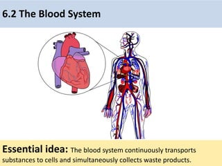
  • 1. 6.2 The Blood System Essential idea: The blood system continuously transports substances to cells and simultaneously collects waste products.
  • 2. Understandings Statement Guidance 6.2 U.1 Arteries convey blood at high pressure from the ventricles to the tissues of the body 6.2 U.2 Arteries have muscle cells and elastic fibers in their walls 6.2 U.3 The muscle and elastic fibers assist in maintaining blood pressure between pump cycles 6.2 U.4 Blood flows through tissues in capillaries. Capillaries have permeable walls that allow exchange of materials between cells in the tissue and the blood in the capillary 6.2 U.5 Veins collect blood at low pressure from the tissues of the body and return it to the atria of the heart. 6.2 U.6 Valves in veins and the heart ensure circulation of blood by preventing backflow. 6.2 U.7 There is a separate circulation for the lungs. 6.2 U.8 The heart beat is initiated by a group of specialized muscle cells in the right atrium called the sinoatrial node 6.2 U.9 The sinoatrial node acts as a pacemaker. 6.2 U.10 The sinoatrial node sends out an electrical signal that stimulates contraction as it is propagated through the walls of the atria and then the walls of the ventricles 6.2 U.11 The heart rate can be increased or decreased by impulses brought to the heart through two nerves from the medulla of the brain. 6.2 U.12 Epinephrine increases the heart rate to prepare for vigorous physical activity
  • 3. Applications and Skills Statement Guidance 6.2 A.1 William Harvey’s discovery of the circulation of the blood with the heart acting as the pump. 6.2 A.2 Pressure changes in the left atrium, left ventricle and aorta during the cardiac cycle. 6.2 A.3 Causes and consequences of occlusion of the coronary arteries. 6.2 S.1 Identification of blood vessels as arteries, capillaries or veins from the structure of their walls. 6.2 S.2 Recognition of the chambers and valves of the heart and the blood vessels connected to it in dissected hearts or in diagrams of heart structure.
  • 4.
  • 5. What are the issues when organizes become multicellular? • Diffusion is not adequate for moving material across more than 1 cell barrier
  • 6. • Circulatory system solves this problem – carries fluids & dissolved material throughout body – cells are never far from body fluid – only a few cells away from blood overcoming the limitations of diffusion
  • 7.
  • 8. There are three main types of blood vessel: Arteries carry high pressure blood away from the heart to tissues that need it Capillaries are very small (< 10 μm diameter) and therefore can penetrate virtually every tissue in the body. Blood moves slowly through them under low pressure providing opportunities for the exchange of substances. Veins carry the low pressure blood back to the heart using valves to ensure blood flows in the correct direction.
  • 9. 6.2 U.1 Arteries convey blood at high pressure from the ventricles to the tissues of the body. AND 6.2 U.2 Arteries have muscle cells and elastic fibers in their walls. AND 6.2 U.3 The muscle and elastic fibers assist in maintaining blood pressure between pump cycles . The structure of arteries• Thick muscular wall and fibrous outer layer help the artery to withstand high pressure • Relatively (to the wall) small lumen maintains high blood pressure. • Muscle contracts to decrease the size of the lumen. This causes an increase blood pressure and therefore maintains high blood pressure between the pulses of high pressure blood travelling from the heart • Elastic fibers stretch to increase the lumen with each pulse of blood. After the pulse of blood passes the fibers recoil decreasing the lumen size and therefore helping to maintain a high blood pressure.
  • 10. 6.2 U.5 Veins collect blood at low pressure from the tissues of the body and return it to the atria of the heart. AND 6.2 U.6 Valves in veins and the heart ensure circulation of blood by preventing backflow. https://commons.wikimedia.org/wiki/File:Venous_valve.svg http://40.media.tumblr.com/tumblr_m0dwjt3WKQ1qzcf71o1_500.jpg Because of the low pressure valves are required to prevent back-flow of the blood and therefore ensure that the blood moves towards to heart. The structure of veins • Veins return blood to the heart for re-circulation. • The large lumen (compared to arteries and the thickness of the wall) means that the blood is under low pressure. • Because there is less pressure to resist the walls of the veins are thinner and less elastic than arteries. They also contain less muscle than the arteries.
  • 11. • Veins carry blood back to the heart. Have thin walls containing valves to prevent the back flow of blood, due to the lower pressure of blood in the veins. • Pressure in veins is low, so veins depend on nearby muscular contractions to move blood along. 6.2.U5 Veins collect blood at low pressure from the tissues of the body and return it to the atria of the heart. AND 6.2.U6 Valves in veins and the heart ensure circulation of blood by preventing backflow.
  • 12. 6.2 U.4 Blood flows through tissues in capillaries. Capillaries have permeable walls that allow exchange of materials between cells in the tissue and the blood in the capillary. The structure of capillaries • Capillaries are the smallest blood vessels and are adapted for the exchange of substances to and from the blood. • This enables tissues to gain nutrients and molecules such as oxygen and to rid themselves of waste material. • Capillaries also allow substances to enter and leave the organism, e.g. gas exchange of oxygen and carbon dioxide in the lungs. Due the the massive number of capillaries present and the small lumen the surface area available for the exchange of substances is very large.
  • 13. 6.2 S.1 Identification of blood vessels as arteries, capillaries or veins from the structure of their walls. a b c d https://www.ouhsc.edu/histology/Text%20Sections/Cardiovascular.html Identify the labelled structures using your understanding of blood vessels.
  • 14. https://www.ouhsc.edu/histology/Text%20Sections/Cardiovascular.html Identify the labelled structures using your understanding of blood vessels. 6.2 S.1 Identification of blood vessels as arteries, capillaries or veins from the structure of their walls.
  • 15. Nature of Science: Theories are regarded as uncertain - William Harvey overturned theories developed by the ancient Greek philosopher Galen on movement of blood in the body. (1.9) Remember a theories are by definition the best accepted explanations and predictions of natural phenomena. Although they are usually thoroughly tested based on evidence and reason theories theories are the only the current best accepted explanation. Theories if successfully questioned can be modified or even rejected/falsified if a better explanation arises. Theories of blood movement around the body • Galen (129 - c216) Blood was not seen to circulate but rather to slowly ebb and flow. Using elusive attractive powers. • Ibn-al-Nafiz (1300) first challenger to Galen idea of blood movement • William Harvey (1578 - 1657) published “De Motu Cordis” in 1628. Even then it took many years for his theory of systemic circulation (similar to modern accepted theory) to succeed Galen’s.
  • 16. Path of blood through the heart • Deoxygenated blood return to the heart to right atrium from the body • The Right Atrium, receives "used blood" from the body. Blood will be pushed through the tricuspid valve to the • Right Ventricle, the chamber which will pump to the lungs through the pulmonic valve to the • Pulmonary Arteries, providing blood to both lungs. Blood is circulated through the lungs where carbon dioxide is removed and oxygen added. It returns through the Systemic Circuit. 6.2 U.7 There is a separate circulation for the lungs.
  • 17. Path of blood through the heart • Oxygenated blood enters the Left Atrium from the lungs • Pulmonary Veins, empty into the Left Atrium. • From the Left Atrium, a chamber which will push the Mitral Valve open. Blood then passes into the • Left Ventricle. Although it doesn't always look like it in drawings done from this angle, this is the largest and most important chamber in the heart. It pumps to the rest of the body. As it pumps, the pressure will close the mitral valve and open the aortic valve, with blood passing through to the Pulmonary Circuit 6.2 U.7 There is a separate circulation for the lungs.
  • 18. 6.2.S.2 Recognition of the chambers and valves of the heart and the blood vessels connected to it in dissected hearts or in diagrams of heart structure a. Atria: thin-walled upper chambers which collect blood returning to the heart Part of the Heart b. Ventricles: thick, muscular lower chambers which pump blood to the lungs and other body tissue. c. Septum (wall): separate the right and left sides of heart to prevent mixing of oxygenated and deoxygenated blood.
  • 19. 6.2.S2 Recognition of the chambers and valves of the heart and the blood vessels connected to it in dissected hearts or in diagrams of heart structure 1. Tricuspid valve /AV valve Valves of the Heart 2. Pulmonary valve 4. Bicuspid valve / AV valve 3. Aortic valve
  • 20. 6.2 S2 Recognition of the chambers and valves of the heart and the blood vessels connected to it in dissected hearts or in diagrams of heart structure. Label the heart diagram
  • 21. 6.2.S2 Recognition of the chambers and valves of the heart and the blood vessels connected to it in dissected hearts or in diagrams of heart structure. Label the heart diagram Now try to label this heart: http://sciencelearn.org.nz/Contexts/See-through-Body/Sci-Media/Animation/Label- the-heart bicuspid valve
  • 22.
  • 23. Pulse • When you take your pulse, you feel (or hear) two changes in pressure. • The first sound is the closing of the AV’s (systolic pressure), the “lub” sound the heart makes. The 2nd sound is the closing of the SV’s (diastolic pressure), the “dub” sound. 6.2 U.8 The heart beat is initiated by a group of specialized muscle cells in the right atrium called the sinoatrial node. 6.2 U.9 The sinoatrial node acts as a pacemaker. 6.2 U.10 The sinoatrial node sends out an electrical signal that stimulates contraction as it is propagated through the walls of the atria and then the walls of the ventricles.
  • 24. Control of the Heart Beat • Beating is due to myogenic muscle. Meaning it is the origin of contraction in the heart and is not controlled externally • The hearts pacemaker (Sinoatrial Node/SA node) which controls the rate of heartbeats is made up of this muscle. • Large groups of these cell cause the atria to contract. • The contraction cause a signal to a group of nerve cells causing the ventricles to contract at the atrioventricular node/AV node. 6.2 U.8 The heart beat is initiated by a group of specialized muscle cells in the right atrium called the sinoatrial node. 6.2 U.9 The sinoatrial node acts as a pacemaker. 6.2 U.10 The sinoatrial node sends out an electrical signal that stimulates contraction as it is propagated through the walls of the atria and then the walls of the ventricles.
  • 25. The control of heart rhythm 6.2 U.8 The heart beat is initiated by a group of specialized muscle cells in the right atrium called the sinoatrial node. 6.2 U.9 The sinoatrial node acts as a pacemaker. 6.2 U.10 The sinoatrial node sends out an electrical signal that stimulates contraction as it is propagated through the walls of the atria and then the walls of the ventricles.
  • 26. The interrelationship of blood flow velocity, cross-sectional area of blood vessels, and blood pressure 6.2 U.8 The heart beat is initiated by a group of specialized muscle cells in the right atrium called the sinoatrial node. 6.2 U.9 The sinoatrial node acts as a pacemaker. 6.2 U.10 The sinoatrial node sends out an electrical signal that stimulates contraction as it is propagated through the walls of the atria and then the walls of the ventricles.
  • 27. 6.2 U.11 The heart rate can be increased or decreased by impulses brought to the heart through two nerves from the medulla of the brain. 6.2 U.12 Epinephrine increases the heart rate to prepare for vigorous physical activity. Control of the Heart Beat • Controlled by the autonomic nervous system response to changing body conditions by speeding up or lowing down breathing • When the level of CO2 are low chemical receptors in the Medulla sends a signal to the Vagus nerve which decreases heart rate. • When the level of CO2 high chemical receptors in the Medulla sends a signal to the Accelerator nerve which increase heart rate. Medulla Vagus Nerve Accelerator Nerve SA Node
  • 28. 6.2 U.11 The heart rate can be increased or decreased by impulses brought to the heart through two nerves from the medulla of the brain. 6.2 U.12 Epinephrine increases the heart rate to prepare for vigorous physical activity. Hormonal Control of Heart Rate • Epinephrine, more commonly known as adrenaline, is a hormone secreted by the medulla of the adrenal glands • Released into the bloodstream, epinephrine causes an increase in heart rate, muscle strength, blood pressure, and sugar metabolism.
  • 29. How does the Heart work? blood from the body blood from the lungs The heart beat begins when the heart muscles relax and blood flows into the atria. STEP ONE
  • 30. The atria then contract and the valves open to allow blood into the ventricles. How does the Heart work? STEP TWO
  • 31. How does the Heart work? The valves close to stop blood flowing backwards. The ventricles contract forcing the blood to the aorta and out of the heart. At the same time, the atria are relaxing and once again filling with blood. The cycle then repeats itself. STEP THREE
  • 32. 6.2 A.2 Pressure changes in the left atrium, left ventricle and aorta during the cardiac cycle. The Cardiac Cycle 1. Right Atria & Ventricles relax and blood flows into the heart from the veins. 2. AV valve open, Right Atria contacts and Ventricle relaxes, blood moves into the ventricle. 3. Pulmonary valve opens, Right Ventricle contracts, blood flows to the lungs. 4. Blood flows from the lung into the left Atria AV valve opens, blood flows into the Left Ventricle. 5. Left Atria contracts blood flows into Left Ventricle. 6. Aortic valve opens, Left Ventricle contracts blood flows into the arteries.
  • 33. 6.2 A.2 Pressure changes in the left atrium, left ventricle and aorta during the cardiac cycle. The Cardiac Cycle Ventricular Pressure 1. Ventricular pressure increases as ventricle contracts. This forces blood into the aorta, increasing aortic pressure. 2. (a) Shows the increase in atrial pressure due to atrial contraction. 3. Shows increase in atrial pressure due to the inflow of blood from the veins (following systole). Ventricular Volume 4. Increase as atrial contraction forces blood into the ventricle 5. Decrease as ventricular contraction forces blood into the aorta 6. Increases as blood returns to the heart following systole. Click on the hyperlink
  • 34. 6.2 A3 Causes and consequences of occlusion of the coronary arteries. 6.3 A1 Causes and consequences of blood clot formation in coronary Arteriosclerosis • Hardening and thickening of the walls of the arteries. • The condition can occur because of fatty deposits on the inner lining of arteries (atherosclerosis). • Calcification of the wall of the arteries, or thickening of the muscular wall of the arteries from chronically elevated blood pressure occurs. • As the deposits of fat and plaque begin to build the lumen of the arteries become reduced. If the plaque frees itself from the wall a blood clot may form. This may lead to coronary thrombosis (reduced blood flow to the heart). • When coronary arteries that supply blood to the heart muscle are effected a shortage of oxygen delivered to the heart itself may lead to a heart attack. Coronary arteries supply heart muscles with oxygen and nutrients
  • 35.
  • 36. 6.2 A3 Causes and consequences of occlusion of the coronary arteries. 6.3 A1 Causes and consequences of blood clot formation in coronary Risk Factors in Coronary Heart Disease Factor Causes Genetics Family history of high cholesterol/blood pressure Age Older people have a greater risk due to a loss in elasticity in arteries Sex Males have a greater risk then females Diet Increases in fat intake lead to higher levels of cholesterol and increase formation of plaque Exercise A lack of exercise increase risk. This is due to a weakening of circulation Obesity Increase in blood pressure which leads to increase plaque formation in the arteries Stress Stress has been linked to increased levels of the hormone Cortisol. Which has been show leads to a increase likelihood of atherosclerosis
  • 37. Essential idea: The lungs are actively ventilated to ensure that gas exchange can occur passively. 6.4 Gas exchange
  • 38. Understandings Statement Guidance 6.4 U.1 Ventilation maintains concentration gradients of oxygen and carbon dioxide between air in alveoli and blood flowing in adjacent capillaries. 6.4 U.2 Type I pneumocytes are extremely thin alveolar cells that are adapted to carry out gas exchange. 6.4 U.3 Type II pneumocytes secrete a solution containing surfactant that creates a moist surface inside the alveoli to prevent the sides of the alveolus adhering to each other by reducing surface tension. 6.4 U.4 Air is carried to the lungs in the trachea and bronchi and then to the alveoli in bronchioles. Students should be able to draw a diagram to show the structure of an alveolus and an adjacent capillary. 6.4 U.5 Muscle contractions cause the pressure changes inside the thorax that force air in and out of the lungs to ventilate them. 6.4 U.6 Different muscles are required for inspiration and expiration because muscles only do work when they contract.
  • 39. Applications and Skills Statement Guidance 6.4 A.1 Causes and consequences of lung cancer. 6.4 A.2 Causes and consequences of emphysema. 6.4 A.3 External and internal intercostal muscles, and diaphragm and abdominal muscles as examples of antagonistic muscle action. 6.4 S.1 Monitoring of ventilation in humans at rest and after mild and vigorous exercise. (Practical 6) Ventilation can either be monitored by simple observation and simple apparatus or by data logging with a spirometer or chest belt and pressure meter. Ventilation rate and tidal volume should be measured, but the terms vital capacity and residual volume are not expected.
  • 40. What is respiration? • External respiration - exchange of O2 & CO2 between respiratory surfaces & the blood [breathing]. • Internal respiration - exchange of O2 & CO2 between the blood & cells. • Cellular respiration - process by which cells use O2 to produce ATP. Gas Exchange in Animals Why do organisms need a respiratory system?
  • 41. CO2 CO2 O2 O2Bloodstream Muscle cells carrying out Cellular Respiration Breathing Glucose + O2 CO2 + H2O + ATP Lungs What is the relationship between respiration and cellular respiration? Why do organisms need a respiratory system?
  • 42. Respiration • (Breathing) provides for the exchange of O2 and CO2 between an organism and its environment Why do organisms need a respiratory system?
  • 43. All respiratory surfaces MUST be: • moist • thin • large enough to meet metabolic needs of organism Challenges: Aquatic organisms - H2O contains only 1/30 of the O2 present in air. Terrestrial organisms - must prevent desiccation of respiratory surface. Why do organisms need a respiratory system? Types of Respiratory Surfaces
  • 44. Structure of the ventilation system a) Trachea b) Cartilage ring support c) Bronchi (plural) Bronchus (single) d) Lung e) Heart f) Sternum g) Rib cage h) Bronchioles j) Alveoli k) Diaphragm 6.4 U.4 Air is carried to the lungs in the trachea and bronchi and then to the alveoli in bronchioles. (Page 78)
  • 45. 6.4 U.4 Air is carried to the lungs in the trachea and bronchi and then to the alveoli in bronchioles. (Page 78)
  • 46. 6.4 U.4 Air is carried to the lungs in the trachea and bronchi and then to the alveoli in bronchioles. (Page 78) Can you use the diagram to produce a flow chart to show the passage of air into the lungs?
  • 47. Review: 6.2 U.11 The heart rate can be increased or decreased by impulses brought to the heart through two nerves from the medulla of the brain. Control of the Heart Beat • Controlled by the autonomic nervous system response to changing body conditions by speeding up or lowing down breathing • When the level of CO2 are low chemical receptors in the Medulla sends a signal to the Vagus nerve which decreases heart rate. • When the level of CO2 high chemical receptors in the Medulla sends a signal to the Accelerator nerve which increase heart rate. Medulla Vagus Nerve Accelerator Nerve SA Node
  • 48. 6.4 A.3 External and internal intercostal muscles, and diaphragm and abdominal muscles as examples of antagonistic muscle action. (Page 78) • The medulla oblongata is involved in regulating many of the bodily processes that are controlled automatically like blood pressure, heart rate and RESPIRATION. • The medulla oblongata detects carbon dioxide (CO2) and Oxygen (O2) levels in the bloodstream and determines what changes need to happen in the body. • It can then send nerve impulses muscles in the ribs and diaphragm, letting them know that they need to either step up their game, or slow down a bit.
  • 49. Ventilation: for gas exchange • The flow of air in and out of the alveoli is called ventilation and has two stages: inspiration (or inhalation) which increases the concentration and expiration (or exhalation) which removes CO2. • For gas exchange to be efficient, high concentration gradients must be maintained in the alveoli. • Lungs are not muscular and cannot ventilate themselves, but instead the whole thorax moves and changes size, due to the action of two sets of muscles: the intercostal muscles and the diaphragm 6.4 U.1 Ventilation maintains concentration gradients of oxygen and carbon dioxide between air in alveoli and blood flowing in adjacent capillaries. (Page 78) Alveoli (air sac)
  • 50. Ventilation of the lungs: Inhale • External intercostal muscles contract, moving ribs up and out • Diaphragm contracts, fattening and moving down • Increased volume in thorax lowering pressure • Air flows into the lungs to balance the pressure change decrease in pressure (draws air inwards) 6.4 A.3 External and internal intercostal muscles, and diaphragm and abdominal muscles as examples of antagonistic muscle action. AND 6.4 U.5 Muscle contractions cause the pressure changes inside the thorax that force air in and out of the lungs to ventilate them. (Page 78)
  • 51. Ventilation of the lungs: Exhale: • Internal intercostal muscles contract, moving ribcage down and in • Abdominal muscles contract pushing the diaphragm up into a dome shape • Decrease in volume of thorax increasing air pressure • Air flows out from the lungs increase in pressure (pushes air outwards) 6.4 A.3 External and internal intercostal muscles, and diaphragm and abdominal muscles as examples of antagonistic muscle action. AND 6.4 U.5 Muscle contractions cause the pressure changes inside the thorax that force air in and out of the lungs to ventilate them. (Page 78)
  • 52. Negative pressure breathing 6.4 A.3 External and internal intercostal muscles, and diaphragm and abdominal muscles as examples of antagonistic muscle action. AND 6.4 U.5 Muscle contractions cause the pressure changes inside the thorax that force air in and out of the lungs to ventilate them. (Page 78)
  • 53. Gas Exchange This is the diffusion of gases (oxygen and carbon dioxide) There are two sites for gas exchange A. Alveoli: Oxygen diffuses into the blood from the alveoli and carbon dioxide diffuses from the blood into the alveoli B. Tissues: Oxygen diffuses from blood into the cells and carbon dioxide diffuses from cells to the blood 6.4 U.1 Ventilation maintains concentration gradients of oxygen and carbon dioxide between air in alveoli and blood flowing in adjacent capillaries. (Page 78)
  • 54. 6.4 U.2 Type I pneumocytes are extremely thin alveolar cells that are adapted to carry out gas exchange. AND (Page 79)
  • 55. 6.4 U.3 Type II pneumocytes secrete a solution containing surfactant that creates a moist surface inside the alveoli to prevent the sides of the alveolus adhering to each other by reducing surface tension. (Page 79)
  • 56. 6.4 U.2 Type I pneumocytes are extremely thin alveolar cells that are adapted to carry out gas exchange. AND 6.4 U.3 Type II pneumocytes secrete a solution containing surfactant that creates a moist surface inside the alveoli to prevent the sides of the alveolus adhering to each other by reducing surface tension. (Page 79) Edited from: https://beyondthedish.files.wordpress.com/2011/11/gas-barrier-small.jpg http://neurobio.drexelmed.edu/education/pil/microanatomy/Cases/01_Quintana/4_Epithelium/Images/alveolarepithelium.jpg Connective tissue Type II pneumocytes • Secrete fluid to moisten the inner surface of the alveolus • Fluid aids diffusion of gases • Fluid contains surfactant to prevent the walls sticking together – maintains the lumen Alveoli walls and gas exchange Type I pneumocytes • A single layer of cells form the walls of an alveolus • Extremely thin – short diffusion distance • Permeable – aids diffusion
  • 57. Emphysema • Disease caused by smoking, lung infections or air pollution. All of which cause an inflammatory response in the lungs. • Protease is released by leukocytes (white blood cells) and inflamed lung tissue. The protease breaks down the connective tissue such as elastin, of the lungs. This results in the destruction of small airways and alveoli. This results in the formation of large air pockets and the breakdown of capillaries • The disease creates a reduction in surface area gas exchange which results shortness of breath, difficulty in breathing, and decreased lung capacity. • Emphysema is a progressive disease with no known cures. There are treatments that can help you manage the disease 6.4 A.2 Causes and consequences of emphysema. (page 79) Symptoms include: • Difficulty breathing • Cough • Loss of appetite • Weight loss
  • 58.
  • 59. Review: 1.6 and U.6 Mutagens, oncogenes and metastasis are involved in the development of primary and secondary tumors. Tumors are abnormal growth of tissue that develop at any stage of life in any part of the body. A cancer is a malignant tumour and is named after the part of the body where the cancer (primary tumour) first develops. Use the links to find out: • most common types of cancer • what causes cancer and associated risk factors • how cancer can be treated http://youtu.be/8BJ8_5Gyhg8 http://www.cancerresearchuk.org/cancer- info/cancerandresearch/all-about-cancer/what-is-cancer/ What causes cancer?
  • 60. 6.4 A.1 Causes and consequences of lung cancer. (Page 78) “Lung cancer is the leading cause of death from cancer in the U.S.” http://www.cancer.gov/types/lung Lung cancer is cancer that starts in the windpipe (trachea), the main airway (bronchus) or the lung tissue. By far the biggest cause of lung cancer is smoking. It causes more than 8 out of 10 cases (86%) including a small proportion caused by exposure to second hand smoke in non smokers (passive smoking). Some other things increase lung cancer risk by a small amount: • Exposure to radon (a radioactive) gas • Air pollution • Previous lung disease • A family history of lung cancer • Past cancer treatment • Previous smoking related cancers • Lowered immunity The symptoms of lung cancer may include: • Being short of breath • Having a cough most of the time • Coughing up phlegm with blood • An ache or pain in the chest or shoulder • Loss of appetite • Tiredness/fatigue • Losing weight Like most cancers lung cancer, if untreated, will end with death. Because detection of lung cancer is difficult it is often only diagnosed in the later stages. As a consequence only 10% of those diagnosed will survive for 5 years. Source: http://www.cancerresearchuk.org/about-cancer/type/lung-cancer/ http://spacecoastdaily.com/wp-content/uploads/2014/01/Cigarette-Smoking-is-Mainly-Causes-of-Lung-Cancer.jpg
  • 61. 6.4 S.1 Monitoring of ventilation in humans at rest and after mild and vigorous exercise. (Practical 6) Monitoring ventilation https://commons.wikimedia.org/wiki/File:DoingSpirometry.JPG https://en.wikipedia.org/wiki/File:Lungvolumes_Updated.png Independent variable: the intensity of exercise Dependent variable: What measure(s) are you using for ventilation? What type of exercise? For how long? How will you make sure that individuals exercise similarly? Frequency of breaths? Spirometer data showing tidal volumes? Ethical and safety concerns: how are you making sure that the experiment is safe? Are participants aware of the risks? Are you seeking written permission from participants? : design an investigation, collect and analyze the data Controlled variables: what are the key factors that need to be measured and kept constant to ensure a fair test? Data analysis: you should be calculating a rate. For example the count of breaths or tidal volume per unit time (s or min)