SlideShare una empresa de Scribd logo
1 de 185
Topic 3: Nucleic Acids
Rosalind Franklin’s X-ray crystallography of DNA
Essential idea: The structure of DNA is ideally suited to
its function
7.1 DNA structure and replication
7.1 S.1 Analysis of results of the Hershey and Chase experiment
providing evidence that DNA is the genetic material.
Hershey and Chase Experiments (1952): Definitive proof that DNA
rather than Protein carries the hereditary information of life
E. Coli bacteriophage: A virus that infects bacteria.
Bacteriophages only contain a protein coat (capsid) and DNA.
They wanted to find out whether the protein or DNA carried the
genetic instructions to make more viruses.
They labeled either the viral proteins or DNA:
– Protein capsid: Labeled with radioactive sulfur (35S)
– DNA: Labeled with radioactive phosphorus (32P)
Radioactive labeled viruses were used to infect cells.
Either Bacteriophage DNA or Proteins Can be
Labeled with Radioactive Elements
7.1 S.1 Analysis of results of the Hershey and Chase experiment
providing evidence that DNA is the genetic material.
Hershey Chase Experiment: DNA is Genetic Material
7.1 S.1 Analysis of results of the Hershey and Chase experiment
providing evidence that DNA is the genetic material.
Hershey and Chase Experiments (1952):
Bacterial cells that were infected with
the two types of bacteriophage, were
then spun down into a pellet
(centrifuged), and examined.
Results:
1. Labeled viral proteins did not enter
infected bacteria (found in
supernatant).
2. Labeled viral DNA did enter bacteria
during viral infection (found in cell
pellet).
Conclusion:
Protein is not necessary to make new
viruses.
DNA is the molecule that carries the
genetic information to make new
viruses!!!!
7.1 S.1 Analysis of results of the Hershey and Chase experiment
providing evidence that DNA is the genetic material.
Rosalind Franklin (1950’s)
• Worked with Maurice Wilkins
• X-ray crystallography = images
of DNA
• Provided measurements on
chemistry of DNA
7.1 A.1 Rosalind Franklin’s and Maurice Wilkins’ investigation of DNA
structure by X-ray diffraction
James Watson & Francis Crick (1953)
• Discovered the double helix
by building models to
conform to Franklin’s X-ray
data and Chargaff’s Rules.
DNA Double Helix
Nitrogenous
Base (A,T,G or C)
“Rungs of ladder”
“Legs of ladder”
Phosphate &
Sugar Backbone
DNA
• Two strands coiled called
a double helix
• Sides made of a pentose
sugar Deoxyribose
bonded to phosphate
(PO4) groups by
phosphodiester bonds
• Center made of nitrogen
bases bonded together
by weak hydrogen bonds
7.1 U.2 DNA structure suggested a mechanism for DNA replication
DNA
• Stands for Deoxyribonucleic acid
• Made up of subunits called
nucleotides
• Nucleotide made of:
1. Phosphate group
2. 5-carbon sugar
3. Nitrogenous base
7.1 U.2 DNA structure suggested a mechanism for DNA replication
DNA Nucleotide
O=P-O
O
Phosphate
Group
N
Nitrogenous base
(A, G, C, or T)
CH2
O
C1
C4
C3 C2
5
Sugar
(deoxyribose)
O
7.1 U.2 DNA structure suggested a mechanism for DNA replication
11
DNA
P
P
P
O
O
O
1
2
3
4
5
5
3
3
5
P
P
P
O
O
O
1
2 3
4
5
5
3
5
3
G C
T A
Hydrogen
Bonds
7.1 U.1 Nucleosomes help to supercoil the DNA
• A nucleosome consists of DNA wrapped around
8 histone proteins (prokaryotic cells lack these
proteins making there DNA “naked).
• The DNA wraps twice around the histone
protein core.
• Another histone protein is attached to the
outside of the DNA strand. It helps maintain
the colloidal structure of the nucleosome.
• DNA, because of its negative charge is
attracted to the positive charge on the amino
acids of the histone proteins.
• Tails of neighboring histones, link up during
chromosomal condensation, causing the
nucleosomes to pull closer together.
• This is part of the supercoiling process that
occurs during mitosis and meiosis
• Supercoiling in general helps regulate
transcription because only certain areas of the
DNA are accessible for the production of mRNA
by transcription. This regulates the production
of a polypeptide. http://pbil.univlyon1.fr/members/sagot/htdocs/tea
m/projects/chromo_net/images/epi.jpg
Nucleosome
8 Histones
DNA Replication
7.1 U.3 DNA polymerases can only add nucleotides to the 3’ end of a primer
Synthesis Phase (S phase)
• S phase during interphase of the cell
cycle
• Nucleus of eukaryotes
Mitosis
-prophase
-metaphase
-anaphase
-telophase
G1 G2
S
phase
interphase
DNA replication takes
place in the S phase.
Mitosis
-prophase
-metaphase
-anaphase
-telophase
G1 G2
S
phase
interphase
DNA replication takes
place in the S phase.
7.1 U.3 DNA polymerases can only add nucleotides to the 3’ end of a primer
Super coiling begin in prophase, making
chromosomes visible for the first time
2.7 S.2 Analysis of Meselson and Stahl’s results to obtain support for the theory of semi-
conservative replication of DNA.
https://upl oad.wikimedia.org/wikipedia/commons/a/a2/DNAreplicationModes.png
Before Meselson and Stahl’s work there were different proposed models for DNA replication.
After their work only semi-conservative replication was found to be biologically significant.
2.7 S.2 Analysis of Meselson and Stahl’s results to obtain support for the theory of semi-
conservative replication of DNA.
Learn about Meselson and Stahl’s
work with DNA to discover the
mechanism of semi-conservative
replication
http://highered.mheducation.com/olcweb/cgi/pluginpop.cg
i?it=swf::535::535::/sites/dl/free/0072437316/120076/bio2
2.swf::Meselson%20and%20Stahl%20Experiment
http://www.nature.com/scitable/topicpage/Semi-Conservative-DNA-Replication-Meselson-and-Stahl-421#
• DNA replication is very specific to the
arrangements of base pairs
• In DNA replication, the strands separate
– Enzymes use each strand as a template to assemble
the new strands
DNA REPLICATION
Parental molecule
of DNA
Both parental strands serve
as templates
Two identical daughter
molecules of DNA
Nucleotides
A
7.1 U.3 DNA polymerases can only add nucleotides to the 3’ end of a primer
1. Helicase: unwinds DNA at origins of replication
2. Initiation proteins separate 2 strands  forms replication bubble
3. Single-Strand Binding Proteins attach and keep the 2 DNA strands
separated and untwisted
4. Primase: puts down RNA primer to start replication
5. DNA polymerase III: adds complimentary bases to leading strand (new
DNA is made 5’  3’)
6. Lagging strand grows in 3’5’ direction by the addition of Okazaki
fragments
7. DNA polymerase I: replaces RNA primers with DNA
8. DNA ligase: seals fragments together
9. DNA gyrase: an enzyme that relieves strain while double-strand DNA is
being unwound by helicase.
7.1 U.5 DNA replication is carried out by a complex system of enzymes. [The proteins
and enzymes involved in DNA replication should include helicase, DNA gyrase, single
strand binding proteins, DNA primase and DNA polymerases I and III.]
Major Steps of DNA Replication:
DNA Gyrase
SSBP
DNA Poly
Lead
Strand
Lagging
Strand
DNA Ligase
RNA Primer
Helicase
Replication Direction
RNA Primase
Helicase The ‘ase’ ending indicates it is an enzyme. Helicase is
DNA’s origin of replication and creates replication forks
2.7 U.2 Helicase unwinds the double helix and separates the two strands by breaking
hydrogen bonds.
• DNA replication begins at the different origins in the 5’ to 3’ direction at
the replication fork.
• RNA primase (is the primer to that starts the process) attaches to the DNA
and adds a small RNA primer to provide a free 3’ OH starting point since DNA
polymerases can only add nucleotides to the 3’ end of a primer
• DNA polymerase III adds free nucleotides in the 5’ to 3’ direction in the
direction of the replication fork.
7.1 U.3 DNA polymerases can only add nucleotides to the 3’ end of a primer
• Begins at Origins of Replication
• Two strands open forming Replication Forks (Y-
shaped region)
• New strands grow at the forks
• The leading strand (copies in one long continuous
piece) begins on the 3’ side and the lagging strand
begins on the 5’ side (copies in small fragments,
which must be pieced together)
Replication
Fork
Parental DNA Molecule
3’
5’
3’
5’
7.1 U.4 DNA replication is continuous on the leading strand and discontinuous on the
lagging strand. [Details of DNA replication differ between prokaryotes and eukaryotes.
Only the prokaryotic system is expected.]
Remember HOW the Carbons Are Numbered!
O
O=P-O
O
Phosphate
Group
N
Nitrogenous base
(A, G, C, or T)
CH2
O
C1
C4
C3 C2
5
Sugar
(deoxyribose)
Leading strand vs. Lagging strand
Primase adds RNA primer is RNA that initiates DNA
synthesis.
DNA Gyrase
7.1 U.3 DNA polymerases can only add nucleotides to the 3’ end of a primer
2.7 U.3 DNA polymerase links nucleotides together to form a new strand, using the pre-
existing strand as a template.
DNA polymerase always moves in a 5’ to 3’ direction
• DNA polymerase catalyzes the covalent phosphodiester bonds
between sugars and phosphate groups forming covalent bonds
DNA polymerase III adds nucleotides in 5’3’
direction on leading strand
2.7 U.1 The replication of DNA is semi-conservative and depends on complementary base pairing.
https://upload.wikimedia.org/wikipedia/commons/3/33/DNA_replication_split_horizontal.svg
1. Each of the nitrogenous bases can only
pair with its partner (A=T and G=C) this is
called complementary base pairing.
2. The two new strands formed will be
identical to the original strand.
2.7.U1 The replication of DNA is semi-conservative and depends on complementary base pairing.
https://upload.wikimedia.org/wikipedia/commons/3/33/DNA_replication_split_horizontal.svg
3. Each new strand contains one original and one new
strand, therefore DNA Replication is said to be a Semi-
Conservative Process.
Replication on leading strand
1. RNA Primase
attaches RNA
Primer
2. DNA Polymerase
attaches
nucleotides in a 5’
to 3’ direction
7.1 U.4 DNA replication is continuous on the leading strand and discontinuous on the
lagging strand. [Details of DNA replication differ between prokaryotes and eukaryotes.
Only the prokaryotic system is expected.]
Replication on lagging
strand 1. RNA Primase attaches
multiple pieces of RNA
Primer
2. DNA Polymerase
attaches nucleotides in a
5’ to 3’ direction in
between the Primer
pieces creating Okazaki
fragments
3. DNA Polymerase
replaces the RNA primer
pieces with DNA
4. Ligase glues the
fragments together
1. Helicase: unwinds DNA at origins of replication Initiation proteins
separate 2 strands  forms replication bubble
2. Single-Strand Binding Proteins attach and keep the 2 DNA strands
separated and untwisted
3. RNA Primase: puts down RNA primer to start replication
4. DNA polymerase III: adds complimentary bases to leading strand (new
DNA is made 5’  3’)
5. Lagging strand grows in 3’5’ direction by the addition of Okazaki
fragments
6. DNA polymerase I: replaces RNA primers with DNA
7. DNA ligase: seals fragments together
8. DNA gyrase: an enzyme that relieves strain while double-strand DNA is
being unwound by helicase.
7.1 U.5 DNA replication is carried out by a complex system of enzymes. [The proteins
and enzymes involved in DNA replication should include helicase, DNA gyrase, single
strand binding proteins, DNA primase and DNA polymerases I and III.]
Major Steps of DNA Replication:
DNA Gyrase
SSBP
DNA Poly
Lead
Strand
Lagging
Strand
DNA Ligase
RNA Primer
Helicase
Replication Direction
RNA Primase
• Dideoxyribonucleotides inhibit DNA
polymerase during replication, thereby stopping
replication from continuing.
• Dideoxyribonucleotides with fluorescent markers,
is incorporated into sequences of DNA, to stop
replication at the point at which they are added.
• This creates different sized fragments with
fluorescent markers that can be separated by gel
electrophoresis and analyzed by comparing the
color of the fluorescence with the fragment
length.
7.1 A.2 Use of nucleotides containing deoxyribonucleic acid to stop DNA replication in
preparation of samples for base sequencing
Problem at the 5’ End
• DNA poly only adds
nucleotides to 3’ end
• No way to complete 5’
ends of daughter strands
• Over many replications,
DNA strands will grow
shorter and shorter
7.1 U.6 Some regions of DNA do not code for proteins but have other important
functions. [The regions of DNA that do not code for proteins should be limited to
regulators of gene expression, introns, telomeres and genes for tRNAs.]
Telomeres: repeated units of short nucleotide sequences
(TTAGGG) at ends of DNA
• Telomeres “cap” ends of DNA to postpone erosion of genes
at ends (TTAGGG)
• Telomerase: enzyme that adds to telomeres
– Eukaryotic germ cells, cancer cells
Telomeres stained
orange at the ends
of mouse
chromosomes
7.1 U.6 Some regions of DNA do not code for proteins but have other important
functions. [The regions of DNA that do not code for proteins should be limited to
regulators of gene expression, introns, telomeres and genes for tRNAs.]
Telomeres & Telomerase
7.1 U.6 Some regions of DNA do not code for proteins but have other important
functions. [The regions of DNA that do not code for proteins should be limited to
regulators of gene expression, introns, telomeres and genes for tRNAs.]
1.6 Cell division
Essential idea: Cell division is essential but must be
controlled.
Why do cells divide:
• Growth: Multicellular organisms
increase their size by increasing
their number of cells through
mitosis
• Asexual reproduction: Certain
eukaryotic organisms may
reproduce asexually by mitosis
(e.g. vegetative reproduction)
• Tissue Repair: Damaged tissue
can recover by replacing dead or
damaged cells
• Embryonic development: A
fertilized egg (zygote) will
undergo mitosis and
differentiation in order to
develop into an embryo
• Cellular division in
eukaryotic cells.
• Chromatin is arranged
into chromosomes.
• Chromosomes double.
• Cell grows in size.
• Cells divide.
• Is cellular cloning.
Cell division
2 phases:
1. Interphase
2. M phase
(mitotic phase)
a. Prophase
b. Metaphase
c. Anaphase
d. Telophase &
cytokinesis
Figure 12.4 The cell cycle
Phases of the Cell Cycle (life cycle of a cell)
Interphase
• The non-dividing phase in a cell
• Lasts about ~ 90% of the cell cycle.
• The cell grows and replicates DNA
preparing for Mitosis.
• There are three periods:
3 periods of Interphase
1. Go – a cell functioning as normal
2. G1 phase – first growth phase
3. S phase- synthesis of DNA
4. G2 phase- 2nd growth phase
Mitosis is a reliable process. Only one error
occurs per 100,000 cell divisions.
1.6 U.4 Interphase is a very active phase of the cell cycle with many
processes occurring in the nucleus and cytoplasm.
1.6 U.4 Interphase is a very active phase of the cell cycle with many processes occurring
in the nucleus and cytoplasm.
Interphase
This when the cell carries out it’s normal functions
Metabolic reactions (e.g. respiration to produce ATP) are
necessary for the life of the cell
Protein synthesis - proteins and enzymes are necessary to allow
cell grow
Organelles numbers are increased to first support the enlarged
cell
DNA is replicated to ensure a second copy is available to enable
mitosis
Cells spend the majority of their time in interphase. It is a
very active phase of the cycle.
Mr
P
O
D
http://botit.botany.wisc.edu/Resources/Botany/Mitosis/Allium/Various%20views/Interphase%20prophase.JPG
1.6 U.5 Cyclins are involved in the control of the cell cycle.
Cyclinsare a family of proteins that control the progression of
cells through the cell cycle
Cells cannot progress to the next
stage of the cell cycle unless the
specific cyclin reaches it threshold.
http://upload.wikimedia.org/wikipedia/commons/thumb/9/99/Protein_CCNE1_PDB_1w98.png/800px-Protein_CCNE1_PDB_1w98.png
Cyclins bind to enzymes called
cyclin-dependent kinases
These kinases then become active and
attach phosphate groups to other proteins
in the cell.
The attachment of phosphate triggers the other proteins to become active
and carry out tasks (specific to one of the phases of the cell cycle).
4
3
2
1
http://upload.wikimedia.org/wikipedia/commons/thumb/9/99/Protein_CCNE1_PDB_1w98.png/800px-Protein_CCNE1_PDB_1w98.png
Triggers cells to
move from G0 to
G1 and from G1
into S phase.
prepares the cell
for DNA
replication in S
phase.
activates DNA
replication inside
the nucleus in S
phase.
promotes the assembly
of the mitotic spindle
and other tasks in the
cytoplasm to prepare
for mitosis.
Progression through parts of the cell cycle are affected in various
ways by specific cyclins
1.6 U.1 Mitosis is division of the nucleus into two genetically identical
daughter nuclei.
http://commons.wikimedia.org/wiki/File:Chromosome.svg
centromere is the part
of a chromosome that
links sister chromatids
Sister chromatids are duplicated
chromosomes attached by a centromere
Get the terminology right centrioles
organize spindle
microtubules
Spindle
microtubules
(also referred to
as spindle
fibers)
In animal cells two centrioles are held by a
protein mass referred to as a centrosome
After anaphase when the sister chromatids
separate they should then be referred to as
chromosomes
It is easy to misuse the terms chromatid and
chromosome. It is even easier to confuse the terms
centromere, centriole and centrosome due to their
similar spelling. Keep the terms clear in your mind
to avoid losing marks.
http://commons.wikimedia.org/wiki/Mitosis#mediaviewer/File:Mitosis_cells_sequence.svg
1.6 U.2 Chromosomes condense by supercoiling during mitosis.
Why supercoil chromosomes? Human cells are on average
10μm in diameter and the
nucelus within each is less
than 5 μm in diameter.
Human chromosomes are
15mm to 85mm (15,000μm
to 85,000 μm) in length.
Chromosomes need to be
stored compactly to fit
within the nuclei of cells.
This problem becomes more
acute during mitosis when
chromosomes need to be
short and compact enough
that they can be separated
and moved to each end of
the cell.
1.6 U.2 Chromosomes condense by supercoiling during mitosis.
How are chromosomes supercoiled?
Strain is placed on a DNA helix by over winding or under winding of the helix
This causes the DNA molecule to coil back on itself becoming shorter and wider
Remember that in eukaryotes proteins called histones form nucleosomes to aid the
process of supercoiling
http://www.maths.uq.edu.au/~infinity/Infinity7/images/supercoiling.gifhttp://vanat.cvm.umn.edu/mMeiosis/images/chromosome-X.jpg
http://highered.mheducation.com/sites/0072495
855/student_view0/chapter2/animation__mitosis
_and_cytokinesis.html
Use the animated tutorials to learn about mitosis
http://www.johnkyrk.com/mitosis.html
http://www.sumanasinc.com/webcontent/animations/content
/mitosis.html
http://outreach.mcb.harvard.edu/animations/cellcycle.
swf
Prophase
• The nucleolus disappears.
• Chromatin condenses into
visible chromosomes.
• There are two sister chromatids
held together by a centromere.
• The mitotic spindle forms in the
cytoplasm. .
1.6 S.1 Identification of phases of mitosis in cells viewed with a microscope or in a
micrograph
Metaphase
• The nuclear envelope
disappears.
• Spindle fibers extend
from each pole to the
cell’s equator.
• Spindle fibers attach to
the centromeres.
Figure 12.3 Chromosome duplication and distribution during mitosis
Anaphase
• Characterized by
movement. It begins when
pairs of sister chromatids
pull apart.
• Sister chromatids move to
opposite poles of the cell.
• Chromosomes look like a
“V” as they are pulled.
• At the end of anaphase, the
two poles have identical
number and types of
chromosomes.
Telophase
• Microtubules elongate the cell.
• Daughter nuclei begin to form at the
two poles.
• Nuclear envelopes re-form.
• Nucleolus reappears.
• Chromatin uncoils.
• The cells cytoplasm begins to pinch.
• It is basically the opposite of
prophase.
1.6 U.3 Cytokinesis occurs after mitosis and is different in plant and animal cells.
mitosis is the division of the nucleus,
cytokinesis is the division of the cytoplasm
to create two cells
Though mitosis is similar for animal and plant cells
cytokinesis is very different.
http://wwwprod.biochem.wisc.edu/biochem/faculty/bednarek/images/figure_color.gif
http://glencoe.mheducation.com/sites/983
4092339/student_view0/chapter10/animati
on_-_cytokinesis.html
http://www.haroldsmithlab.com/images/pg_HeLa_cell_division.jpg
Figure 12.8 Cytokinesis in animal and plant cells
1.6 S.1 Identification of phases of mitosis in cells viewed with a microscope or in a
micrograph. 1.6 S.2 Determination of a mitotic index from a micrograph.
http://www.nuffieldfoundation.org/practical-biology/investigating-mitosis-allium-root-tip-squash
A very good, well explained lab outline for creating slides and calculating the
mitotic index.
http://www.biology.arizona.edu/cell_bio/activities/cell_cycle/cell_cycle.html
An excellent online
alternative if resources don’t
permit students to create
and view their own slides
1.6 U.6 Mutagens, oncogenes and metastasis are involved in the development of
primary and secondary tumors.
Tumors are abnormal growth of tissue that develop at any stage of life in any part
of the body.
A cancer is a malignant tumour and is named after the part of the body where the
cancer (primary tumour) first develops. Use the links to find out:
• most common types of cancer
• what causes cancer and associated risk factors
• how cancer can be treated
1.6 U.6 Mutagens, oncogenes and metastasis are involved in the development of
primary and secondary tumors.
mutation in a oncogene
If a mutation occurs in an oncogenes it can become cancerous. In normal cells
oncogenes control of the cell cycle and cell division.
http://en.wikipedia.org/wiki/Oncogene#mediaviewer/File:Oncogenes_illustration.jpg
uncontrolled cell division
tumor formation
malfunction in the control
of the cell cycle
1.6 U.6 Mutagens, oncogenes and metastasis are involved in the development of
primary and secondary tumors.
Mutagens are agents that
cause gene mutations. Not all
mutations result in cancers,
but anything that causes a
mutation has the potential to
cause a cancer.
Mutagens can be:
• chemicals that cause
mutations are referred to as
carcinogens
• high energy radiation such
as X-rays
• short-wave ultraviolet light
• Some viruses
A mutation is a change in an organisms genetic code. A mutation/change in the
base sequence of a certain genes can result in cancer.
http://en.wikipedia.org/wiki/Oncogene#mediaviewer/File:Oncogenes_illustration.jpg
1.6 U.6 Mutagens, oncogenes and metastasis are involved in the development of
primary and secondary tumors.
Factors (other than exposure to
mutagens) that increase the
probability of tumour
development include:
• The vast number of cells in a
human body – the greater
the number of cells the
greater the chance of a
mutation.
• The longer a life span the
greater the chance of a
mutation.
Several mutations must occur in the same cell for it to become a tumour
causing cell. The probability of this happening in a single cell is extremely small.
http://en.wikipedia.org/wiki/Oncogene#mediaviewer/File:Oncogenes_illustration.jpg
1.6 A.1 The correlation between smoking and incidence of cancers.
http://en.wikipedia.org/wiki/File:Smoking_lung_cancer.png
There are many other similar
surveys in different countries,
with different demographics
that show similar results.
Along with lung cancer,
cancers of mouth and throat
are very common as these
areas are in direct contact with
the smoke too. It might
surprise you that the following
cancers are also more
common in smokers:
• Head and neck
• Bladder
• Kidneys
• Breast
• Pancreas
• Colon
a. Describe the relationship shown.
b. What type of correlation is shown
c. How strong is the correlation? Justify your answer by discussing the evidence.
d. The correlation shown here is lagged. A lag is a time gap between the factors. Estimate the
size of the lag between cigarette consumption and lung cancer death.
http://en.wikipedia.org/wiki/File:Smoking_lung_cancer.png
a. Describe the relationship shown.
b. What type of correlation is shown
c. How strong is the correlation? Justify your answer by discussing the evidence.
d. The correlation shown here is lagged. A lag is a time gap between the factors. Estimate
the size of the lag between cigarette consumption and lung cancer death.
There are many other similar surveys
in different countries, with different
demographics that show similar
results. Along with lung cancer,
cancers of mouth and throat are
very common as these areas are in
direct contact with the smoke too. It
might surprise you that the following
cancers are also more common in
smokers:
• Head and neck
• Bladder
• Kidneys
• Breast
• Pancreas
• Colon
Essential idea: Information stored as a code in DNA is
copied onto mRNA
7.2 Transcription & Gene Expression
http://www.knowingforsure.com/wp-content/uploads/2015/01/Traits.jpg
Trait vs Fate
2.7 U.4 Transcription is the synthesis of mRNA copied from the DNA base sequences by
RNA polymerase.
2.7 U.5 Translation is the synthesis of polypeptides on ribosomes.
Q - What is the purpose of transcription and translation?
A - Gene expression the processes of create a polypeptides which
in turns folds to become a protein. Proteins carry out many essential
functions in cells. For more detail review 2.4.U7.
Rhodopsin - A
Light absorbing
pigment
Rubisco
• Full name ribulose bisphosphate carboxylase
• Enzyme - catalyzes the reaction that fixes carbon
dioxide from the atmosphere
• Provides the source of carbon from which all
carbon compounds, required by living organisms,
are produced.
• Found in high concentrations in leaves and algal
cells
http://upload.wikimedia.org/wikipedia/commons/b/b0/Mint-leaves-2007.jpg
2.4 A.1 Rubisco, insulin, immunoglobulins, rhodopsin, collagen and spider silk as examples of the
range of protein functions.
Collagen
• A number of different forms
• All are rope-like proteins made of three polypeptides wound
together.
• About a quarter of all protein in the human body is collagen
• Forms a mesh of fibers in skin and in blood vessel walls that resists
tearing.
• Gives strength to tendons, ligaments, skin and blood vessel walls.
• Forms part of teeth and bones, helps to prevent cracks and fractures
to bones and teeth https://en.wikipedia.org/wiki/Tooth_(human)#med
iaviewer/File:Teeth_by_David_Shankbone.jpg
2.4 A.1 Rubisco, insulin, immunoglobulins, rhodopsin, collagen and spider silk as
examples of the range of protein functions.
spider silk
• Different types of silk with
different functions
• Dragline silk is stronger
than steel and tougher than
Kevlar
• When first made it contains
regions where the
polypeptide forms parallel
arrays (bottom)
• Some regions seem like a
disordered tangle (middle)
• When the stretched the
polypeptide gradually
extends, making the silk
extensible and very
resistant to breaking.
https://en.wikipedia.org/wiki/Spider_silk#mediaviewer/File:Structure_of_spider_silk_thread_Modified.svg
2.4 A.1 Rubisco, insulin, immunoglobulins, rhodopsin, collagen and spider silk as
examples of the range of protein functions.
2.7 U.4 Transcription is the synthesis of mRNA copied from the DNA base sequences by
RNA polymerase 2.7 U.5 Translation is the synthesis of polypeptides on ribosomes.
http://learn.genetics.utah.edu/content/molecules/transcribe/
7.1 U.1 Nucleosomes help to supercoil the DNA. 7.2 U.5 Gene expression is regulated by
proteins that bind to specific base sequences in DNA.
• A nucleosome consists of DNA wrapped around 8
histone proteins (prokaryotic cells lack these
proteins making there DNA “naked).
• The DNA wraps twice around the histone protein
core.
• Another histone protein is attached to the outside
of the DNA strand. It helps maintain the colloidal
structure of the nucleosome.
• DNA, because of its negative charge is attracted to
the positive charge on the amino acids of the
histone proteins.
• Tails of neighboring histones, link up during
chromosomal condensation, causing the
nucleosomes to pull closer together.
• This is part of the supercoiling process that occurs
during mitosis and meiosis
• Supercoiling in general helps regulate transcription
because only certain areas of the DNA are
accessible for the production of mRNA by
transcription. This regulates the production of a
polypeptide. http://pbil.univlyon1.fr/members/sagot/htdocs/tea
m/projects/chromo_net/images/epi.jpg
MethylationAcetylation
7.2 U.2 Nucleosomes help to regulate transcription in eukaryotes.
• Supercoiling helps regulate
transcription, one supercoiling
modification is through the
modification of the histone tails.
• Acetylation Acetyl groups can be
added to the positively charged
histone tails, they become negative
and that repels the negatively
charged DNA. This opens up the
nucleosome so the DNA is not as
close to the histone anymore
causing gene expression
• Methylation Methyl group is non
polar which causes DNA remains
tightly packed and transcription is
inhibited.
7.2 S.1 Analysis of changes in the DNA methylation patterns.
http://i.dailymail.co.uk/i/pix/2008/09/12/article-1054890-
029CF17900000578-854_233x364.jpg
• Another way gene expression can be controlled is
through methylation (adding a methyl CH3 group) to the histone
proteins.
• Methylation of the histone proteins inhibites transcription of the gene
• The amount of methylation can vary over an organisms lifetime and
can be affected by environmental factors
7.2 U.6 The environment of a cell and of an organism has an impact on
gene expression.
The impact gene expression
Morphogenic Effect (Aging affects) the accumulation of damage
cells over a lifetime; decreases the capacity to maintain
homeostasis
7.2 U.2 Nucleosomes help to regulate transcription in eukaryotes.
Epigenetics
• The changes related to gene
expression or cellular
phenotype of without changes
to the nucleotide sequence of
the genome.
• Examples of mechanisms that
produce such changes are DNA
methylation and histone
modification of the
nucleosomes, each of which
alters how genes are expressed
without altering the
underlying DNA sequence.
Trait vs Fate
2.7.U4 Transcription is the synthesis of mRNA copied from the DNA base sequences by
RNA polymerase.
Step one: Transcription the process by which an RNA sequence is produced from a
DNA template:
Gene expression is the constructions of a protein from the DNA using RNA.
RNA with the help of ribosomes constructs a protein from amino acids
7.2 A.1 The promoter as an example of non-coding DNA with a function.
• The promoter region is a
DNA sequence that
initiates transcription and
is an example of non-
coding DNA that plays a
role in gene expression.
This promoter region is
called the TATA box.
• The promoter sequence is
located near the start site
of transcription and is
where the RNA
polymerase binds in order
for transcription to take
place.
• DNA always is copied in a
5’ to 3’ direction.
http://study.com/cimages/multimages/16/junk_dna_1.jpg
• The enzyme RNA polymerase binds to a site on the DNA at the start of a gene (The sequence of
DNA that is transcribed into RNA is called a gene).
• RNA polymerase separates the DNA strands and synthesizes a complementary RNA copy from
the antisense DNA strand.
2.7 U.4 Transcription is the synthesis of mRNA copied from the DNA base sequences by RNA
polymerase.
7.2 U.3 Eukaryotic cells modify mRNA after transcription.
http://i.dailymail.co.uk/i/pix/2008/09/12/article-1054890-
029CF17900000578-854_233x364.jpg
a) The gene has a promotor
region and a terminator
region
b) Transcription requires the
presence of a regulator
protein from another gene
(possible from another
chromosome).
c) The RNA polymerase can
now bind to the promotor
and begin the transcription
of the gene.
d) The mRNA is transcribed
including introns
e) The completed mRNA which
will require post
transcriptional modification
to remove the introns.
click4biology
TATA box
7.2 U.1 Transcription occurs in a 5’ to 3’ direction. [RNA polymerase adds the 5´ end of
the free RNA nucleotide to the 3´ end of the growing mRNA molecule.]
• Transcription occurs in a 5’ to 3’
direction where the 5’ end of the free RNA
nucleotide is added to the 3’ end of the
RNA molecule that is being synthesized.
Consisting of 3 stages called
1. Initiation RNA polymerase binds to the
promoter with the help of specific binding
proteins
2. Elongation transcription machinery needs
to move histones out of the way, unwinding
DNA, allowing RNA Polymerase to synthesis of
a new RNA strand in the 5′ to 3′ direction
3. Termination RNA synthesis will continue
along the DNA template strand until the
polymerase encounters a signal that tells it to
stop
7.1 U.6 Some regions of DNA do not code for proteins but have other important
functions. [The regions of DNA that do not code for proteins should be limited to
regulators of gene expression, introns, telomeres and genes for tRNAs.]
• There are many areas of DNA containing
repetitive sequences, especially in
eukaryotic DNA, in humans it makes up
between 24-37% of our genome. These
repetitive areas usually occurs near the
ends of chromosomes.
• Introns are non-coded regions, exons
are coded areas for a protein, these
two areas must be separated and
introns must be removed before leaving
the nucleus for protein synthesis to take
place.
*Spliceosome are enzymes constructed
from RNA and are used in eukaryotic cell
to remove these introns.
http://www.phschool.com/science/biology_place/bio
coach/images/transcription/eusplice.gif
Post Transcriptional Modification
Pre mRNA
Mature mRNA
7.1 U.6 Some regions of DNA do not code for proteins but have other important
functions. [The regions of DNA that do not code for proteins should be limited to
regulators of gene expression, introns, telomeres and genes for tRNAs.]
• In addition to introns,
some repetitive sequences
are called telomeres, these
areas protect DNA during
replication. Telomeres are
the caps at the end of each
strand of DNA that protect
our chromosomes, like the
plastic tips at the end of
shoelace They prevents the
loss of genes near the end
of the chromosomes.
Telomeres stained green and red at
the ends of the chromosomes
http://www.newswise.com/images/uplo
ads/2010/09/28/karlsederhr.jpg
7.2 U.4 Splicing of mRNA increases the number of different proteins an
organism can produce
1. Promotor region
2. Free Nucleotide
Phosphates
3. Addition of Nucleotides
to the new mRNA
4. Early mRNA
5. Early mRNA showing
introns (non-coding)
6.Introns removed by the
enzyme spliceosome
allowing exons to combine
7. Mature mRNA ready for
translation
8. mRNA going to
cytoplasm.
click4biology
7.2 U.4 Splicing of mRNA increases the number of different proteins an organism can
produce.
https://commons.wikimedia.org/wiki/File:DNA_alternative_splicing.gif
The splicing process above can happen in different ways to the same gene, particular
exons (of a gene) may be included within or excluded from mature mRNA
Multiple proteins produced by a single gene. Each proteins produced will vary in
it’s biological function. An example of this is the IgM gene which produces
different immunoglobulins (antibodies) to fight different pathogens.
• The variable nature of the Short tandem repeats (STR) regions that are analyzed for
forensic testing intensifies the discrimination between one DNA profile and another.
Theses sections have high rates of mutations and change frequently. Forensic science
takes advantage of the population's variability in STR lengths, enabling scientists to
distinguish one DNA sample from another. For example, the likelihood that any two
individuals (except identical twins) will have the same 13-loci DNA profile can be as low
as 1 in 1 billion or less.
7.1 A.3 Tandem repeats are used in DNA profiling.
• Short tandem repeats (STRs), also known as variable tandem repeats (VNTRs) are regions of
noncoding DNA that contain repeats of the same nucleotide sequence. These short repeats
show variations between individuals in terms of the number of times the sequences is
repeated.
Example
• CATACATACATACATACATACATACATA
repeated 7 times for one individual.
However, in another individual, 11
times
CATACATACATACATACATACATACATACATA
CATACATACATA.
• Used in DNA profiling used in crime
scene investigations, genealogical and
paternity tests.
7.2 U.6 The environment of a cell and of an organism has an
impact on gene expression.
• The environment, as well as the organism's internal
world, which includes such factors as its hormones
and metabolism can have an impact on gene
expression
• Temperature and light are external conditions which
can affect gene expression in certain organisms.
• As an example, Himalayan rabbits carry the gene,
which is required for the development of pigments in
the fur, skin, and eyes, and whose expression is
regulated by temperature
• Specifically, a gene called the C gene is inactive
above 35°C, and it is maximally active from 15°C to
25°C. This temperature regulation of gene expression
produces rabbits with a distinctive coat coloring.
• In the warm weather no pigments fur is white
• In low temperature the rabbit's extremities (i.e.,
the ears, tip of the nose, and feet), where the,
the C gene actively produces pigment, making
these parts of the animal black.
http://upload.wikimedia.org/wikipedia/commons/0/06/Kr%C
3%B3liki_kalifornijskie_666.jpg
http://upload.wikimedia.org/wikipedia/en/8/81/Kostya2.jpg
Essential idea: Information transferred from DNA to mRNA is
translated into an amino acid sequence.
7.3 Translation
Section of Titin, our largest known protein
http://circ.ahajournals.org/content/124/8/876/F2.large.jpg
Components of Translation
1. mRNA = message
2. tRNA = interpreter
3. Ribosome = site of translation
7.3 U.1 Initiation of translation involves assembly of the components that carry out the
process.
2.7 U.5 Translation is the synthesis of polypeptides on ribosomes.
http://www.nature.com/scitable/topicpage/ribosomes-transcription-and-translation-14120660
Translation is the process of protein synthesis in which the genetic information encoded
in mRNA is translated into a sequence of amino acids in a polypeptide chain
A ribosome is composed of two halves, a large and a
small subunit. During translation, ribosomal subunits
assemble together like a sandwich on the strand of
mRNA:
• Each subunit is composed of RNA molecules and
proteins
• The small subunit binds to the mRNA
• The large subunit has binding sites for tRNAs and
also catalyzes peptide bonds between amino
acids
Ribosomes
Active sites:
• A site: holds AA to be
added
• P site: holds growing
polypeptide chain
• E site: exit site for
tRNA
7.3 U.1 Initiation of translation involves assembly of the components that carry out the
process.
7.3 S.2 The use of molecular visualization software to analyse the structure of eukaryotic
ribosomes and a tRNA molecule.
D-Loop
Ribosome
sight
recognition
T-Loop
Ribosome
sight
recognition
Acceptor End
With the help of ATP this is
the site of attachment of
The amino acid
7.3 S.2 The use of molecular visualization software to analyse the structure of eukaryotic
ribosomes and a tRNA molecule.
D-Loop
Ribosome
sight
recognition
T-Loop
Ribosome
sight
recognition
Acceptor End
With the help of ATP this is
the site of attachment of
The amino acid
7.3 A.1 tRNA activating enzymes illustrate enzyme–substrate specificity and the role of
phosphorylation.
Building tRNA binds with a specific
amino acid is a catalyzed reaction
1. tRNA-activating enzyme
2. ATP binds to the enzyme.
3. Specific amino acid binds to the
acceptor site(ACC) on the tRNA
molecule.
Building tRNA binds with a specific amino acid
7.3 A.1 tRNA-activating enzymes illustrate enzyme–substrate specificity and the role of
phosphorylation.
http://www.phschool.com/science/biology_place/biocoach/translation/addani.html
tRNA
• Transcribed in nucleus
• Specific to each amino acid
• Transfer Amino Acids to
ribosomes
• Anticodon: pairs with
complementary mRNA codon
• Base-pairing rules between 3rd
base of codon & anticodon are
not as strict.
7.3 U.1 Initiation of translation involves assembly of the components that carry out the
process.
Translation stages:
Initiation, Elongation and Termination
• Translation occurs in the 5' to 3' direction along the mRNA
A. Initiation begins with the attachment of the ribosome to the mRNA using the start codon
AUG
B. Elongation The ribosome moves one codon along the mRNA (in a 5’ – 3’ direction):
• The tRNA in the P site is moved to the E site and then released
• The tRNA in the A site is moved into P site
C. Termination occurs at the STOP codon (UGA, UAG or UAA).
• a release factor attaches to the A site
• the polypeptide chain is released
7.3 U.1 Initiation of translation involves assembly of the components that carry out the
process.
The role of RNA in Protein Synthesis
• 3 Types of RNA molecules in the steps from gene
to protein:
1. Messenger RNA (mRNA), Makes a
complimentary copy of DNA in the form of RNA.
Length varies depending on the gene sequence
2. Transfer RNA (tRNA) carries amino acid to the site
of synthesis.
3. Ribosomal RNA (rRNA), stabilizes the site of
synthesis
7.3 U.1 Initiation of translation involves assembly of the components that carry out the
process.
7.3 U.2 Synthesis of the polypeptide involves a repeated cycle of events.
• Step one of translation is the small
and large sub units of the ribosome
come together between the mRNA
sequence
• Step two tRNA (carrying
methionine (Met), the start
code) attaches to the mRNA at the
A site
• Step three the first tRNA moves to
the P, a second tRNA located at
the A. The two amino acids
form a peptide bond.
7.3 U.2 Synthesis of the polypeptide involves a repeated cycle of events.
• The two amino acids are
joined together through
a condensation reaction that
creates a peptide bond
between the two amino
acids.
• Step Four The
ribosome moves along
the mRNA one codon
shifting the tRNA that
was attached to
methionine to the E site.
• The tRNA is released back
into the cytoplasm from the E
site, allowing it to pick up
another amino acid
(methionine) to build another
polypeptide.
7.3 U.2 Synthesis of the polypeptide involves a repeated cycle of events.
• Another tRNA moves into the
empty A site bringing the next
amino acid corresponding to
themRNA codon.
• Again, the amino acid is attached
to the polypeptide forming a
peptide bond, the ribosome slides
across one codon and tRNA at the
P site moves into the E site
releasing it back into the
cytoplasm.
• The ribosome continues to move
along the mRNA adding amino
acids to the polypeptide chain.
• This process continues until a stop
codon is reached.
7.3 U.3 Disassembly of the components follows termination of translation.
• Termination begins
when 1 of the 3 stop
codons
 UAA
 UGA
 UAG
moves into the A site.
• These tRNA have no
attached amino acids.
• When the stop codon
is reached
the ribosome
dissociates and the
polypeptide is
released.
https://www.youtube.com/watch?v=G2yovIdpTVk
Watch the animation about the process of translation.
2.7 U.6 The amino acid sequence of polypeptides is determined by mRNA according to
the genetic code.
The central dogma of genetics
Messenger RNA (mRNA): A transcript copy of a gene used
to encode a polypeptide
• The length of mRNA molecules varies – 23,000 different
genes, the average length for mammals is approximately
2,200 nucleotides (this translates to approximately 730
amino acids in the average polypeptide but can vary
dependent on the protein that is made)
• Only certain genes in a genome need to be expressed
depending on:
• Cell specialism
• Environment
• Therefore not all genes (are transcribed) and translated
• If a cell needs to produce a lot of a certain protein (e.g. β
cells in the pancreas specialize in secreting insulin to
control blood sugar) then many copies of the required
mRNA are created.
2.7 U.7 Codons of three bases on mRNA correspond to one amino acid in a
polypeptide.
The genetic code is the set of rules
by which information encoded in
mRNA sequences is converted into
proteins (amino acid sequences) by
living cells
• Codons are a triplet of bases
which encodes a particular
amino acid
• As there are four bases, there
are 64 different codon
combinations (4 x 4 x 4 = 64)
• The codons can translate for 20
amino acids
based on
Amino acids are carried by transfer RNA (tRNA)
The anti-codons on tRNA are complementary to the codons on mRNA
• Different codons can translate for the same amino acid (e.g. GAU and GAC both translate for Aspartate) therefore
the genetic code is said to be degenerate
• The order of the codons determines the amino acid sequence for a protein
• The coding region always starts with a START codon (AUG) therefore the first amino acid in all polypeptides is
Methionine
• The coding region of mRNA terminates with a STOP codon - the STOP codon does not add an amino acid –
instead it causes the release of the polypeptide
2.7 S.1 Use a table of the genetic code to deduce which codon(s) corresponds to which amino acid.
2.7 S.3 Use a table of mRNA codons and their corresponding amino acids to deduce the sequence
of amino acids coded by a short mRNA strand of known base sequence.
2.7 S.4 Deducing the DNA base sequence for the mRNA strand.
The diagram summarizes the
process of protein synthesis.
You should be able to use a
section of genetic code,
transcribe and translate it to
deduce the polypeptide
synthesized.
2.7.S1, 2.7.S3, 2.7.S4
Practice transcribing and translating using the learn genetics tutorial.
http://learn.genetics.utah.edu/content/molecules/transcribe/
2.7 S.1, 2.7 S.3, 2.7 S.4
Now use this table to answer the questions on the next slide
n.b. You just have to be able to use the table. You do not have to memorize which
codon translates to which amino acid.
2.7 S.1, 2.7 S.3, 2.7 S.4
1. Deduce the codon(s) that translate for Aspartate.
2. If mRNA contains the base sequence CUGACUAGGUCCGGA
a. deduce the amino acid sequence of the polypeptide translated.
b. deduce the base sequence of the DNA antisense strand from
which the mRNA was transcribed.
3. If mRNA contains the base sequence ACUAAC deduce the base
sequence of the DNA sense strand.
2.7 S.1, 2.7 S.3, 2.7 S.4
1. Deduce the codon(s) that translate for Aspartate.
2. If mRNA contains the base sequence CUGACUAGGUCCGGA
a. deduce the amino acid sequence of the polypeptide translated.
b. deduce the base sequence of the DNA antisense strand from
which the mRNA was transcribed.
3. If mRNA contains the base sequence ACUAAC deduce the base
sequence of the DNA sense strand.
(the sense strand is the template for the mRNA the only change is
that uracil is replaced by thymine)
ACTAAC
GACTGATCCAGGCCT (the antisense strand is complementary to the mRNA,
but remember that uracil is replaced by thymine)
GAU, GAC
Leucine + Threonine + Lysine + Arginine + Serine + Glycine
2.7 S.1 Use a table of the genetic code to deduce which codon(s) corresponds to which
amino acid.
2.7 S.1, 2.7 S.3, 2.7 S.4
2.7 S.1 Use a table of the genetic code to deduce which codon(s) corresponds to which
amino acid.
2.7 S.1, 2.7 S.3, 2.7 S.4
2.7 S.1 Use a table of the genetic code to deduce which codon(s) corresponds to which
amino acid.
7.3 U.4 Free ribosomes synthesize proteins for use primarily within the cell.
https://www.studyblue.com/notes/note/n/molecular-exam-3/deck/2630328
• Free ribosomes in the
cytoplasm synthesize
proteins that will be
used inside the cell in
the cytoplasm,
mitochondria and
chloroplasts (in
autotrophs)
Ribosomes effect in translation
• Ribosome are found in Prokaryotes (70's) and Eukaryotes (80's).
Including P and A sites. START codons and STOP codons begin and
termination translation.
Polyribosome= Polysomes
• Multiple ribosomes on the same mRNA at the same time.
• All ribosome move 5' to 3' in sequence.
• In protein synthesis polyribosomes increase the quantity of
polypeptide synthesized.
7.3 S.1 Identification of polysomes in electron micrographs of
prokaryotes and eukaryotes.
7.3 U.4 Free ribosomes synthesize proteins for use primarily within the cell.
7.3 S.1 Identification of polysomes in electron micrographs of prokaryotes and
eukaryotes.
• In prokaryotes,
several ribosomes
can attach
themselves to the
growing mRNA
chains to form a
polysome while
the mRNA chains
are still attached
to the DNA
7.3 S.1 Identification of polysomes in electron micrographs of prokaryotes and
eukaryotes.
• In eukaryotes, the mRNA
detaches from the DNA
and is then transported
through pores in the
nuclear envelope to the
ribosomes in the
cytoplasm. Once in the
cytosol, eukaryote mRNA
can also form polysomes
7.3 U.5 Bound ribosomes synthesize proteins primarily for secretion or for use in
lysosomes.
• Ribosomes attached to ER create
proteins that are secreted from the
cell by exocytosis or are used in
lysosomes.
• Proteins that are destined to be
used in lysosomes, ER, Golgi
Apparatus, the plasma membrane
or secreted by the cell are made by
ribosomes bound by the
endoplasmic reticulum
• Ribosomes that become bound to
the ER are directed here by a signal
sequence that is part of that
specific polypeptide
• This signal sequence on the
polypeptide binds to a signal
recognition protein (SRP)
• The SRP guides the polypeptide and
ribosome to the ER where it binds
to an SRP receptor
http://herbmitchell.info/Figure.4-8-Synthesissecretoryprotein.jpg
7.3 U.6 Translation can occur immediately after transcription in prokaryotes due to the
absence of a nuclear membrane.
• Since prokaryotic DNA is not compartmentalized into a nucleus, once transcription
begins creating a strand of mRNA, translation can begin immediately as the mRNA
strand is created
• In eukaryotes, the completed mRNA has to be transported from the nucleus,
through the nuclear pore to the ribosome on the ER or in the cytosol
http://www.mun.ca/biology/scarr/iGen3_05-09_Figure-L.jpg
Prokaryotes vs. Eukaryotes
Prokaryotes Eukaryotes
• Transcription and
translation both in
cytoplasm
• DNA/RNA in cytoplasm
• RNA poly binds directly to
promoter
• Transcription makes mRNA
(not processed)
• No introns
• Transcription in nucleus;
translation in cytoplasm
• DNA in nucleus, RNA travels
in/out nucleus
• RNA poly binds to TATA box
& transcription factors
• Transcription makes pre-
mRNA  RNA processing 
final mRNA
• Exons, introns (cut out)
7.3 U.6 Translation can occur immediately after transcription in prokaryotes due to the
absence of a nuclear membrane.
Structure of Proteins
The complex structure of
proteins is explained by
referring to 4 levels of
organization
A. Primary
B. Secondary
C. Tertiary
D. Quaternary
http://upload.wikimedia.org/wikipedia/commons/2/26/225_Peptide_Bond-01.jpg
Structure of Proteins
Primary structure:
• The order/ number of amino acids in a polypeptide chain.
• Linear shape (no internal bonding)
7.3 U.7 The sequence and number of amino acids in the polypeptide is the primary
structure.
7.3 U.8 The secondary structure is the formation of alpha helices and beta pleated
sheets stabilized by hydrogen bonding
Secondary Structure:
Hydrogen bonding causes
The primary structure of the
polypeptide to fold and coil
Into some characteristic
ways:
• Alpha Helix
• Beta pleated sheets
Beta-pleated sheets:
• Flat, zig-zag structure
• A number of chains which are hydrogen bonded together
• Forms a sheet
Example: Fibers in in silk
7.3 U.8 The secondary structure is the formation of alpha helices and beta pleated
sheets stabilized by hydrogen bonding
• Tertiary structure is the three-dimensional conformation
of a polypeptide.
• The polypeptide folds just after it is formed in
translation.
• The shape is maintained by intermolecular bonds
7.3 U.9 The tertiary structure is the further folding of the polypeptide stabilized by
interactions between R groups.
http://cnx.org/resources/36c08f3ac1c144763610fa69fbb9e278/Figure_03_04_08.jpg
7.3 U.10 The quaternary structure exists in proteins with more than one polypeptide
chain.
• Quaternary structure is the linking together of two or more
polypeptides to form a single protein.
• The protein structure below has 4 different polypeptide chains.
http://www.topsan.org/@api/deki/files/6029/=EK5976M_Fig3Comparisons.png
Conserved sequence: a
homologous sequence of DNA
that is identical across all
members of a species.
Bioinformatics: uses computer
databases to store and analyze
gene & protein sequences from
large amounts of data collected
from sequencing genes of
various organisms
Faster, more powerful computers allow scientist to identify
conserved sequences & genes by looking for patterns and
homologous sequences within organisms’ genome. If a
sequence is homologous across species or individuals of a
species, it usually has a functional role. Eg. It codes for a
protein (a gene).
7.3 S.2 The use of molecular visualization software to analyze the structure of eukaryotic
ribosomes and a tRNA molecule.
2.5 Enzymes
Essential idea: Enzymes control the metabolism of the cell.
http://cdn.instructables.com/F7F/38MA/HAFHKT7I/F7F38MAHAFHKT7I.LARGE.jpg
Below is an enzymatic reaction browning, which
may protect the developing seeds from pathogens
8.1 Metabolism (AHL)
Essential idea: Metabolic reactions are regulated in response to the
cell’s needs.
https://mediaeatout.files.wordpress.com/2013/11/candidates-eating-obama-sized.jpg
2.5 U.1 Enzymes have an active site to which specific substrates bind.
2.5 U.2 Enzyme catalysis involves molecular motion and the collision of substrates with
the active site.
Enzyme: A globular protein
that increases the rate of a
biochemical reaction by
lowering the activation
energy threshold (i.e. a
biological catalyst). The
energy need for chemical
reactions to occur
http://www.northland.cc.mn.us/biology/biology1111/animations/enzyme.swf
Use the animation to find out more about enzymes and how they work.
A good alternative is How Enzymes Work from McGraw and Hill
http://highered.mheducation.com/sites/0072495855/student_view0/chapter2/animation__how_enzymes_work.html
8.1 U.2 Enzymes lower the activation energy of the chemical reactions that they
catalyze.
• The substrate binds to the enzymes’ active site and the active site is altered reaching
the transition state (the enzyme-substrate complex).
• Due to the binding the bonds in the substrate molecule are stressed/become less
stable.
• Binding to an enzyme lowers the overall energy needed for a reaction to occur.
• The activation energy of the reaction is then becomes reduced.
How do enzymes lower the activation energy of a reaction?
http://en.wikipedia.org/wiki/Image:Induced_fit_diagram.png
• Enzymes are protein catalysts that enormously speed
up reactions. They often have an “-ase” ending to their
name.
– e.g., hexokinase, catalase, peptidase, mutase
• They are not themselves changed (except for a brief
period of time) and are the same before and after a
reaction.
• Enzymes:
1. Lower the activation energy: this is the MOST important
characteristic
2. Do not add or remove energy from a reaction
3. Do not change the equilibrium for a reaction
4. Are reused over and over
2.5 U.1 Enzymes have an active site to which specific substrates bind.
Activation Energy is the energy need to get a reaction started.
Enzymes decrease the barrier to starting a reaction. This energy is
called the activation energy, or the energy required for a reaction to
start.
8.1 U.2 Enzymes lower the activation energy of the chemical reactions that they
catalyzed.
Enzymes Lower EA
Lock & Key Hypothesis
a) Large globular protein enzyme
b) Active Site where the substrate combines to the enzyme
c)Substrate which fits the active site
d) Activated complex The substrate is weakened to allow the reaction.
e) Unchanged enzyme/ re-used at low concentrations
f) Product of the reaction
* Polar regions on the enzyme’s active site help bring the enzyme and the
product together
Induced Fit Hypothesis
Many enzymes catalyze more then one
reaction, this is due to Hydrogen
bonding (weak IMF’) holding the active
site together.
a. As the substrate approaches, polar
regions of the enzyme’s active site
attract the substrate.
b. Enzyme activation site allows for a
shape change or induce a fit.
Causing a chemical change
c. This stress reduces the activation
energy or the reaction returns the
enzyme to its original shape
2.5 U.2 Enzyme catalysis involves molecular motion and the collision of substrates with
the active site.
http://www.kscience.co.uk/animations/model.swf
The simulation from KScience
allows you to both see enzyme
kinetics happening and secondly
how it is affected by different
factors
• Two substances must have the
proper alignment and
energy (in the form of motion)
to create a chemical
reaction
• The direction and movement is
constantly changing and is
random
• Collisions occur at random
between the substrate and
enzyme
• Successful reactions only
occur if the substrate and the
active site of the enzyme are
correctly aligned and the
collide with sufficient KE
2.5 U.2 Enzyme catalysis involves molecular motion and the collision of substrates with
the active site.
• Most enzyme reactions
occur when the substrates
are dissolved in water
• All molecules dissolved in
water are in random
motion, with each molecule
moving separately
• If not immobilized the
enzyme can move too,
however enzymes tend be
larger than the substrate(s)
and therefore move more
slowly
Maltase
2.5 U.5 Immobilized enzymes are widely used in industry.
http://pubs.rsc.org/services/images/RSCpubs.ePlatform.Service.FreeContent.ImageService.svc/ImageService/Articleimage/2013/CS/c3cs35506c/c3cs35506c-f1.gif
Advantages of enzyme immobilization:
• Concentration of substrate can be increased as the enzyme is not dissolved – this
increases the rate of reaction
• Recycled enzymes can be used many times, immobilized enzymes are easy to separate
from the reaction mixture, resulting in a cost saving.
• Separation of the products is straight forward (this also means that the the reaction can
stopped at the correct time).
• Stability of the enzyme to changes in temperature and pH is increased reducing the rate
of degradation, again resulting in a cost saving.
Enzymes used in industry are usually
immobilized. They are attached to a material
so that their movement is restricted.
Common ways of doing this are:
• Aggregations of enzymes bonded together
• Attached to surfaces, e.g. glass
• Entrapped in gels, e.g. alginate gel beads
2.5 U.5 Immobilized enzymes are widely used in industry.
Common uses of enzymes in industry include:
http://pubs.rsc.org/services/images/RSCpubs.ePlatform.Service.FreeContent.ImageService.svc/ImageService/Articleimage/2013/CS/c3cs35506c/c3cs35506c-f1.gif
1.Detergents
2.Biofuels
3.Textiles
4.Brewing
5.Medicine &
Biotechnology
6.Juice yield
7.Paper production
1. Detergents contain proteases
and lipases to help breakdown
protein and fat stains https://i1.ytimg.com/vi/lQ6fCZgYc8g/hqdefault.jpg
2. Enzymes are used to breakdown
the starch in grains into biofuels
that can be combusted
http://chromblog.thermoscientific.com/Portals/49739/images/biofuel9.jpg
http://greenodin.com/wp-content/uploads/2014/12/GreenOdin-GO-Biodiesel-Van-1200.png
3. In the textiles industry enzymes help in the processing of
fibers, e.g. polishing cloth to make it appear more shiny
4. In the brewing industry
enzymes help a number of
processes including the
clarification of the beerhttp://www.brewreviewcrew.com/cans-vs-bottles-fight/
5. In Medicine & Biotechnology enzymes are
widely used in everything from diagnostic tests
tests to contact lens cleaners to cutting DNA in
genetic engineering.
http://www.medwesteye.com/wp-content/uploads/2014/12/learn-the-proper-care-of-contact-lenses.jpg
6. Enzymes are widely used in the food industry, e.g.
• fruit juice, pectin to increase the juice yield from
fruit
• Fructose is used as a sweetener, it is converted from
glucose by isomerase
• Rennin is used to help in cheese productionhttps://theramblingreed.files.wordpress.com/2014/08/img_6338.jpg
7. Paper production uses enzymes
to helping in the pulping of wood
http://i00.i.aliimg.com/img/pb/479/389/262/1281752366918_hz-cnmyalibaba-web2_15708.jpg
Optimal environmental
condition(s) favor the most
active enzyme conformation.
• Temperature
• pH
• Concentration of
substrate/enzyme
• Cofactors
• Enzyme inhibitors
• Allosteric regulation
(noncompetitive)
• Ionic concentration
2.5 U.3 Temperature, pH and substrate concentration affect the rate of activity of
enzymes.
1. The Effect Of Temperature
(a) •Increase Kinetic energy of substrate
and enzyme.
•Increased chance of collisions
•Low temperatures has a low rate of
reaction
(b) •Optimum temperature = maximum
rate of reaction
•Balance between enzyme stability
and kinetic energy of reactants
(c) •Rapid decrease in the rate of reaction
•High temperatures destabilize the
enzyme molecule
•Enzyme is denatured
Denaturing is a structural change in a protein that results in a loss
(usually permanent) of its biological properties.
• Enzymes are globular proteins
• Enzymes have tertiary structure
• Tertiary structure is maintained by hydrogen, ionic and covalent bonds
• Shape of the active site is maintained by hydrogen, ionic and covalent bonds
The bonds within enzymes (and proteins) has an increasing
strength of:
Hydrogen
Ionic
Covalent
2.5 U.4 Enzymes can be denatured.
Proteins found in the
egg white include
albumins, globulins and
mucoproteins
Under intense heat,
hydrogen bonds that
formed during the
secondary structure
are broken
The proteins then lose their
shapes, thus changing their
functions
By cooking it, you have effectively denatured the egg
What happens when you cook an egg?
2. The Effect Of pH
(a) Decrease in pH (increase in H+)
• H+ interact with exposed R groups
on active site.
• Enzyme active site changes shape
• Specificity reduced
• Decrease in rate of reaction
(b) Optimum rate of reaction for the pH=
(d)
• Active site structure and structure
specific to the complementary shape
of the substrate.
• Successful activated complex and
therefore reactions occur.
(c) Increase in pH (increase in OH-)
• Increase in base (OH-) concentration
• Enzyme active site changes shape
• Specificity reduced
• Decrease in rate of reaction
Denaturation of an
enzyme by a change in pH
• If there is a deviation from
the optimal pH the hydrogen
sulfide bridges break and the
enzyme loses shape.
• Loss of the activation site
shape leads to loss of
function
Denaturation of an enzyme by a change in pH
• Enzymes have an optimum pH at which they achieve their maximum rate of
reaction
• Pepsin has an optimal pH of 2 at (a)
• Amylase has an optimal pH of (c)
• pH affects the charge of the amino acids of the active site
• This changes the properties of the active site
• e.g. carboxyl R group will be uncharged COOH at low pH but COO- at high pH.
3. The effect of
substrate concentration
(a) •Increase conc. of substrate molecules
•Increased chance of collision with
enzyme
•Greater chance of forming activated
complex
•Increase in rate of reaction
(b) •Rate begins to level
•Active sites beginning to become
saturated with substrate (fully occupied)
•New substrate must wait for previous
reaction to complete and the product to
exit the active site
(c) •Full saturation of the active sites by
substrate
•Rate becomes constant for further
increases in substrate concentration.
• Metabolism: the sum total of all
chemical reactions that occur
within an organism.
• Metabolic Pathway
sequences of biochemical reactions
that occur in all living cells
Two types of metabolic pathways
1. Linear Metabolic Pathways:
•Chemical changes in living things
often occurring with a number of
intermediate stages.
•Each stage has its own enzyme.
•Catabolic pathways breakdown
molecules
•Anabolic pathways build up
molecules
8.1 U.1 Metabolic pathways consist of chains and cycles of enzyme- catalyzed reactions.
The first step of cellular respiration
Glycolysis
2. Cyclic Metabolic Pathways:
•The initial substrate is fed into the
cycle.
• Enzyme (a) combines the
regenerated Intermediate 4 to
catalyzes the production of
intermediate 1
• Enzyme (b) converts
intermediate 1 to intermediate 2
• Enzyme (c) converts
intermediate 2 to intermediate
3. The product is formed and
removed.
• Enzyme (d) converts
intermediate 3 to intermediate 4
and the cycle repeats.
Second step of cellular respiration
Krebs Cycle
• Inhibitors are substances that reduce or completely stop the action of an
enzyme
• Inhibition can act on the active site (competitive) or on another region of
the enzyme molecule(non-competitive). The competition in the former
being for the active site of the enzyme. Competitive blocks the active site
and non-competitive attaches to another site on the enzyme and changes
the shape of the active site of the enzyme
8.1 U.3 Enzyme inhibitors can be competitive or non-competitive.
Non-Competitive inhibition
Competitive Inhibition
8.1 U.3 Enzyme inhibitors can be competitive or non-competitive.
8.1 S.2 Distinguishing different types of inhibition from graphs at specified substrate
concentration.
https://wikispaces.psu.edu/download/attachments/46924781/image-6.jpg
Rate of reaction is reduced
Features of competitive inhibitors
When the concentration of substrate
begins to exceed the amount of
inhibitor, the maximum rate of the
uninhibited enzyme can be achieved.
However, it takes a much higher
concentration of substrate to achieve
this maximum rate.
https://wikispaces.psu.edu/download/attachments/46924781/image-6.jpg
Rate of reaction is reduced
Features of non-competitive inhibitors
It takes approximately the same
concentration of enzyme to reach the
maximum rate, but the maximum
rate is lower than the uninhibited
enzyme.
• The binding of the non-competitive inhibitor prevents
some of the enzymes from being able to react regardless
of substrate concentration.
• Those enzymes that do not bind inhibitors follow the
same pattern as the normal enzyme.
8.1 S.2 Distinguishing different types of inhibition from graphs at specified substrate
concentration.
8.1 A.1 End-product inhibition of the pathway that converts threonine to isoleucine.
http://www.uic.edu/classes/bios/bios100/lecturesf04am/feedback-inh.gif
Isoleucine from threonine
• The synthesis of
isoleucine from threonine
in a series of five enzyme-
catalysed steps
• As the concentration of
isoleucine increases,
some of it binds to the
allosteric site the enzyme
threonine deaminase acts
as a non-competitive
inhibitor.
• The pathway is then
turned off, regulating
isoleucine production.
Isoleucine in the body is responsible for some of
the following: energy levels, sugar levels,
hemoglobin production. The amino acid has
been know to assist in wound healing,
simulating immune function and promoting the
secretion of several important hormones.
8.1 A.1 End-product inhibition of the pathway that converts threonine to isoleucine.
8.1 A.2 Use of databases to identify potential new anti-malarial drugs.
http://upload.wikimedia.org/wikipedia/commons/0/02/Mosquito_bite4.jpg
• Malaria is a disease caused by the pathogen Plasmodium
falciparum.
• This protozoan uses mosquitoes as a host as well as
humans and hence can be passed on by mosquito bites
• In one study, approx. 300,000 chemicals were screened against
a chloroquine-sensitive 3D7 strain and the chloroquine-resistant
K1 strain of P. falciparum.
• Other related and unrelated organisms, including human cell
lines, were also screened.
• (19) new chemicals that inhibit the enzymes normally targeted
by anti-malarial drugs were identified
• Additionally (15) chemicals that bind to malarial proteins were
identified – this can help in the location of P. falciparum
• These results indicate possible new directions for drug research.
Increasing drug resistance to anti-malarial drugs has
lead to the use of bioinformatics and
chemogenomics to try and identify new drugs.
8.1 A.2 Use of databases to identify potential new anti-malarial drugs.
• Sometimes when a chemical binds to a target
site, it can significantly alter metabolic activity.
• Massive libraries of chemicals are tested
individually on a range of related organisms.
• For each organism a range of target sites are
identified.
• A range of chemicals which are known to work
on those sites are tested.
Bioinformatics is an approach whereby multiple research groups can add
information to a database enabling other groups to query the database.
Bioinformatics has facilitated research into metabolic
pathways is referred to as chemogenomics.
8.1 S.1 Calculating and plotting rates of reaction from raw experimental results.
The rate of reaction can be calculated using the formula:
Rate of reaction (s-1) = 1 / time taken (s)
Time taken in enzyme experiments this is commonly the time to reach a measurable end
point or when a standard event, caused by the enzyme reaction, has come to pass. This is
usually measured by the effects of the accumulation of product, but can as easily be
measured by the disappearance of substrates.
http://www.scienceexperimentsforkids.us/wp-content/uploads/2011/08/hydrogen-experiments-for-kids-3-img.jpg
Use the results from it or data from one of your
enzyme inhibition labs to calculate the rate of
reaction.
Enzyme inhibition can be investigated using these two
outlines by Science & Plants for Schools:
• The effect of end product, phosphate, upon the
enzyme phosphatase
• The inhibition of catechol oxidase by lead
Lactose Intolerance
• Lactose (milk sugar) can cause allergies in some people.
• This is often because they are unable to produce the enzyme lactase
in sufficient quantities.
• Most people produce less lactase as they get older. After all, we
don’t live off milk once we have been weaned.
• In some regions such as Europe, a mutation has allowed lactose
production to continue into adulthood. This mutation is not present
in people who are lactose intolerant
2.5 A.1 Methods of production of lactose-free milk and its advantages.
Green High Tolerance
Red Low Tolerance
Global estimates of lactose intolerance.
How can we cope with lactose intolerance?
• Take a lactase
supplement. These
are produced
industrially using the
Aspergillus niger
fungus
• Drink lactose free
milk. Milk treated
with lactase
(produced by A.
niger) and essentially
‘pre-digested’ before
being packaged.
2.5 A.1 Methods of production of lactose-free milk and its advantages.
Other uses of lactose free milk:
• As a means to increase the sweetness of milk (glucose and
galactose are sweeter in flavor), thus negating the need for
artificial sweeteners
• As a way of reducing the crystallization of ice-creams
(glucose and galactose are more soluble than lactose)
• As a means of shortening the production time for yogurts or
cheese (bacteria ferment glucose and galactose more readily
than lactose)
Production of Lactose-free milk
• Lactase obtained from commonly from yeast
(bacteria is an alternative)
• Lactase is bound to the surface of alginate beads
• Milk is passed (repeatedly) over the beads
• The lactose is broken down into glucose and
galactose
• The immobilized enzyme remains to be used
again and does not affect the quality of the
lactose free milk
2.5 S.1 Design of experiments to test the effect of temperature, pH and substrate
concentration on the activity of enzymes.
2.5 S.2 Experimental investigation of a factor affecting enzyme activity. (Practical 3)
Possible research questions, what are you going to investigate (independent variable)?
• What is the effect of substrate concentration?
• What is the effect of temperature?
• What is the effect of pH?
• Which type of yeast has a higher concentration of catalase?
Important things to consider:
• How are you going to vary the mass/volume/concentration of your variable?
• What units will you be measuring your variable in?
• Have you chosen an effect range or values to answer your question?
• Are the concentrations/chemicals you are using safe to handle?
Catalase is one of the most widespread enzymes. It catalyzes the conversion of
hydrogen peroxide, a toxic by-product of metabolism, into water and oxygen.
How are you going to measure your results (dependent variable)?
• Are you measuring the increase of a product or the disappearance of a substrate?
• Are you measuring directly (e.g. testing for the concentration of the product) or
indirectly (change in pH)?
• What equipment will you be using to measure your results?
• What are the units and uncertainty given both the equipment and how you choose
to use it?
• What time period do you need to run the experiment for? How fast is the enzyme
action likely to be?
• How many repeats will you need to make sure your results are reliable?
2.5 S.1 Design of experiments to test the effect of temperature, pH and substrate
concentration on the activity of enzymes.
2.5 S.2 Experimental investigation of a factor affecting enzyme activity. (Practical 3)
How are you going to make sure it is a fair test (control variables)?
• What variables other than your independent variable could affect the results?
• Why would these variables affect the results?
• How will you ensure each is kept constant and monitored?
• What level should they be kept constant at? If a control variable is too far from it’s
optimum then it could limit the enzyme action and no change would be seen in the
results.
• If a variable cannot be controlled it should still be discussed and considered as an
uncontrolled variable.
Safety and ethics:
• Are you using any equipment that may cause you or others harm? What steps have you taken to
minimize this risk?
• If you intend to use animals have you first considered alternative subjects?
• If you still intend to use animals are subjects have you ensured both:
o no harm comes to them as a result of the experiment
o The experiment does not induce stress or conditions beyond that normally found in their
natural environment
2.5 S.1 Design of experiments to test the effect of temperature, pH and substrate
concentration on the activity of enzymes.
2.5 S.2 Experimental investigation of a factor affecting enzyme activity. (Practical 3)
Bibliography /
Acknowledgments

Más contenido relacionado

La actualidad más candente

La actualidad más candente (20)

7.1 dna & replication
7.1 dna & replication7.1 dna & replication
7.1 dna & replication
 
HELIX-LOOP-HELIX, HELIX-TURN-HELIX
HELIX-LOOP-HELIX, HELIX-TURN-HELIXHELIX-LOOP-HELIX, HELIX-TURN-HELIX
HELIX-LOOP-HELIX, HELIX-TURN-HELIX
 
Structure and Function of Nucleic Acids
Structure and Function of Nucleic AcidsStructure and Function of Nucleic Acids
Structure and Function of Nucleic Acids
 
Prokaryotic and eukaryotic dna replication with their clinical applications
Prokaryotic and eukaryotic dna replication with their clinical applicationsProkaryotic and eukaryotic dna replication with their clinical applications
Prokaryotic and eukaryotic dna replication with their clinical applications
 
Erwin chargaff
Erwin chargaffErwin chargaff
Erwin chargaff
 
Chemistry of nucleic acids
Chemistry of nucleic acidsChemistry of nucleic acids
Chemistry of nucleic acids
 
DNA structure
DNA structureDNA structure
DNA structure
 
Dna in basic
Dna in basicDna in basic
Dna in basic
 
Dna replication slide
Dna replication slideDna replication slide
Dna replication slide
 
DNA Replication
 DNA Replication DNA Replication
DNA Replication
 
structure types and function of RNA
structure types and function of RNAstructure types and function of RNA
structure types and function of RNA
 
IB Biology 2.7 & 7.1 Slides: DNA Replication
IB Biology 2.7 & 7.1 Slides: DNA ReplicationIB Biology 2.7 & 7.1 Slides: DNA Replication
IB Biology 2.7 & 7.1 Slides: DNA Replication
 
DNA double helix
DNA double helixDNA double helix
DNA double helix
 
Dna replication
Dna replication Dna replication
Dna replication
 
DNA Transcription- Part-1
DNA Transcription- Part-1DNA Transcription- Part-1
DNA Transcription- Part-1
 
Types of DNA
Types of DNATypes of DNA
Types of DNA
 
Dna replication
Dna replicationDna replication
Dna replication
 
Nucleic acid
Nucleic acidNucleic acid
Nucleic acid
 
Structure of dna
Structure of dnaStructure of dna
Structure of dna
 
Different Forms of DNA
Different Forms of DNA Different Forms of DNA
Different Forms of DNA
 

Similar a Topic 3: Nucleic Acid

Dna replication and importance of its inhibition pdf
Dna replication and importance of its inhibition pdfDna replication and importance of its inhibition pdf
Dna replication and importance of its inhibition pdfssuserf4e856
 
Prokaryotic DNA replication
Prokaryotic DNA replication   Prokaryotic DNA replication
Prokaryotic DNA replication Vishrut Ghare
 
DNA Replication
DNA ReplicationDNA Replication
DNA ReplicationSalmaAjmal
 
Dna replication in eukaryotes
Dna replication in eukaryotesDna replication in eukaryotes
Dna replication in eukaryotesAhmad Raza
 
Dna structure & replication
Dna  structure & replicationDna  structure & replication
Dna structure & replicationshiv chaudhary
 
THE_MOLECULAR_BASICS_OF_IN⁸HERITANCE.ppt
THE_MOLECULAR_BASICS_OF_IN⁸HERITANCE.pptTHE_MOLECULAR_BASICS_OF_IN⁸HERITANCE.ppt
THE_MOLECULAR_BASICS_OF_IN⁸HERITANCE.pptChisamaSichone1
 
DNA structure replication transcription translation
DNA structure replication transcription translationDNA structure replication transcription translation
DNA structure replication transcription translationAman Ullah
 
Dna replication b.pharm
Dna replication b.pharmDna replication b.pharm
Dna replication b.pharmKamlesh Yadav
 
replicationfinal.ppt
replicationfinal.pptreplicationfinal.ppt
replicationfinal.pptCYBEROCTAPAS
 
Dna replication, transcription and translation
Dna replication, transcription and translationDna replication, transcription and translation
Dna replication, transcription and translationAshfaq Ahmad
 
DNA and RNA , Structure, Functions, Types, difference, Similarities, Protein ...
DNA and RNA , Structure, Functions, Types, difference, Similarities, Protein ...DNA and RNA , Structure, Functions, Types, difference, Similarities, Protein ...
DNA and RNA , Structure, Functions, Types, difference, Similarities, Protein ...AKSHAYMAGAR17
 
7.2 dna replication
7.2 dna replication7.2 dna replication
7.2 dna replicationcartlidge
 

Similar a Topic 3: Nucleic Acid (20)

Dna replication and importance of its inhibition pdf
Dna replication and importance of its inhibition pdfDna replication and importance of its inhibition pdf
Dna replication and importance of its inhibition pdf
 
Prokaryotic DNA replication
Prokaryotic DNA replication   Prokaryotic DNA replication
Prokaryotic DNA replication
 
Dna replication.botany
Dna replication.botanyDna replication.botany
Dna replication.botany
 
DNA Replication
DNA ReplicationDNA Replication
DNA Replication
 
Dna replication in eukaryotes
Dna replication in eukaryotesDna replication in eukaryotes
Dna replication in eukaryotes
 
Microbial genetics lectures 4, 5, and 6
Microbial genetics lectures 4, 5, and 6Microbial genetics lectures 4, 5, and 6
Microbial genetics lectures 4, 5, and 6
 
Presentation.pptx
Presentation.pptxPresentation.pptx
Presentation.pptx
 
Dna and replication
Dna and replication Dna and replication
Dna and replication
 
Dna structure & replication
Dna  structure & replicationDna  structure & replication
Dna structure & replication
 
THE_MOLECULAR_BASICS_OF_IN⁸HERITANCE.ppt
THE_MOLECULAR_BASICS_OF_IN⁸HERITANCE.pptTHE_MOLECULAR_BASICS_OF_IN⁸HERITANCE.ppt
THE_MOLECULAR_BASICS_OF_IN⁸HERITANCE.ppt
 
DNA structure replication transcription translation
DNA structure replication transcription translationDNA structure replication transcription translation
DNA structure replication transcription translation
 
DNA , the molecular basis of inheritance
DNA , the molecular basis of inheritanceDNA , the molecular basis of inheritance
DNA , the molecular basis of inheritance
 
Dna replication b.pharm
Dna replication b.pharmDna replication b.pharm
Dna replication b.pharm
 
DNA replication1.ppt
DNA replication1.pptDNA replication1.ppt
DNA replication1.ppt
 
Chemistry of nucleic acids
Chemistry of nucleic acidsChemistry of nucleic acids
Chemistry of nucleic acids
 
replicationfinal.ppt
replicationfinal.pptreplicationfinal.ppt
replicationfinal.ppt
 
DNA replication
DNA replication DNA replication
DNA replication
 
Dna replication, transcription and translation
Dna replication, transcription and translationDna replication, transcription and translation
Dna replication, transcription and translation
 
DNA and RNA , Structure, Functions, Types, difference, Similarities, Protein ...
DNA and RNA , Structure, Functions, Types, difference, Similarities, Protein ...DNA and RNA , Structure, Functions, Types, difference, Similarities, Protein ...
DNA and RNA , Structure, Functions, Types, difference, Similarities, Protein ...
 
7.2 dna replication
7.2 dna replication7.2 dna replication
7.2 dna replication
 

Más de Bob Smullen

1. complete stats notes
1. complete stats notes1. complete stats notes
1. complete stats notesBob Smullen
 
Endocrine System
Endocrine SystemEndocrine System
Endocrine SystemBob Smullen
 
IB Biology Human Reproduction
IB Biology Human Reproduction IB Biology Human Reproduction
IB Biology Human Reproduction Bob Smullen
 
Topic 8: Ecology Option C Part 2
Topic 8: Ecology Option C Part 2Topic 8: Ecology Option C Part 2
Topic 8: Ecology Option C Part 2Bob Smullen
 
Topic 8: Ecology Option C Part 1
Topic 8: Ecology Option C Part 1Topic 8: Ecology Option C Part 1
Topic 8: Ecology Option C Part 1Bob Smullen
 
Topic 7: Plant Science
Topic 7: Plant ScienceTopic 7: Plant Science
Topic 7: Plant ScienceBob Smullen
 
Topic 6: Evolution
Topic 6: Evolution Topic 6: Evolution
Topic 6: Evolution Bob Smullen
 
Topic Five: Genetics
Topic Five: GeneticsTopic Five: Genetics
Topic Five: GeneticsBob Smullen
 
Topic 2: Biochemistry
Topic 2: Biochemistry Topic 2: Biochemistry
Topic 2: Biochemistry Bob Smullen
 
6.5 neurons and synapses
6.5 neurons and synapses 6.5 neurons and synapses
6.5 neurons and synapses Bob Smullen
 
Immune system hl
Immune system hlImmune system hl
Immune system hlBob Smullen
 
6.2 and 6.4 transport and respiration
6.2 and 6.4  transport and respiration6.2 and 6.4  transport and respiration
6.2 and 6.4 transport and respirationBob Smullen
 
6.1 digestion HL Year One
6.1 digestion HL Year One6.1 digestion HL Year One
6.1 digestion HL Year OneBob Smullen
 
Ecology Optional Topic Student Notes
Ecology Optional Topic Student NotesEcology Optional Topic Student Notes
Ecology Optional Topic Student NotesBob Smullen
 
C.6 nitrogen cycle .pptx
C.6 nitrogen cycle .pptxC.6 nitrogen cycle .pptx
C.6 nitrogen cycle .pptxBob Smullen
 
C.5 population.pptx
C.5 population.pptxC.5 population.pptx
C.5 population.pptxBob Smullen
 

Más de Bob Smullen (20)

1. complete stats notes
1. complete stats notes1. complete stats notes
1. complete stats notes
 
Topic One: Cell
Topic One: CellTopic One: Cell
Topic One: Cell
 
Endocrine System
Endocrine SystemEndocrine System
Endocrine System
 
IB Biology Human Reproduction
IB Biology Human Reproduction IB Biology Human Reproduction
IB Biology Human Reproduction
 
Topic 8: Ecology Option C Part 2
Topic 8: Ecology Option C Part 2Topic 8: Ecology Option C Part 2
Topic 8: Ecology Option C Part 2
 
Topic 8: Ecology Option C Part 1
Topic 8: Ecology Option C Part 1Topic 8: Ecology Option C Part 1
Topic 8: Ecology Option C Part 1
 
Topic 7: Plant Science
Topic 7: Plant ScienceTopic 7: Plant Science
Topic 7: Plant Science
 
Topic 6: Evolution
Topic 6: Evolution Topic 6: Evolution
Topic 6: Evolution
 
Topic Five: Genetics
Topic Five: GeneticsTopic Five: Genetics
Topic Five: Genetics
 
Topic 2: Biochemistry
Topic 2: Biochemistry Topic 2: Biochemistry
Topic 2: Biochemistry
 
11.3 kidney
11.3 kidney11.3 kidney
11.3 kidney
 
11.2 Movement
11.2 Movement 11.2 Movement
11.2 Movement
 
6.5 neurons and synapses
6.5 neurons and synapses 6.5 neurons and synapses
6.5 neurons and synapses
 
Immune system hl
Immune system hlImmune system hl
Immune system hl
 
Ia headings
Ia headingsIa headings
Ia headings
 
6.2 and 6.4 transport and respiration
6.2 and 6.4  transport and respiration6.2 and 6.4  transport and respiration
6.2 and 6.4 transport and respiration
 
6.1 digestion HL Year One
6.1 digestion HL Year One6.1 digestion HL Year One
6.1 digestion HL Year One
 
Ecology Optional Topic Student Notes
Ecology Optional Topic Student NotesEcology Optional Topic Student Notes
Ecology Optional Topic Student Notes
 
C.6 nitrogen cycle .pptx
C.6 nitrogen cycle .pptxC.6 nitrogen cycle .pptx
C.6 nitrogen cycle .pptx
 
C.5 population.pptx
C.5 population.pptxC.5 population.pptx
C.5 population.pptx
 

Último

Advanced Views - Calendar View in Odoo 17
Advanced Views - Calendar View in Odoo 17Advanced Views - Calendar View in Odoo 17
Advanced Views - Calendar View in Odoo 17Celine George
 
How to Make a Pirate ship Primary Education.pptx
How to Make a Pirate ship Primary Education.pptxHow to Make a Pirate ship Primary Education.pptx
How to Make a Pirate ship Primary Education.pptxmanuelaromero2013
 
1029-Danh muc Sach Giao Khoa khoi 6.pdf
1029-Danh muc Sach Giao Khoa khoi  6.pdf1029-Danh muc Sach Giao Khoa khoi  6.pdf
1029-Danh muc Sach Giao Khoa khoi 6.pdfQucHHunhnh
 
BASLIQ CURRENT LOOKBOOK LOOKBOOK(1) (1).pdf
BASLIQ CURRENT LOOKBOOK  LOOKBOOK(1) (1).pdfBASLIQ CURRENT LOOKBOOK  LOOKBOOK(1) (1).pdf
BASLIQ CURRENT LOOKBOOK LOOKBOOK(1) (1).pdfSoniaTolstoy
 
Interactive Powerpoint_How to Master effective communication
Interactive Powerpoint_How to Master effective communicationInteractive Powerpoint_How to Master effective communication
Interactive Powerpoint_How to Master effective communicationnomboosow
 
mini mental status format.docx
mini    mental       status     format.docxmini    mental       status     format.docx
mini mental status format.docxPoojaSen20
 
URLs and Routing in the Odoo 17 Website App
URLs and Routing in the Odoo 17 Website AppURLs and Routing in the Odoo 17 Website App
URLs and Routing in the Odoo 17 Website AppCeline George
 
Separation of Lanthanides/ Lanthanides and Actinides
Separation of Lanthanides/ Lanthanides and ActinidesSeparation of Lanthanides/ Lanthanides and Actinides
Separation of Lanthanides/ Lanthanides and ActinidesFatimaKhan178732
 
Grant Readiness 101 TechSoup and Remy Consulting
Grant Readiness 101 TechSoup and Remy ConsultingGrant Readiness 101 TechSoup and Remy Consulting
Grant Readiness 101 TechSoup and Remy ConsultingTechSoup
 
A Critique of the Proposed National Education Policy Reform
A Critique of the Proposed National Education Policy ReformA Critique of the Proposed National Education Policy Reform
A Critique of the Proposed National Education Policy ReformChameera Dedduwage
 
Organic Name Reactions for the students and aspirants of Chemistry12th.pptx
Organic Name Reactions  for the students and aspirants of Chemistry12th.pptxOrganic Name Reactions  for the students and aspirants of Chemistry12th.pptx
Organic Name Reactions for the students and aspirants of Chemistry12th.pptxVS Mahajan Coaching Centre
 
The Most Excellent Way | 1 Corinthians 13
The Most Excellent Way | 1 Corinthians 13The Most Excellent Way | 1 Corinthians 13
The Most Excellent Way | 1 Corinthians 13Steve Thomason
 
Industrial Policy - 1948, 1956, 1973, 1977, 1980, 1991
Industrial Policy - 1948, 1956, 1973, 1977, 1980, 1991Industrial Policy - 1948, 1956, 1973, 1977, 1980, 1991
Industrial Policy - 1948, 1956, 1973, 1977, 1980, 1991RKavithamani
 
Nutritional Needs Presentation - HLTH 104
Nutritional Needs Presentation - HLTH 104Nutritional Needs Presentation - HLTH 104
Nutritional Needs Presentation - HLTH 104misteraugie
 
CARE OF CHILD IN INCUBATOR..........pptx
CARE OF CHILD IN INCUBATOR..........pptxCARE OF CHILD IN INCUBATOR..........pptx
CARE OF CHILD IN INCUBATOR..........pptxGaneshChakor2
 
Contemporary philippine arts from the regions_PPT_Module_12 [Autosaved] (1).pptx
Contemporary philippine arts from the regions_PPT_Module_12 [Autosaved] (1).pptxContemporary philippine arts from the regions_PPT_Module_12 [Autosaved] (1).pptx
Contemporary philippine arts from the regions_PPT_Module_12 [Autosaved] (1).pptxRoyAbrique
 
Web & Social Media Analytics Previous Year Question Paper.pdf
Web & Social Media Analytics Previous Year Question Paper.pdfWeb & Social Media Analytics Previous Year Question Paper.pdf
Web & Social Media Analytics Previous Year Question Paper.pdfJayanti Pande
 

Último (20)

Advanced Views - Calendar View in Odoo 17
Advanced Views - Calendar View in Odoo 17Advanced Views - Calendar View in Odoo 17
Advanced Views - Calendar View in Odoo 17
 
How to Make a Pirate ship Primary Education.pptx
How to Make a Pirate ship Primary Education.pptxHow to Make a Pirate ship Primary Education.pptx
How to Make a Pirate ship Primary Education.pptx
 
1029-Danh muc Sach Giao Khoa khoi 6.pdf
1029-Danh muc Sach Giao Khoa khoi  6.pdf1029-Danh muc Sach Giao Khoa khoi  6.pdf
1029-Danh muc Sach Giao Khoa khoi 6.pdf
 
BASLIQ CURRENT LOOKBOOK LOOKBOOK(1) (1).pdf
BASLIQ CURRENT LOOKBOOK  LOOKBOOK(1) (1).pdfBASLIQ CURRENT LOOKBOOK  LOOKBOOK(1) (1).pdf
BASLIQ CURRENT LOOKBOOK LOOKBOOK(1) (1).pdf
 
Interactive Powerpoint_How to Master effective communication
Interactive Powerpoint_How to Master effective communicationInteractive Powerpoint_How to Master effective communication
Interactive Powerpoint_How to Master effective communication
 
mini mental status format.docx
mini    mental       status     format.docxmini    mental       status     format.docx
mini mental status format.docx
 
URLs and Routing in the Odoo 17 Website App
URLs and Routing in the Odoo 17 Website AppURLs and Routing in the Odoo 17 Website App
URLs and Routing in the Odoo 17 Website App
 
Separation of Lanthanides/ Lanthanides and Actinides
Separation of Lanthanides/ Lanthanides and ActinidesSeparation of Lanthanides/ Lanthanides and Actinides
Separation of Lanthanides/ Lanthanides and Actinides
 
Grant Readiness 101 TechSoup and Remy Consulting
Grant Readiness 101 TechSoup and Remy ConsultingGrant Readiness 101 TechSoup and Remy Consulting
Grant Readiness 101 TechSoup and Remy Consulting
 
Mattingly "AI & Prompt Design: Structured Data, Assistants, & RAG"
Mattingly "AI & Prompt Design: Structured Data, Assistants, & RAG"Mattingly "AI & Prompt Design: Structured Data, Assistants, & RAG"
Mattingly "AI & Prompt Design: Structured Data, Assistants, & RAG"
 
A Critique of the Proposed National Education Policy Reform
A Critique of the Proposed National Education Policy ReformA Critique of the Proposed National Education Policy Reform
A Critique of the Proposed National Education Policy Reform
 
Organic Name Reactions for the students and aspirants of Chemistry12th.pptx
Organic Name Reactions  for the students and aspirants of Chemistry12th.pptxOrganic Name Reactions  for the students and aspirants of Chemistry12th.pptx
Organic Name Reactions for the students and aspirants of Chemistry12th.pptx
 
The Most Excellent Way | 1 Corinthians 13
The Most Excellent Way | 1 Corinthians 13The Most Excellent Way | 1 Corinthians 13
The Most Excellent Way | 1 Corinthians 13
 
Industrial Policy - 1948, 1956, 1973, 1977, 1980, 1991
Industrial Policy - 1948, 1956, 1973, 1977, 1980, 1991Industrial Policy - 1948, 1956, 1973, 1977, 1980, 1991
Industrial Policy - 1948, 1956, 1973, 1977, 1980, 1991
 
Nutritional Needs Presentation - HLTH 104
Nutritional Needs Presentation - HLTH 104Nutritional Needs Presentation - HLTH 104
Nutritional Needs Presentation - HLTH 104
 
INDIA QUIZ 2024 RLAC DELHI UNIVERSITY.pptx
INDIA QUIZ 2024 RLAC DELHI UNIVERSITY.pptxINDIA QUIZ 2024 RLAC DELHI UNIVERSITY.pptx
INDIA QUIZ 2024 RLAC DELHI UNIVERSITY.pptx
 
CARE OF CHILD IN INCUBATOR..........pptx
CARE OF CHILD IN INCUBATOR..........pptxCARE OF CHILD IN INCUBATOR..........pptx
CARE OF CHILD IN INCUBATOR..........pptx
 
Contemporary philippine arts from the regions_PPT_Module_12 [Autosaved] (1).pptx
Contemporary philippine arts from the regions_PPT_Module_12 [Autosaved] (1).pptxContemporary philippine arts from the regions_PPT_Module_12 [Autosaved] (1).pptx
Contemporary philippine arts from the regions_PPT_Module_12 [Autosaved] (1).pptx
 
Web & Social Media Analytics Previous Year Question Paper.pdf
Web & Social Media Analytics Previous Year Question Paper.pdfWeb & Social Media Analytics Previous Year Question Paper.pdf
Web & Social Media Analytics Previous Year Question Paper.pdf
 
Mattingly "AI & Prompt Design: The Basics of Prompt Design"
Mattingly "AI & Prompt Design: The Basics of Prompt Design"Mattingly "AI & Prompt Design: The Basics of Prompt Design"
Mattingly "AI & Prompt Design: The Basics of Prompt Design"
 

Topic 3: Nucleic Acid

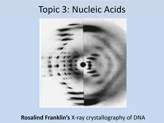
  • 1. Topic 3: Nucleic Acids Rosalind Franklin’s X-ray crystallography of DNA
  • 2. Essential idea: The structure of DNA is ideally suited to its function 7.1 DNA structure and replication
  • 3. 7.1 S.1 Analysis of results of the Hershey and Chase experiment providing evidence that DNA is the genetic material. Hershey and Chase Experiments (1952): Definitive proof that DNA rather than Protein carries the hereditary information of life E. Coli bacteriophage: A virus that infects bacteria. Bacteriophages only contain a protein coat (capsid) and DNA. They wanted to find out whether the protein or DNA carried the genetic instructions to make more viruses. They labeled either the viral proteins or DNA: – Protein capsid: Labeled with radioactive sulfur (35S) – DNA: Labeled with radioactive phosphorus (32P) Radioactive labeled viruses were used to infect cells.
  • 4. Either Bacteriophage DNA or Proteins Can be Labeled with Radioactive Elements 7.1 S.1 Analysis of results of the Hershey and Chase experiment providing evidence that DNA is the genetic material.
  • 5. Hershey Chase Experiment: DNA is Genetic Material 7.1 S.1 Analysis of results of the Hershey and Chase experiment providing evidence that DNA is the genetic material.
  • 6. Hershey and Chase Experiments (1952): Bacterial cells that were infected with the two types of bacteriophage, were then spun down into a pellet (centrifuged), and examined. Results: 1. Labeled viral proteins did not enter infected bacteria (found in supernatant). 2. Labeled viral DNA did enter bacteria during viral infection (found in cell pellet). Conclusion: Protein is not necessary to make new viruses. DNA is the molecule that carries the genetic information to make new viruses!!!! 7.1 S.1 Analysis of results of the Hershey and Chase experiment providing evidence that DNA is the genetic material.
  • 7. Rosalind Franklin (1950’s) • Worked with Maurice Wilkins • X-ray crystallography = images of DNA • Provided measurements on chemistry of DNA 7.1 A.1 Rosalind Franklin’s and Maurice Wilkins’ investigation of DNA structure by X-ray diffraction James Watson & Francis Crick (1953) • Discovered the double helix by building models to conform to Franklin’s X-ray data and Chargaff’s Rules.
  • 8. DNA Double Helix Nitrogenous Base (A,T,G or C) “Rungs of ladder” “Legs of ladder” Phosphate & Sugar Backbone DNA • Two strands coiled called a double helix • Sides made of a pentose sugar Deoxyribose bonded to phosphate (PO4) groups by phosphodiester bonds • Center made of nitrogen bases bonded together by weak hydrogen bonds 7.1 U.2 DNA structure suggested a mechanism for DNA replication
  • 9. DNA • Stands for Deoxyribonucleic acid • Made up of subunits called nucleotides • Nucleotide made of: 1. Phosphate group 2. 5-carbon sugar 3. Nitrogenous base 7.1 U.2 DNA structure suggested a mechanism for DNA replication
  • 10. DNA Nucleotide O=P-O O Phosphate Group N Nitrogenous base (A, G, C, or T) CH2 O C1 C4 C3 C2 5 Sugar (deoxyribose) O 7.1 U.2 DNA structure suggested a mechanism for DNA replication
  • 12. 7.1 U.1 Nucleosomes help to supercoil the DNA • A nucleosome consists of DNA wrapped around 8 histone proteins (prokaryotic cells lack these proteins making there DNA “naked). • The DNA wraps twice around the histone protein core. • Another histone protein is attached to the outside of the DNA strand. It helps maintain the colloidal structure of the nucleosome. • DNA, because of its negative charge is attracted to the positive charge on the amino acids of the histone proteins. • Tails of neighboring histones, link up during chromosomal condensation, causing the nucleosomes to pull closer together. • This is part of the supercoiling process that occurs during mitosis and meiosis • Supercoiling in general helps regulate transcription because only certain areas of the DNA are accessible for the production of mRNA by transcription. This regulates the production of a polypeptide. http://pbil.univlyon1.fr/members/sagot/htdocs/tea m/projects/chromo_net/images/epi.jpg
  • 14. DNA Replication 7.1 U.3 DNA polymerases can only add nucleotides to the 3’ end of a primer
  • 15. Synthesis Phase (S phase) • S phase during interphase of the cell cycle • Nucleus of eukaryotes Mitosis -prophase -metaphase -anaphase -telophase G1 G2 S phase interphase DNA replication takes place in the S phase. Mitosis -prophase -metaphase -anaphase -telophase G1 G2 S phase interphase DNA replication takes place in the S phase. 7.1 U.3 DNA polymerases can only add nucleotides to the 3’ end of a primer Super coiling begin in prophase, making chromosomes visible for the first time
  • 16. 2.7 S.2 Analysis of Meselson and Stahl’s results to obtain support for the theory of semi- conservative replication of DNA. https://upl oad.wikimedia.org/wikipedia/commons/a/a2/DNAreplicationModes.png Before Meselson and Stahl’s work there were different proposed models for DNA replication. After their work only semi-conservative replication was found to be biologically significant.
  • 17. 2.7 S.2 Analysis of Meselson and Stahl’s results to obtain support for the theory of semi- conservative replication of DNA. Learn about Meselson and Stahl’s work with DNA to discover the mechanism of semi-conservative replication http://highered.mheducation.com/olcweb/cgi/pluginpop.cg i?it=swf::535::535::/sites/dl/free/0072437316/120076/bio2 2.swf::Meselson%20and%20Stahl%20Experiment http://www.nature.com/scitable/topicpage/Semi-Conservative-DNA-Replication-Meselson-and-Stahl-421#
  • 18. • DNA replication is very specific to the arrangements of base pairs • In DNA replication, the strands separate – Enzymes use each strand as a template to assemble the new strands DNA REPLICATION Parental molecule of DNA Both parental strands serve as templates Two identical daughter molecules of DNA Nucleotides A 7.1 U.3 DNA polymerases can only add nucleotides to the 3’ end of a primer
  • 19. 1. Helicase: unwinds DNA at origins of replication 2. Initiation proteins separate 2 strands  forms replication bubble 3. Single-Strand Binding Proteins attach and keep the 2 DNA strands separated and untwisted 4. Primase: puts down RNA primer to start replication 5. DNA polymerase III: adds complimentary bases to leading strand (new DNA is made 5’  3’) 6. Lagging strand grows in 3’5’ direction by the addition of Okazaki fragments 7. DNA polymerase I: replaces RNA primers with DNA 8. DNA ligase: seals fragments together 9. DNA gyrase: an enzyme that relieves strain while double-strand DNA is being unwound by helicase. 7.1 U.5 DNA replication is carried out by a complex system of enzymes. [The proteins and enzymes involved in DNA replication should include helicase, DNA gyrase, single strand binding proteins, DNA primase and DNA polymerases I and III.] Major Steps of DNA Replication:
  • 20. DNA Gyrase SSBP DNA Poly Lead Strand Lagging Strand DNA Ligase RNA Primer Helicase Replication Direction RNA Primase
  • 21. Helicase The ‘ase’ ending indicates it is an enzyme. Helicase is DNA’s origin of replication and creates replication forks 2.7 U.2 Helicase unwinds the double helix and separates the two strands by breaking hydrogen bonds.
  • 22. • DNA replication begins at the different origins in the 5’ to 3’ direction at the replication fork. • RNA primase (is the primer to that starts the process) attaches to the DNA and adds a small RNA primer to provide a free 3’ OH starting point since DNA polymerases can only add nucleotides to the 3’ end of a primer • DNA polymerase III adds free nucleotides in the 5’ to 3’ direction in the direction of the replication fork. 7.1 U.3 DNA polymerases can only add nucleotides to the 3’ end of a primer
  • 23. • Begins at Origins of Replication • Two strands open forming Replication Forks (Y- shaped region) • New strands grow at the forks • The leading strand (copies in one long continuous piece) begins on the 3’ side and the lagging strand begins on the 5’ side (copies in small fragments, which must be pieced together) Replication Fork Parental DNA Molecule 3’ 5’ 3’ 5’ 7.1 U.4 DNA replication is continuous on the leading strand and discontinuous on the lagging strand. [Details of DNA replication differ between prokaryotes and eukaryotes. Only the prokaryotic system is expected.]
  • 24. Remember HOW the Carbons Are Numbered! O O=P-O O Phosphate Group N Nitrogenous base (A, G, C, or T) CH2 O C1 C4 C3 C2 5 Sugar (deoxyribose)
  • 25. Leading strand vs. Lagging strand
  • 26. Primase adds RNA primer is RNA that initiates DNA synthesis. DNA Gyrase 7.1 U.3 DNA polymerases can only add nucleotides to the 3’ end of a primer
  • 27. 2.7 U.3 DNA polymerase links nucleotides together to form a new strand, using the pre- existing strand as a template. DNA polymerase always moves in a 5’ to 3’ direction • DNA polymerase catalyzes the covalent phosphodiester bonds between sugars and phosphate groups forming covalent bonds
  • 28. DNA polymerase III adds nucleotides in 5’3’ direction on leading strand
  • 29. 2.7 U.1 The replication of DNA is semi-conservative and depends on complementary base pairing. https://upload.wikimedia.org/wikipedia/commons/3/33/DNA_replication_split_horizontal.svg 1. Each of the nitrogenous bases can only pair with its partner (A=T and G=C) this is called complementary base pairing. 2. The two new strands formed will be identical to the original strand.
  • 30. 2.7.U1 The replication of DNA is semi-conservative and depends on complementary base pairing. https://upload.wikimedia.org/wikipedia/commons/3/33/DNA_replication_split_horizontal.svg 3. Each new strand contains one original and one new strand, therefore DNA Replication is said to be a Semi- Conservative Process.
  • 31. Replication on leading strand 1. RNA Primase attaches RNA Primer 2. DNA Polymerase attaches nucleotides in a 5’ to 3’ direction 7.1 U.4 DNA replication is continuous on the leading strand and discontinuous on the lagging strand. [Details of DNA replication differ between prokaryotes and eukaryotes. Only the prokaryotic system is expected.]
  • 32. Replication on lagging strand 1. RNA Primase attaches multiple pieces of RNA Primer 2. DNA Polymerase attaches nucleotides in a 5’ to 3’ direction in between the Primer pieces creating Okazaki fragments 3. DNA Polymerase replaces the RNA primer pieces with DNA 4. Ligase glues the fragments together
  • 33. 1. Helicase: unwinds DNA at origins of replication Initiation proteins separate 2 strands  forms replication bubble 2. Single-Strand Binding Proteins attach and keep the 2 DNA strands separated and untwisted 3. RNA Primase: puts down RNA primer to start replication 4. DNA polymerase III: adds complimentary bases to leading strand (new DNA is made 5’  3’) 5. Lagging strand grows in 3’5’ direction by the addition of Okazaki fragments 6. DNA polymerase I: replaces RNA primers with DNA 7. DNA ligase: seals fragments together 8. DNA gyrase: an enzyme that relieves strain while double-strand DNA is being unwound by helicase. 7.1 U.5 DNA replication is carried out by a complex system of enzymes. [The proteins and enzymes involved in DNA replication should include helicase, DNA gyrase, single strand binding proteins, DNA primase and DNA polymerases I and III.] Major Steps of DNA Replication:
  • 34. DNA Gyrase SSBP DNA Poly Lead Strand Lagging Strand DNA Ligase RNA Primer Helicase Replication Direction RNA Primase
  • 35. • Dideoxyribonucleotides inhibit DNA polymerase during replication, thereby stopping replication from continuing. • Dideoxyribonucleotides with fluorescent markers, is incorporated into sequences of DNA, to stop replication at the point at which they are added. • This creates different sized fragments with fluorescent markers that can be separated by gel electrophoresis and analyzed by comparing the color of the fluorescence with the fragment length. 7.1 A.2 Use of nucleotides containing deoxyribonucleic acid to stop DNA replication in preparation of samples for base sequencing
  • 36. Problem at the 5’ End • DNA poly only adds nucleotides to 3’ end • No way to complete 5’ ends of daughter strands • Over many replications, DNA strands will grow shorter and shorter 7.1 U.6 Some regions of DNA do not code for proteins but have other important functions. [The regions of DNA that do not code for proteins should be limited to regulators of gene expression, introns, telomeres and genes for tRNAs.]
  • 37. Telomeres: repeated units of short nucleotide sequences (TTAGGG) at ends of DNA • Telomeres “cap” ends of DNA to postpone erosion of genes at ends (TTAGGG) • Telomerase: enzyme that adds to telomeres – Eukaryotic germ cells, cancer cells Telomeres stained orange at the ends of mouse chromosomes 7.1 U.6 Some regions of DNA do not code for proteins but have other important functions. [The regions of DNA that do not code for proteins should be limited to regulators of gene expression, introns, telomeres and genes for tRNAs.]
  • 38. Telomeres & Telomerase 7.1 U.6 Some regions of DNA do not code for proteins but have other important functions. [The regions of DNA that do not code for proteins should be limited to regulators of gene expression, introns, telomeres and genes for tRNAs.]
  • 39. 1.6 Cell division Essential idea: Cell division is essential but must be controlled.
  • 40. Why do cells divide: • Growth: Multicellular organisms increase their size by increasing their number of cells through mitosis • Asexual reproduction: Certain eukaryotic organisms may reproduce asexually by mitosis (e.g. vegetative reproduction) • Tissue Repair: Damaged tissue can recover by replacing dead or damaged cells • Embryonic development: A fertilized egg (zygote) will undergo mitosis and differentiation in order to develop into an embryo
  • 41. • Cellular division in eukaryotic cells. • Chromatin is arranged into chromosomes. • Chromosomes double. • Cell grows in size. • Cells divide. • Is cellular cloning. Cell division
  • 42. 2 phases: 1. Interphase 2. M phase (mitotic phase) a. Prophase b. Metaphase c. Anaphase d. Telophase & cytokinesis Figure 12.4 The cell cycle Phases of the Cell Cycle (life cycle of a cell)
  • 43. Interphase • The non-dividing phase in a cell • Lasts about ~ 90% of the cell cycle. • The cell grows and replicates DNA preparing for Mitosis. • There are three periods: 3 periods of Interphase 1. Go – a cell functioning as normal 2. G1 phase – first growth phase 3. S phase- synthesis of DNA 4. G2 phase- 2nd growth phase Mitosis is a reliable process. Only one error occurs per 100,000 cell divisions. 1.6 U.4 Interphase is a very active phase of the cell cycle with many processes occurring in the nucleus and cytoplasm.
  • 44. 1.6 U.4 Interphase is a very active phase of the cell cycle with many processes occurring in the nucleus and cytoplasm. Interphase This when the cell carries out it’s normal functions Metabolic reactions (e.g. respiration to produce ATP) are necessary for the life of the cell Protein synthesis - proteins and enzymes are necessary to allow cell grow Organelles numbers are increased to first support the enlarged cell DNA is replicated to ensure a second copy is available to enable mitosis Cells spend the majority of their time in interphase. It is a very active phase of the cycle. Mr P O D http://botit.botany.wisc.edu/Resources/Botany/Mitosis/Allium/Various%20views/Interphase%20prophase.JPG
  • 45. 1.6 U.5 Cyclins are involved in the control of the cell cycle. Cyclinsare a family of proteins that control the progression of cells through the cell cycle Cells cannot progress to the next stage of the cell cycle unless the specific cyclin reaches it threshold. http://upload.wikimedia.org/wikipedia/commons/thumb/9/99/Protein_CCNE1_PDB_1w98.png/800px-Protein_CCNE1_PDB_1w98.png Cyclins bind to enzymes called cyclin-dependent kinases These kinases then become active and attach phosphate groups to other proteins in the cell. The attachment of phosphate triggers the other proteins to become active and carry out tasks (specific to one of the phases of the cell cycle). 4 3 2 1
  • 46. http://upload.wikimedia.org/wikipedia/commons/thumb/9/99/Protein_CCNE1_PDB_1w98.png/800px-Protein_CCNE1_PDB_1w98.png Triggers cells to move from G0 to G1 and from G1 into S phase. prepares the cell for DNA replication in S phase. activates DNA replication inside the nucleus in S phase. promotes the assembly of the mitotic spindle and other tasks in the cytoplasm to prepare for mitosis. Progression through parts of the cell cycle are affected in various ways by specific cyclins
  • 47. 1.6 U.1 Mitosis is division of the nucleus into two genetically identical daughter nuclei. http://commons.wikimedia.org/wiki/File:Chromosome.svg centromere is the part of a chromosome that links sister chromatids Sister chromatids are duplicated chromosomes attached by a centromere Get the terminology right centrioles organize spindle microtubules Spindle microtubules (also referred to as spindle fibers) In animal cells two centrioles are held by a protein mass referred to as a centrosome After anaphase when the sister chromatids separate they should then be referred to as chromosomes It is easy to misuse the terms chromatid and chromosome. It is even easier to confuse the terms centromere, centriole and centrosome due to their similar spelling. Keep the terms clear in your mind to avoid losing marks. http://commons.wikimedia.org/wiki/Mitosis#mediaviewer/File:Mitosis_cells_sequence.svg
  • 48. 1.6 U.2 Chromosomes condense by supercoiling during mitosis. Why supercoil chromosomes? Human cells are on average 10μm in diameter and the nucelus within each is less than 5 μm in diameter. Human chromosomes are 15mm to 85mm (15,000μm to 85,000 μm) in length. Chromosomes need to be stored compactly to fit within the nuclei of cells. This problem becomes more acute during mitosis when chromosomes need to be short and compact enough that they can be separated and moved to each end of the cell.
  • 49. 1.6 U.2 Chromosomes condense by supercoiling during mitosis. How are chromosomes supercoiled? Strain is placed on a DNA helix by over winding or under winding of the helix This causes the DNA molecule to coil back on itself becoming shorter and wider Remember that in eukaryotes proteins called histones form nucleosomes to aid the process of supercoiling http://www.maths.uq.edu.au/~infinity/Infinity7/images/supercoiling.gifhttp://vanat.cvm.umn.edu/mMeiosis/images/chromosome-X.jpg
  • 50. http://highered.mheducation.com/sites/0072495 855/student_view0/chapter2/animation__mitosis _and_cytokinesis.html Use the animated tutorials to learn about mitosis http://www.johnkyrk.com/mitosis.html http://www.sumanasinc.com/webcontent/animations/content /mitosis.html http://outreach.mcb.harvard.edu/animations/cellcycle. swf
  • 51. Prophase • The nucleolus disappears. • Chromatin condenses into visible chromosomes. • There are two sister chromatids held together by a centromere. • The mitotic spindle forms in the cytoplasm. . 1.6 S.1 Identification of phases of mitosis in cells viewed with a microscope or in a micrograph
  • 52. Metaphase • The nuclear envelope disappears. • Spindle fibers extend from each pole to the cell’s equator. • Spindle fibers attach to the centromeres.
  • 53. Figure 12.3 Chromosome duplication and distribution during mitosis
  • 54. Anaphase • Characterized by movement. It begins when pairs of sister chromatids pull apart. • Sister chromatids move to opposite poles of the cell. • Chromosomes look like a “V” as they are pulled. • At the end of anaphase, the two poles have identical number and types of chromosomes.
  • 55. Telophase • Microtubules elongate the cell. • Daughter nuclei begin to form at the two poles. • Nuclear envelopes re-form. • Nucleolus reappears. • Chromatin uncoils. • The cells cytoplasm begins to pinch. • It is basically the opposite of prophase.
  • 56. 1.6 U.3 Cytokinesis occurs after mitosis and is different in plant and animal cells. mitosis is the division of the nucleus, cytokinesis is the division of the cytoplasm to create two cells Though mitosis is similar for animal and plant cells cytokinesis is very different. http://wwwprod.biochem.wisc.edu/biochem/faculty/bednarek/images/figure_color.gif http://glencoe.mheducation.com/sites/983 4092339/student_view0/chapter10/animati on_-_cytokinesis.html http://www.haroldsmithlab.com/images/pg_HeLa_cell_division.jpg
  • 57. Figure 12.8 Cytokinesis in animal and plant cells
  • 58. 1.6 S.1 Identification of phases of mitosis in cells viewed with a microscope or in a micrograph. 1.6 S.2 Determination of a mitotic index from a micrograph. http://www.nuffieldfoundation.org/practical-biology/investigating-mitosis-allium-root-tip-squash A very good, well explained lab outline for creating slides and calculating the mitotic index. http://www.biology.arizona.edu/cell_bio/activities/cell_cycle/cell_cycle.html An excellent online alternative if resources don’t permit students to create and view their own slides
  • 59. 1.6 U.6 Mutagens, oncogenes and metastasis are involved in the development of primary and secondary tumors. Tumors are abnormal growth of tissue that develop at any stage of life in any part of the body. A cancer is a malignant tumour and is named after the part of the body where the cancer (primary tumour) first develops. Use the links to find out: • most common types of cancer • what causes cancer and associated risk factors • how cancer can be treated
  • 60. 1.6 U.6 Mutagens, oncogenes and metastasis are involved in the development of primary and secondary tumors. mutation in a oncogene If a mutation occurs in an oncogenes it can become cancerous. In normal cells oncogenes control of the cell cycle and cell division. http://en.wikipedia.org/wiki/Oncogene#mediaviewer/File:Oncogenes_illustration.jpg uncontrolled cell division tumor formation malfunction in the control of the cell cycle
  • 61. 1.6 U.6 Mutagens, oncogenes and metastasis are involved in the development of primary and secondary tumors. Mutagens are agents that cause gene mutations. Not all mutations result in cancers, but anything that causes a mutation has the potential to cause a cancer. Mutagens can be: • chemicals that cause mutations are referred to as carcinogens • high energy radiation such as X-rays • short-wave ultraviolet light • Some viruses A mutation is a change in an organisms genetic code. A mutation/change in the base sequence of a certain genes can result in cancer. http://en.wikipedia.org/wiki/Oncogene#mediaviewer/File:Oncogenes_illustration.jpg
  • 62. 1.6 U.6 Mutagens, oncogenes and metastasis are involved in the development of primary and secondary tumors. Factors (other than exposure to mutagens) that increase the probability of tumour development include: • The vast number of cells in a human body – the greater the number of cells the greater the chance of a mutation. • The longer a life span the greater the chance of a mutation. Several mutations must occur in the same cell for it to become a tumour causing cell. The probability of this happening in a single cell is extremely small. http://en.wikipedia.org/wiki/Oncogene#mediaviewer/File:Oncogenes_illustration.jpg
  • 63. 1.6 A.1 The correlation between smoking and incidence of cancers. http://en.wikipedia.org/wiki/File:Smoking_lung_cancer.png There are many other similar surveys in different countries, with different demographics that show similar results. Along with lung cancer, cancers of mouth and throat are very common as these areas are in direct contact with the smoke too. It might surprise you that the following cancers are also more common in smokers: • Head and neck • Bladder • Kidneys • Breast • Pancreas • Colon
  • 64. a. Describe the relationship shown. b. What type of correlation is shown c. How strong is the correlation? Justify your answer by discussing the evidence. d. The correlation shown here is lagged. A lag is a time gap between the factors. Estimate the size of the lag between cigarette consumption and lung cancer death.
  • 65. http://en.wikipedia.org/wiki/File:Smoking_lung_cancer.png a. Describe the relationship shown. b. What type of correlation is shown c. How strong is the correlation? Justify your answer by discussing the evidence. d. The correlation shown here is lagged. A lag is a time gap between the factors. Estimate the size of the lag between cigarette consumption and lung cancer death. There are many other similar surveys in different countries, with different demographics that show similar results. Along with lung cancer, cancers of mouth and throat are very common as these areas are in direct contact with the smoke too. It might surprise you that the following cancers are also more common in smokers: • Head and neck • Bladder • Kidneys • Breast • Pancreas • Colon
  • 66. Essential idea: Information stored as a code in DNA is copied onto mRNA 7.2 Transcription & Gene Expression http://www.knowingforsure.com/wp-content/uploads/2015/01/Traits.jpg Trait vs Fate
  • 67. 2.7 U.4 Transcription is the synthesis of mRNA copied from the DNA base sequences by RNA polymerase. 2.7 U.5 Translation is the synthesis of polypeptides on ribosomes. Q - What is the purpose of transcription and translation? A - Gene expression the processes of create a polypeptides which in turns folds to become a protein. Proteins carry out many essential functions in cells. For more detail review 2.4.U7. Rhodopsin - A Light absorbing pigment
  • 68. Rubisco • Full name ribulose bisphosphate carboxylase • Enzyme - catalyzes the reaction that fixes carbon dioxide from the atmosphere • Provides the source of carbon from which all carbon compounds, required by living organisms, are produced. • Found in high concentrations in leaves and algal cells http://upload.wikimedia.org/wikipedia/commons/b/b0/Mint-leaves-2007.jpg 2.4 A.1 Rubisco, insulin, immunoglobulins, rhodopsin, collagen and spider silk as examples of the range of protein functions.
  • 69. Collagen • A number of different forms • All are rope-like proteins made of three polypeptides wound together. • About a quarter of all protein in the human body is collagen • Forms a mesh of fibers in skin and in blood vessel walls that resists tearing. • Gives strength to tendons, ligaments, skin and blood vessel walls. • Forms part of teeth and bones, helps to prevent cracks and fractures to bones and teeth https://en.wikipedia.org/wiki/Tooth_(human)#med iaviewer/File:Teeth_by_David_Shankbone.jpg 2.4 A.1 Rubisco, insulin, immunoglobulins, rhodopsin, collagen and spider silk as examples of the range of protein functions.
  • 70. spider silk • Different types of silk with different functions • Dragline silk is stronger than steel and tougher than Kevlar • When first made it contains regions where the polypeptide forms parallel arrays (bottom) • Some regions seem like a disordered tangle (middle) • When the stretched the polypeptide gradually extends, making the silk extensible and very resistant to breaking. https://en.wikipedia.org/wiki/Spider_silk#mediaviewer/File:Structure_of_spider_silk_thread_Modified.svg 2.4 A.1 Rubisco, insulin, immunoglobulins, rhodopsin, collagen and spider silk as examples of the range of protein functions.
  • 71. 2.7 U.4 Transcription is the synthesis of mRNA copied from the DNA base sequences by RNA polymerase 2.7 U.5 Translation is the synthesis of polypeptides on ribosomes. http://learn.genetics.utah.edu/content/molecules/transcribe/
  • 72. 7.1 U.1 Nucleosomes help to supercoil the DNA. 7.2 U.5 Gene expression is regulated by proteins that bind to specific base sequences in DNA. • A nucleosome consists of DNA wrapped around 8 histone proteins (prokaryotic cells lack these proteins making there DNA “naked). • The DNA wraps twice around the histone protein core. • Another histone protein is attached to the outside of the DNA strand. It helps maintain the colloidal structure of the nucleosome. • DNA, because of its negative charge is attracted to the positive charge on the amino acids of the histone proteins. • Tails of neighboring histones, link up during chromosomal condensation, causing the nucleosomes to pull closer together. • This is part of the supercoiling process that occurs during mitosis and meiosis • Supercoiling in general helps regulate transcription because only certain areas of the DNA are accessible for the production of mRNA by transcription. This regulates the production of a polypeptide. http://pbil.univlyon1.fr/members/sagot/htdocs/tea m/projects/chromo_net/images/epi.jpg
  • 74. 7.2 U.2 Nucleosomes help to regulate transcription in eukaryotes. • Supercoiling helps regulate transcription, one supercoiling modification is through the modification of the histone tails. • Acetylation Acetyl groups can be added to the positively charged histone tails, they become negative and that repels the negatively charged DNA. This opens up the nucleosome so the DNA is not as close to the histone anymore causing gene expression • Methylation Methyl group is non polar which causes DNA remains tightly packed and transcription is inhibited.
  • 75. 7.2 S.1 Analysis of changes in the DNA methylation patterns. http://i.dailymail.co.uk/i/pix/2008/09/12/article-1054890- 029CF17900000578-854_233x364.jpg • Another way gene expression can be controlled is through methylation (adding a methyl CH3 group) to the histone proteins. • Methylation of the histone proteins inhibites transcription of the gene • The amount of methylation can vary over an organisms lifetime and can be affected by environmental factors
  • 76. 7.2 U.6 The environment of a cell and of an organism has an impact on gene expression. The impact gene expression Morphogenic Effect (Aging affects) the accumulation of damage cells over a lifetime; decreases the capacity to maintain homeostasis
  • 77. 7.2 U.2 Nucleosomes help to regulate transcription in eukaryotes. Epigenetics • The changes related to gene expression or cellular phenotype of without changes to the nucleotide sequence of the genome. • Examples of mechanisms that produce such changes are DNA methylation and histone modification of the nucleosomes, each of which alters how genes are expressed without altering the underlying DNA sequence. Trait vs Fate
  • 78. 2.7.U4 Transcription is the synthesis of mRNA copied from the DNA base sequences by RNA polymerase. Step one: Transcription the process by which an RNA sequence is produced from a DNA template: Gene expression is the constructions of a protein from the DNA using RNA. RNA with the help of ribosomes constructs a protein from amino acids
  • 79. 7.2 A.1 The promoter as an example of non-coding DNA with a function. • The promoter region is a DNA sequence that initiates transcription and is an example of non- coding DNA that plays a role in gene expression. This promoter region is called the TATA box. • The promoter sequence is located near the start site of transcription and is where the RNA polymerase binds in order for transcription to take place. • DNA always is copied in a 5’ to 3’ direction. http://study.com/cimages/multimages/16/junk_dna_1.jpg
  • 80. • The enzyme RNA polymerase binds to a site on the DNA at the start of a gene (The sequence of DNA that is transcribed into RNA is called a gene). • RNA polymerase separates the DNA strands and synthesizes a complementary RNA copy from the antisense DNA strand. 2.7 U.4 Transcription is the synthesis of mRNA copied from the DNA base sequences by RNA polymerase.
  • 81. 7.2 U.3 Eukaryotic cells modify mRNA after transcription. http://i.dailymail.co.uk/i/pix/2008/09/12/article-1054890- 029CF17900000578-854_233x364.jpg a) The gene has a promotor region and a terminator region b) Transcription requires the presence of a regulator protein from another gene (possible from another chromosome). c) The RNA polymerase can now bind to the promotor and begin the transcription of the gene. d) The mRNA is transcribed including introns e) The completed mRNA which will require post transcriptional modification to remove the introns. click4biology TATA box
  • 82. 7.2 U.1 Transcription occurs in a 5’ to 3’ direction. [RNA polymerase adds the 5´ end of the free RNA nucleotide to the 3´ end of the growing mRNA molecule.] • Transcription occurs in a 5’ to 3’ direction where the 5’ end of the free RNA nucleotide is added to the 3’ end of the RNA molecule that is being synthesized. Consisting of 3 stages called 1. Initiation RNA polymerase binds to the promoter with the help of specific binding proteins 2. Elongation transcription machinery needs to move histones out of the way, unwinding DNA, allowing RNA Polymerase to synthesis of a new RNA strand in the 5′ to 3′ direction 3. Termination RNA synthesis will continue along the DNA template strand until the polymerase encounters a signal that tells it to stop
  • 83. 7.1 U.6 Some regions of DNA do not code for proteins but have other important functions. [The regions of DNA that do not code for proteins should be limited to regulators of gene expression, introns, telomeres and genes for tRNAs.] • There are many areas of DNA containing repetitive sequences, especially in eukaryotic DNA, in humans it makes up between 24-37% of our genome. These repetitive areas usually occurs near the ends of chromosomes. • Introns are non-coded regions, exons are coded areas for a protein, these two areas must be separated and introns must be removed before leaving the nucleus for protein synthesis to take place. *Spliceosome are enzymes constructed from RNA and are used in eukaryotic cell to remove these introns. http://www.phschool.com/science/biology_place/bio coach/images/transcription/eusplice.gif
  • 85. 7.1 U.6 Some regions of DNA do not code for proteins but have other important functions. [The regions of DNA that do not code for proteins should be limited to regulators of gene expression, introns, telomeres and genes for tRNAs.] • In addition to introns, some repetitive sequences are called telomeres, these areas protect DNA during replication. Telomeres are the caps at the end of each strand of DNA that protect our chromosomes, like the plastic tips at the end of shoelace They prevents the loss of genes near the end of the chromosomes. Telomeres stained green and red at the ends of the chromosomes http://www.newswise.com/images/uplo ads/2010/09/28/karlsederhr.jpg
  • 86. 7.2 U.4 Splicing of mRNA increases the number of different proteins an organism can produce 1. Promotor region 2. Free Nucleotide Phosphates 3. Addition of Nucleotides to the new mRNA 4. Early mRNA 5. Early mRNA showing introns (non-coding) 6.Introns removed by the enzyme spliceosome allowing exons to combine 7. Mature mRNA ready for translation 8. mRNA going to cytoplasm. click4biology
  • 87. 7.2 U.4 Splicing of mRNA increases the number of different proteins an organism can produce. https://commons.wikimedia.org/wiki/File:DNA_alternative_splicing.gif The splicing process above can happen in different ways to the same gene, particular exons (of a gene) may be included within or excluded from mature mRNA Multiple proteins produced by a single gene. Each proteins produced will vary in it’s biological function. An example of this is the IgM gene which produces different immunoglobulins (antibodies) to fight different pathogens.
  • 88. • The variable nature of the Short tandem repeats (STR) regions that are analyzed for forensic testing intensifies the discrimination between one DNA profile and another. Theses sections have high rates of mutations and change frequently. Forensic science takes advantage of the population's variability in STR lengths, enabling scientists to distinguish one DNA sample from another. For example, the likelihood that any two individuals (except identical twins) will have the same 13-loci DNA profile can be as low as 1 in 1 billion or less. 7.1 A.3 Tandem repeats are used in DNA profiling.
  • 89. 7.1 A.3 Tandem repeats are used in DNA profiling. • Short tandem repeats (STRs), also known as variable tandem repeats (VNTRs) are regions of noncoding DNA that contain repeats of the same nucleotide sequence. These short repeats show variations between individuals in terms of the number of times the sequences is repeated. Example • CATACATACATACATACATACATACATA repeated 7 times for one individual. However, in another individual, 11 times CATACATACATACATACATACATACATACATA CATACATACATA. • Used in DNA profiling used in crime scene investigations, genealogical and paternity tests.
  • 90. 7.2 U.6 The environment of a cell and of an organism has an impact on gene expression. • The environment, as well as the organism's internal world, which includes such factors as its hormones and metabolism can have an impact on gene expression • Temperature and light are external conditions which can affect gene expression in certain organisms. • As an example, Himalayan rabbits carry the gene, which is required for the development of pigments in the fur, skin, and eyes, and whose expression is regulated by temperature • Specifically, a gene called the C gene is inactive above 35°C, and it is maximally active from 15°C to 25°C. This temperature regulation of gene expression produces rabbits with a distinctive coat coloring. • In the warm weather no pigments fur is white • In low temperature the rabbit's extremities (i.e., the ears, tip of the nose, and feet), where the, the C gene actively produces pigment, making these parts of the animal black. http://upload.wikimedia.org/wikipedia/commons/0/06/Kr%C 3%B3liki_kalifornijskie_666.jpg http://upload.wikimedia.org/wikipedia/en/8/81/Kostya2.jpg
  • 91. Essential idea: Information transferred from DNA to mRNA is translated into an amino acid sequence. 7.3 Translation Section of Titin, our largest known protein http://circ.ahajournals.org/content/124/8/876/F2.large.jpg
  • 92. Components of Translation 1. mRNA = message 2. tRNA = interpreter 3. Ribosome = site of translation 7.3 U.1 Initiation of translation involves assembly of the components that carry out the process.
  • 93. 2.7 U.5 Translation is the synthesis of polypeptides on ribosomes. http://www.nature.com/scitable/topicpage/ribosomes-transcription-and-translation-14120660 Translation is the process of protein synthesis in which the genetic information encoded in mRNA is translated into a sequence of amino acids in a polypeptide chain A ribosome is composed of two halves, a large and a small subunit. During translation, ribosomal subunits assemble together like a sandwich on the strand of mRNA: • Each subunit is composed of RNA molecules and proteins • The small subunit binds to the mRNA • The large subunit has binding sites for tRNAs and also catalyzes peptide bonds between amino acids
  • 94. Ribosomes Active sites: • A site: holds AA to be added • P site: holds growing polypeptide chain • E site: exit site for tRNA 7.3 U.1 Initiation of translation involves assembly of the components that carry out the process.
  • 95. 7.3 S.2 The use of molecular visualization software to analyse the structure of eukaryotic ribosomes and a tRNA molecule. D-Loop Ribosome sight recognition T-Loop Ribosome sight recognition Acceptor End With the help of ATP this is the site of attachment of The amino acid
  • 96. 7.3 S.2 The use of molecular visualization software to analyse the structure of eukaryotic ribosomes and a tRNA molecule. D-Loop Ribosome sight recognition T-Loop Ribosome sight recognition Acceptor End With the help of ATP this is the site of attachment of The amino acid
  • 97. 7.3 A.1 tRNA activating enzymes illustrate enzyme–substrate specificity and the role of phosphorylation. Building tRNA binds with a specific amino acid is a catalyzed reaction 1. tRNA-activating enzyme 2. ATP binds to the enzyme. 3. Specific amino acid binds to the acceptor site(ACC) on the tRNA molecule. Building tRNA binds with a specific amino acid
  • 98. 7.3 A.1 tRNA-activating enzymes illustrate enzyme–substrate specificity and the role of phosphorylation. http://www.phschool.com/science/biology_place/biocoach/translation/addani.html
  • 99. tRNA • Transcribed in nucleus • Specific to each amino acid • Transfer Amino Acids to ribosomes • Anticodon: pairs with complementary mRNA codon • Base-pairing rules between 3rd base of codon & anticodon are not as strict. 7.3 U.1 Initiation of translation involves assembly of the components that carry out the process.
  • 100. Translation stages: Initiation, Elongation and Termination • Translation occurs in the 5' to 3' direction along the mRNA A. Initiation begins with the attachment of the ribosome to the mRNA using the start codon AUG B. Elongation The ribosome moves one codon along the mRNA (in a 5’ – 3’ direction): • The tRNA in the P site is moved to the E site and then released • The tRNA in the A site is moved into P site C. Termination occurs at the STOP codon (UGA, UAG or UAA). • a release factor attaches to the A site • the polypeptide chain is released 7.3 U.1 Initiation of translation involves assembly of the components that carry out the process.
  • 101. The role of RNA in Protein Synthesis • 3 Types of RNA molecules in the steps from gene to protein: 1. Messenger RNA (mRNA), Makes a complimentary copy of DNA in the form of RNA. Length varies depending on the gene sequence 2. Transfer RNA (tRNA) carries amino acid to the site of synthesis. 3. Ribosomal RNA (rRNA), stabilizes the site of synthesis 7.3 U.1 Initiation of translation involves assembly of the components that carry out the process.
  • 102. 7.3 U.2 Synthesis of the polypeptide involves a repeated cycle of events. • Step one of translation is the small and large sub units of the ribosome come together between the mRNA sequence • Step two tRNA (carrying methionine (Met), the start code) attaches to the mRNA at the A site • Step three the first tRNA moves to the P, a second tRNA located at the A. The two amino acids form a peptide bond.
  • 103. 7.3 U.2 Synthesis of the polypeptide involves a repeated cycle of events. • The two amino acids are joined together through a condensation reaction that creates a peptide bond between the two amino acids. • Step Four The ribosome moves along the mRNA one codon shifting the tRNA that was attached to methionine to the E site. • The tRNA is released back into the cytoplasm from the E site, allowing it to pick up another amino acid (methionine) to build another polypeptide.
  • 104. 7.3 U.2 Synthesis of the polypeptide involves a repeated cycle of events. • Another tRNA moves into the empty A site bringing the next amino acid corresponding to themRNA codon. • Again, the amino acid is attached to the polypeptide forming a peptide bond, the ribosome slides across one codon and tRNA at the P site moves into the E site releasing it back into the cytoplasm. • The ribosome continues to move along the mRNA adding amino acids to the polypeptide chain. • This process continues until a stop codon is reached.
  • 105. 7.3 U.3 Disassembly of the components follows termination of translation. • Termination begins when 1 of the 3 stop codons  UAA  UGA  UAG moves into the A site. • These tRNA have no attached amino acids. • When the stop codon is reached the ribosome dissociates and the polypeptide is released.
  • 107.
  • 108. 2.7 U.6 The amino acid sequence of polypeptides is determined by mRNA according to the genetic code. The central dogma of genetics Messenger RNA (mRNA): A transcript copy of a gene used to encode a polypeptide • The length of mRNA molecules varies – 23,000 different genes, the average length for mammals is approximately 2,200 nucleotides (this translates to approximately 730 amino acids in the average polypeptide but can vary dependent on the protein that is made) • Only certain genes in a genome need to be expressed depending on: • Cell specialism • Environment • Therefore not all genes (are transcribed) and translated • If a cell needs to produce a lot of a certain protein (e.g. β cells in the pancreas specialize in secreting insulin to control blood sugar) then many copies of the required mRNA are created.
  • 109. 2.7 U.7 Codons of three bases on mRNA correspond to one amino acid in a polypeptide. The genetic code is the set of rules by which information encoded in mRNA sequences is converted into proteins (amino acid sequences) by living cells • Codons are a triplet of bases which encodes a particular amino acid • As there are four bases, there are 64 different codon combinations (4 x 4 x 4 = 64) • The codons can translate for 20 amino acids based on Amino acids are carried by transfer RNA (tRNA) The anti-codons on tRNA are complementary to the codons on mRNA • Different codons can translate for the same amino acid (e.g. GAU and GAC both translate for Aspartate) therefore the genetic code is said to be degenerate • The order of the codons determines the amino acid sequence for a protein • The coding region always starts with a START codon (AUG) therefore the first amino acid in all polypeptides is Methionine • The coding region of mRNA terminates with a STOP codon - the STOP codon does not add an amino acid – instead it causes the release of the polypeptide
  • 110. 2.7 S.1 Use a table of the genetic code to deduce which codon(s) corresponds to which amino acid. 2.7 S.3 Use a table of mRNA codons and their corresponding amino acids to deduce the sequence of amino acids coded by a short mRNA strand of known base sequence. 2.7 S.4 Deducing the DNA base sequence for the mRNA strand. The diagram summarizes the process of protein synthesis. You should be able to use a section of genetic code, transcribe and translate it to deduce the polypeptide synthesized.
  • 111. 2.7.S1, 2.7.S3, 2.7.S4 Practice transcribing and translating using the learn genetics tutorial. http://learn.genetics.utah.edu/content/molecules/transcribe/
  • 112. 2.7 S.1, 2.7 S.3, 2.7 S.4 Now use this table to answer the questions on the next slide n.b. You just have to be able to use the table. You do not have to memorize which codon translates to which amino acid.
  • 113. 2.7 S.1, 2.7 S.3, 2.7 S.4 1. Deduce the codon(s) that translate for Aspartate. 2. If mRNA contains the base sequence CUGACUAGGUCCGGA a. deduce the amino acid sequence of the polypeptide translated. b. deduce the base sequence of the DNA antisense strand from which the mRNA was transcribed. 3. If mRNA contains the base sequence ACUAAC deduce the base sequence of the DNA sense strand.
  • 114. 1. Deduce the codon(s) that translate for Aspartate. 2. If mRNA contains the base sequence CUGACUAGGUCCGGA a. deduce the amino acid sequence of the polypeptide translated. b. deduce the base sequence of the DNA antisense strand from which the mRNA was transcribed. 3. If mRNA contains the base sequence ACUAAC deduce the base sequence of the DNA sense strand. 2.7 S.1, 2.7 S.3, 2.7 S.4
  • 115. 2.7 S.1, 2.7 S.3, 2.7 S.4 1. Deduce the codon(s) that translate for Aspartate. 2. If mRNA contains the base sequence CUGACUAGGUCCGGA a. deduce the amino acid sequence of the polypeptide translated. b. deduce the base sequence of the DNA antisense strand from which the mRNA was transcribed. 3. If mRNA contains the base sequence ACUAAC deduce the base sequence of the DNA sense strand. (the sense strand is the template for the mRNA the only change is that uracil is replaced by thymine) ACTAAC GACTGATCCAGGCCT (the antisense strand is complementary to the mRNA, but remember that uracil is replaced by thymine) GAU, GAC Leucine + Threonine + Lysine + Arginine + Serine + Glycine
  • 116. 2.7 S.1 Use a table of the genetic code to deduce which codon(s) corresponds to which amino acid.
  • 117. 2.7 S.1, 2.7 S.3, 2.7 S.4
  • 118. 2.7 S.1 Use a table of the genetic code to deduce which codon(s) corresponds to which amino acid.
  • 119. 2.7 S.1, 2.7 S.3, 2.7 S.4
  • 120. 2.7 S.1 Use a table of the genetic code to deduce which codon(s) corresponds to which amino acid.
  • 121. 7.3 U.4 Free ribosomes synthesize proteins for use primarily within the cell. https://www.studyblue.com/notes/note/n/molecular-exam-3/deck/2630328 • Free ribosomes in the cytoplasm synthesize proteins that will be used inside the cell in the cytoplasm, mitochondria and chloroplasts (in autotrophs)
  • 122. Ribosomes effect in translation • Ribosome are found in Prokaryotes (70's) and Eukaryotes (80's). Including P and A sites. START codons and STOP codons begin and termination translation. Polyribosome= Polysomes • Multiple ribosomes on the same mRNA at the same time. • All ribosome move 5' to 3' in sequence. • In protein synthesis polyribosomes increase the quantity of polypeptide synthesized. 7.3 S.1 Identification of polysomes in electron micrographs of prokaryotes and eukaryotes.
  • 123. 7.3 U.4 Free ribosomes synthesize proteins for use primarily within the cell.
  • 124. 7.3 S.1 Identification of polysomes in electron micrographs of prokaryotes and eukaryotes. • In prokaryotes, several ribosomes can attach themselves to the growing mRNA chains to form a polysome while the mRNA chains are still attached to the DNA
  • 125. 7.3 S.1 Identification of polysomes in electron micrographs of prokaryotes and eukaryotes. • In eukaryotes, the mRNA detaches from the DNA and is then transported through pores in the nuclear envelope to the ribosomes in the cytoplasm. Once in the cytosol, eukaryote mRNA can also form polysomes
  • 126. 7.3 U.5 Bound ribosomes synthesize proteins primarily for secretion or for use in lysosomes. • Ribosomes attached to ER create proteins that are secreted from the cell by exocytosis or are used in lysosomes. • Proteins that are destined to be used in lysosomes, ER, Golgi Apparatus, the plasma membrane or secreted by the cell are made by ribosomes bound by the endoplasmic reticulum • Ribosomes that become bound to the ER are directed here by a signal sequence that is part of that specific polypeptide • This signal sequence on the polypeptide binds to a signal recognition protein (SRP) • The SRP guides the polypeptide and ribosome to the ER where it binds to an SRP receptor http://herbmitchell.info/Figure.4-8-Synthesissecretoryprotein.jpg
  • 127. 7.3 U.6 Translation can occur immediately after transcription in prokaryotes due to the absence of a nuclear membrane. • Since prokaryotic DNA is not compartmentalized into a nucleus, once transcription begins creating a strand of mRNA, translation can begin immediately as the mRNA strand is created • In eukaryotes, the completed mRNA has to be transported from the nucleus, through the nuclear pore to the ribosome on the ER or in the cytosol http://www.mun.ca/biology/scarr/iGen3_05-09_Figure-L.jpg
  • 128. Prokaryotes vs. Eukaryotes Prokaryotes Eukaryotes • Transcription and translation both in cytoplasm • DNA/RNA in cytoplasm • RNA poly binds directly to promoter • Transcription makes mRNA (not processed) • No introns • Transcription in nucleus; translation in cytoplasm • DNA in nucleus, RNA travels in/out nucleus • RNA poly binds to TATA box & transcription factors • Transcription makes pre- mRNA  RNA processing  final mRNA • Exons, introns (cut out) 7.3 U.6 Translation can occur immediately after transcription in prokaryotes due to the absence of a nuclear membrane.
  • 129. Structure of Proteins The complex structure of proteins is explained by referring to 4 levels of organization A. Primary B. Secondary C. Tertiary D. Quaternary http://upload.wikimedia.org/wikipedia/commons/2/26/225_Peptide_Bond-01.jpg
  • 130. Structure of Proteins Primary structure: • The order/ number of amino acids in a polypeptide chain. • Linear shape (no internal bonding) 7.3 U.7 The sequence and number of amino acids in the polypeptide is the primary structure.
  • 131. 7.3 U.8 The secondary structure is the formation of alpha helices and beta pleated sheets stabilized by hydrogen bonding Secondary Structure: Hydrogen bonding causes The primary structure of the polypeptide to fold and coil Into some characteristic ways: • Alpha Helix • Beta pleated sheets
  • 132. Beta-pleated sheets: • Flat, zig-zag structure • A number of chains which are hydrogen bonded together • Forms a sheet Example: Fibers in in silk 7.3 U.8 The secondary structure is the formation of alpha helices and beta pleated sheets stabilized by hydrogen bonding
  • 133. • Tertiary structure is the three-dimensional conformation of a polypeptide. • The polypeptide folds just after it is formed in translation. • The shape is maintained by intermolecular bonds 7.3 U.9 The tertiary structure is the further folding of the polypeptide stabilized by interactions between R groups. http://cnx.org/resources/36c08f3ac1c144763610fa69fbb9e278/Figure_03_04_08.jpg
  • 134. 7.3 U.10 The quaternary structure exists in proteins with more than one polypeptide chain. • Quaternary structure is the linking together of two or more polypeptides to form a single protein. • The protein structure below has 4 different polypeptide chains. http://www.topsan.org/@api/deki/files/6029/=EK5976M_Fig3Comparisons.png
  • 135. Conserved sequence: a homologous sequence of DNA that is identical across all members of a species. Bioinformatics: uses computer databases to store and analyze gene & protein sequences from large amounts of data collected from sequencing genes of various organisms Faster, more powerful computers allow scientist to identify conserved sequences & genes by looking for patterns and homologous sequences within organisms’ genome. If a sequence is homologous across species or individuals of a species, it usually has a functional role. Eg. It codes for a protein (a gene). 7.3 S.2 The use of molecular visualization software to analyze the structure of eukaryotic ribosomes and a tRNA molecule.
  • 136. 2.5 Enzymes Essential idea: Enzymes control the metabolism of the cell. http://cdn.instructables.com/F7F/38MA/HAFHKT7I/F7F38MAHAFHKT7I.LARGE.jpg Below is an enzymatic reaction browning, which may protect the developing seeds from pathogens
  • 137. 8.1 Metabolism (AHL) Essential idea: Metabolic reactions are regulated in response to the cell’s needs. https://mediaeatout.files.wordpress.com/2013/11/candidates-eating-obama-sized.jpg
  • 138. 2.5 U.1 Enzymes have an active site to which specific substrates bind. 2.5 U.2 Enzyme catalysis involves molecular motion and the collision of substrates with the active site. Enzyme: A globular protein that increases the rate of a biochemical reaction by lowering the activation energy threshold (i.e. a biological catalyst). The energy need for chemical reactions to occur http://www.northland.cc.mn.us/biology/biology1111/animations/enzyme.swf Use the animation to find out more about enzymes and how they work. A good alternative is How Enzymes Work from McGraw and Hill http://highered.mheducation.com/sites/0072495855/student_view0/chapter2/animation__how_enzymes_work.html
  • 139. 8.1 U.2 Enzymes lower the activation energy of the chemical reactions that they catalyze. • The substrate binds to the enzymes’ active site and the active site is altered reaching the transition state (the enzyme-substrate complex). • Due to the binding the bonds in the substrate molecule are stressed/become less stable. • Binding to an enzyme lowers the overall energy needed for a reaction to occur. • The activation energy of the reaction is then becomes reduced. How do enzymes lower the activation energy of a reaction? http://en.wikipedia.org/wiki/Image:Induced_fit_diagram.png
  • 140. • Enzymes are protein catalysts that enormously speed up reactions. They often have an “-ase” ending to their name. – e.g., hexokinase, catalase, peptidase, mutase • They are not themselves changed (except for a brief period of time) and are the same before and after a reaction. • Enzymes: 1. Lower the activation energy: this is the MOST important characteristic 2. Do not add or remove energy from a reaction 3. Do not change the equilibrium for a reaction 4. Are reused over and over 2.5 U.1 Enzymes have an active site to which specific substrates bind.
  • 141. Activation Energy is the energy need to get a reaction started. Enzymes decrease the barrier to starting a reaction. This energy is called the activation energy, or the energy required for a reaction to start. 8.1 U.2 Enzymes lower the activation energy of the chemical reactions that they catalyzed.
  • 143. Lock & Key Hypothesis a) Large globular protein enzyme b) Active Site where the substrate combines to the enzyme c)Substrate which fits the active site d) Activated complex The substrate is weakened to allow the reaction. e) Unchanged enzyme/ re-used at low concentrations f) Product of the reaction * Polar regions on the enzyme’s active site help bring the enzyme and the product together
  • 144. Induced Fit Hypothesis Many enzymes catalyze more then one reaction, this is due to Hydrogen bonding (weak IMF’) holding the active site together. a. As the substrate approaches, polar regions of the enzyme’s active site attract the substrate. b. Enzyme activation site allows for a shape change or induce a fit. Causing a chemical change c. This stress reduces the activation energy or the reaction returns the enzyme to its original shape
  • 145. 2.5 U.2 Enzyme catalysis involves molecular motion and the collision of substrates with the active site. http://www.kscience.co.uk/animations/model.swf The simulation from KScience allows you to both see enzyme kinetics happening and secondly how it is affected by different factors • Two substances must have the proper alignment and energy (in the form of motion) to create a chemical reaction • The direction and movement is constantly changing and is random • Collisions occur at random between the substrate and enzyme • Successful reactions only occur if the substrate and the active site of the enzyme are correctly aligned and the collide with sufficient KE
  • 146. 2.5 U.2 Enzyme catalysis involves molecular motion and the collision of substrates with the active site. • Most enzyme reactions occur when the substrates are dissolved in water • All molecules dissolved in water are in random motion, with each molecule moving separately • If not immobilized the enzyme can move too, however enzymes tend be larger than the substrate(s) and therefore move more slowly
  • 148. 2.5 U.5 Immobilized enzymes are widely used in industry. http://pubs.rsc.org/services/images/RSCpubs.ePlatform.Service.FreeContent.ImageService.svc/ImageService/Articleimage/2013/CS/c3cs35506c/c3cs35506c-f1.gif Advantages of enzyme immobilization: • Concentration of substrate can be increased as the enzyme is not dissolved – this increases the rate of reaction • Recycled enzymes can be used many times, immobilized enzymes are easy to separate from the reaction mixture, resulting in a cost saving. • Separation of the products is straight forward (this also means that the the reaction can stopped at the correct time). • Stability of the enzyme to changes in temperature and pH is increased reducing the rate of degradation, again resulting in a cost saving. Enzymes used in industry are usually immobilized. They are attached to a material so that their movement is restricted. Common ways of doing this are: • Aggregations of enzymes bonded together • Attached to surfaces, e.g. glass • Entrapped in gels, e.g. alginate gel beads
  • 149. 2.5 U.5 Immobilized enzymes are widely used in industry. Common uses of enzymes in industry include: http://pubs.rsc.org/services/images/RSCpubs.ePlatform.Service.FreeContent.ImageService.svc/ImageService/Articleimage/2013/CS/c3cs35506c/c3cs35506c-f1.gif 1.Detergents 2.Biofuels 3.Textiles 4.Brewing 5.Medicine & Biotechnology 6.Juice yield 7.Paper production
  • 150. 1. Detergents contain proteases and lipases to help breakdown protein and fat stains https://i1.ytimg.com/vi/lQ6fCZgYc8g/hqdefault.jpg
  • 151. 2. Enzymes are used to breakdown the starch in grains into biofuels that can be combusted http://chromblog.thermoscientific.com/Portals/49739/images/biofuel9.jpg http://greenodin.com/wp-content/uploads/2014/12/GreenOdin-GO-Biodiesel-Van-1200.png
  • 152. 3. In the textiles industry enzymes help in the processing of fibers, e.g. polishing cloth to make it appear more shiny
  • 153. 4. In the brewing industry enzymes help a number of processes including the clarification of the beerhttp://www.brewreviewcrew.com/cans-vs-bottles-fight/
  • 154. 5. In Medicine & Biotechnology enzymes are widely used in everything from diagnostic tests tests to contact lens cleaners to cutting DNA in genetic engineering. http://www.medwesteye.com/wp-content/uploads/2014/12/learn-the-proper-care-of-contact-lenses.jpg
  • 155. 6. Enzymes are widely used in the food industry, e.g. • fruit juice, pectin to increase the juice yield from fruit • Fructose is used as a sweetener, it is converted from glucose by isomerase • Rennin is used to help in cheese productionhttps://theramblingreed.files.wordpress.com/2014/08/img_6338.jpg
  • 156. 7. Paper production uses enzymes to helping in the pulping of wood http://i00.i.aliimg.com/img/pb/479/389/262/1281752366918_hz-cnmyalibaba-web2_15708.jpg
  • 157. Optimal environmental condition(s) favor the most active enzyme conformation. • Temperature • pH • Concentration of substrate/enzyme • Cofactors • Enzyme inhibitors • Allosteric regulation (noncompetitive) • Ionic concentration 2.5 U.3 Temperature, pH and substrate concentration affect the rate of activity of enzymes.
  • 158. 1. The Effect Of Temperature (a) •Increase Kinetic energy of substrate and enzyme. •Increased chance of collisions •Low temperatures has a low rate of reaction (b) •Optimum temperature = maximum rate of reaction •Balance between enzyme stability and kinetic energy of reactants (c) •Rapid decrease in the rate of reaction •High temperatures destabilize the enzyme molecule •Enzyme is denatured
  • 159. Denaturing is a structural change in a protein that results in a loss (usually permanent) of its biological properties. • Enzymes are globular proteins • Enzymes have tertiary structure • Tertiary structure is maintained by hydrogen, ionic and covalent bonds • Shape of the active site is maintained by hydrogen, ionic and covalent bonds The bonds within enzymes (and proteins) has an increasing strength of: Hydrogen Ionic Covalent 2.5 U.4 Enzymes can be denatured.
  • 160. Proteins found in the egg white include albumins, globulins and mucoproteins Under intense heat, hydrogen bonds that formed during the secondary structure are broken The proteins then lose their shapes, thus changing their functions By cooking it, you have effectively denatured the egg What happens when you cook an egg?
  • 161. 2. The Effect Of pH (a) Decrease in pH (increase in H+) • H+ interact with exposed R groups on active site. • Enzyme active site changes shape • Specificity reduced • Decrease in rate of reaction (b) Optimum rate of reaction for the pH= (d) • Active site structure and structure specific to the complementary shape of the substrate. • Successful activated complex and therefore reactions occur. (c) Increase in pH (increase in OH-) • Increase in base (OH-) concentration • Enzyme active site changes shape • Specificity reduced • Decrease in rate of reaction
  • 162. Denaturation of an enzyme by a change in pH • If there is a deviation from the optimal pH the hydrogen sulfide bridges break and the enzyme loses shape. • Loss of the activation site shape leads to loss of function
  • 163. Denaturation of an enzyme by a change in pH • Enzymes have an optimum pH at which they achieve their maximum rate of reaction • Pepsin has an optimal pH of 2 at (a) • Amylase has an optimal pH of (c) • pH affects the charge of the amino acids of the active site • This changes the properties of the active site • e.g. carboxyl R group will be uncharged COOH at low pH but COO- at high pH.
  • 164. 3. The effect of substrate concentration (a) •Increase conc. of substrate molecules •Increased chance of collision with enzyme •Greater chance of forming activated complex •Increase in rate of reaction (b) •Rate begins to level •Active sites beginning to become saturated with substrate (fully occupied) •New substrate must wait for previous reaction to complete and the product to exit the active site (c) •Full saturation of the active sites by substrate •Rate becomes constant for further increases in substrate concentration.
  • 165. • Metabolism: the sum total of all chemical reactions that occur within an organism. • Metabolic Pathway sequences of biochemical reactions that occur in all living cells Two types of metabolic pathways 1. Linear Metabolic Pathways: •Chemical changes in living things often occurring with a number of intermediate stages. •Each stage has its own enzyme. •Catabolic pathways breakdown molecules •Anabolic pathways build up molecules 8.1 U.1 Metabolic pathways consist of chains and cycles of enzyme- catalyzed reactions. The first step of cellular respiration Glycolysis
  • 166. 2. Cyclic Metabolic Pathways: •The initial substrate is fed into the cycle. • Enzyme (a) combines the regenerated Intermediate 4 to catalyzes the production of intermediate 1 • Enzyme (b) converts intermediate 1 to intermediate 2 • Enzyme (c) converts intermediate 2 to intermediate 3. The product is formed and removed. • Enzyme (d) converts intermediate 3 to intermediate 4 and the cycle repeats.
  • 167. Second step of cellular respiration Krebs Cycle
  • 168. • Inhibitors are substances that reduce or completely stop the action of an enzyme • Inhibition can act on the active site (competitive) or on another region of the enzyme molecule(non-competitive). The competition in the former being for the active site of the enzyme. Competitive blocks the active site and non-competitive attaches to another site on the enzyme and changes the shape of the active site of the enzyme 8.1 U.3 Enzyme inhibitors can be competitive or non-competitive.
  • 169. Non-Competitive inhibition Competitive Inhibition 8.1 U.3 Enzyme inhibitors can be competitive or non-competitive.
  • 170. 8.1 S.2 Distinguishing different types of inhibition from graphs at specified substrate concentration. https://wikispaces.psu.edu/download/attachments/46924781/image-6.jpg Rate of reaction is reduced Features of competitive inhibitors When the concentration of substrate begins to exceed the amount of inhibitor, the maximum rate of the uninhibited enzyme can be achieved. However, it takes a much higher concentration of substrate to achieve this maximum rate.
  • 171. https://wikispaces.psu.edu/download/attachments/46924781/image-6.jpg Rate of reaction is reduced Features of non-competitive inhibitors It takes approximately the same concentration of enzyme to reach the maximum rate, but the maximum rate is lower than the uninhibited enzyme. • The binding of the non-competitive inhibitor prevents some of the enzymes from being able to react regardless of substrate concentration. • Those enzymes that do not bind inhibitors follow the same pattern as the normal enzyme. 8.1 S.2 Distinguishing different types of inhibition from graphs at specified substrate concentration.
  • 172. 8.1 A.1 End-product inhibition of the pathway that converts threonine to isoleucine. http://www.uic.edu/classes/bios/bios100/lecturesf04am/feedback-inh.gif Isoleucine from threonine • The synthesis of isoleucine from threonine in a series of five enzyme- catalysed steps • As the concentration of isoleucine increases, some of it binds to the allosteric site the enzyme threonine deaminase acts as a non-competitive inhibitor. • The pathway is then turned off, regulating isoleucine production.
  • 173. Isoleucine in the body is responsible for some of the following: energy levels, sugar levels, hemoglobin production. The amino acid has been know to assist in wound healing, simulating immune function and promoting the secretion of several important hormones. 8.1 A.1 End-product inhibition of the pathway that converts threonine to isoleucine.
  • 174. 8.1 A.2 Use of databases to identify potential new anti-malarial drugs. http://upload.wikimedia.org/wikipedia/commons/0/02/Mosquito_bite4.jpg • Malaria is a disease caused by the pathogen Plasmodium falciparum. • This protozoan uses mosquitoes as a host as well as humans and hence can be passed on by mosquito bites
  • 175. • In one study, approx. 300,000 chemicals were screened against a chloroquine-sensitive 3D7 strain and the chloroquine-resistant K1 strain of P. falciparum. • Other related and unrelated organisms, including human cell lines, were also screened. • (19) new chemicals that inhibit the enzymes normally targeted by anti-malarial drugs were identified • Additionally (15) chemicals that bind to malarial proteins were identified – this can help in the location of P. falciparum • These results indicate possible new directions for drug research. Increasing drug resistance to anti-malarial drugs has lead to the use of bioinformatics and chemogenomics to try and identify new drugs. 8.1 A.2 Use of databases to identify potential new anti-malarial drugs.
  • 176. • Sometimes when a chemical binds to a target site, it can significantly alter metabolic activity. • Massive libraries of chemicals are tested individually on a range of related organisms. • For each organism a range of target sites are identified. • A range of chemicals which are known to work on those sites are tested. Bioinformatics is an approach whereby multiple research groups can add information to a database enabling other groups to query the database. Bioinformatics has facilitated research into metabolic pathways is referred to as chemogenomics.
  • 177. 8.1 S.1 Calculating and plotting rates of reaction from raw experimental results. The rate of reaction can be calculated using the formula: Rate of reaction (s-1) = 1 / time taken (s) Time taken in enzyme experiments this is commonly the time to reach a measurable end point or when a standard event, caused by the enzyme reaction, has come to pass. This is usually measured by the effects of the accumulation of product, but can as easily be measured by the disappearance of substrates. http://www.scienceexperimentsforkids.us/wp-content/uploads/2011/08/hydrogen-experiments-for-kids-3-img.jpg Use the results from it or data from one of your enzyme inhibition labs to calculate the rate of reaction. Enzyme inhibition can be investigated using these two outlines by Science & Plants for Schools: • The effect of end product, phosphate, upon the enzyme phosphatase • The inhibition of catechol oxidase by lead
  • 178. Lactose Intolerance • Lactose (milk sugar) can cause allergies in some people. • This is often because they are unable to produce the enzyme lactase in sufficient quantities. • Most people produce less lactase as they get older. After all, we don’t live off milk once we have been weaned. • In some regions such as Europe, a mutation has allowed lactose production to continue into adulthood. This mutation is not present in people who are lactose intolerant 2.5 A.1 Methods of production of lactose-free milk and its advantages.
  • 179. Green High Tolerance Red Low Tolerance Global estimates of lactose intolerance.
  • 180. How can we cope with lactose intolerance? • Take a lactase supplement. These are produced industrially using the Aspergillus niger fungus • Drink lactose free milk. Milk treated with lactase (produced by A. niger) and essentially ‘pre-digested’ before being packaged.
  • 181. 2.5 A.1 Methods of production of lactose-free milk and its advantages. Other uses of lactose free milk: • As a means to increase the sweetness of milk (glucose and galactose are sweeter in flavor), thus negating the need for artificial sweeteners • As a way of reducing the crystallization of ice-creams (glucose and galactose are more soluble than lactose) • As a means of shortening the production time for yogurts or cheese (bacteria ferment glucose and galactose more readily than lactose) Production of Lactose-free milk • Lactase obtained from commonly from yeast (bacteria is an alternative) • Lactase is bound to the surface of alginate beads • Milk is passed (repeatedly) over the beads • The lactose is broken down into glucose and galactose • The immobilized enzyme remains to be used again and does not affect the quality of the lactose free milk
  • 182. 2.5 S.1 Design of experiments to test the effect of temperature, pH and substrate concentration on the activity of enzymes. 2.5 S.2 Experimental investigation of a factor affecting enzyme activity. (Practical 3) Possible research questions, what are you going to investigate (independent variable)? • What is the effect of substrate concentration? • What is the effect of temperature? • What is the effect of pH? • Which type of yeast has a higher concentration of catalase? Important things to consider: • How are you going to vary the mass/volume/concentration of your variable? • What units will you be measuring your variable in? • Have you chosen an effect range or values to answer your question? • Are the concentrations/chemicals you are using safe to handle? Catalase is one of the most widespread enzymes. It catalyzes the conversion of hydrogen peroxide, a toxic by-product of metabolism, into water and oxygen.
  • 183. How are you going to measure your results (dependent variable)? • Are you measuring the increase of a product or the disappearance of a substrate? • Are you measuring directly (e.g. testing for the concentration of the product) or indirectly (change in pH)? • What equipment will you be using to measure your results? • What are the units and uncertainty given both the equipment and how you choose to use it? • What time period do you need to run the experiment for? How fast is the enzyme action likely to be? • How many repeats will you need to make sure your results are reliable? 2.5 S.1 Design of experiments to test the effect of temperature, pH and substrate concentration on the activity of enzymes. 2.5 S.2 Experimental investigation of a factor affecting enzyme activity. (Practical 3)
  • 184. How are you going to make sure it is a fair test (control variables)? • What variables other than your independent variable could affect the results? • Why would these variables affect the results? • How will you ensure each is kept constant and monitored? • What level should they be kept constant at? If a control variable is too far from it’s optimum then it could limit the enzyme action and no change would be seen in the results. • If a variable cannot be controlled it should still be discussed and considered as an uncontrolled variable. Safety and ethics: • Are you using any equipment that may cause you or others harm? What steps have you taken to minimize this risk? • If you intend to use animals have you first considered alternative subjects? • If you still intend to use animals are subjects have you ensured both: o no harm comes to them as a result of the experiment o The experiment does not induce stress or conditions beyond that normally found in their natural environment 2.5 S.1 Design of experiments to test the effect of temperature, pH and substrate concentration on the activity of enzymes. 2.5 S.2 Experimental investigation of a factor affecting enzyme activity. (Practical 3)