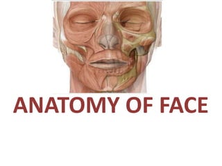
Anatomy of face
•
29 recomendaciones•6,289 vistas

Plastic Surgery Classes: Anatomy of face

Recomendados
Más contenido relacionado
La actualidad más candente
La actualidad más candente (20)
Similar a Anatomy of face
Similar a Anatomy of face (20)
Más de Dr Sourabh Shankar Chakraborty
Más de Dr Sourabh Shankar Chakraborty (20)
Último
Último (20)
Anatomy of face
- 2. Contents • Introduction • Boundaries of face • Layers of face • Symmetry of face • Aesthetic regions of face • Bones • Muscles of face • Nerve supply • Blood supply & venous drainage • Lymphatic drainage • Applied anatomy
- 3. Introduction • Very vascular • Due to rich vascularity face blush and blanch. • Facial skin is rich in sebaceous gland and sweat gland. • Wounds of face bleed profusely but heal rapidly. • Sebaceous glands keep the skin oily but also cause acne in adult. • No deep fascia is present in the face.
- 4. Boundaries of Face Superiorly Inferiorly Each side • To the hairline • Chin • Base of mandible • Auricle Note: forehead is common to both scalp & face.
- 7. Aesthetic Regions of Face
- 8. Bones of Face • The facial skeleton consists of 14 stationary bones and the mandible. • These 14 bones form the basic shape of the face, and are responsible for providing attachments for muscles that make the jaw move and control facial expressions.
- 10. Bones of the Face
- 11. Muscles of Face General Characters • Called muscle of facial expression and lie in superficial fascia. • Develop from mesoderm of 2nd pharyngeal arch. • Supplied by the Facial nerve. • Act as closers and openers of facial orifices.
- 12. Muscles of Face General Characters • Surround the facial openings. • Originate from bone or other muscle. • Pass in the superficial fascia. • Attach to the facial skin or other facial muscle.
- 13. Muscles of Face Classification: 1. Muscles of the eyelid 2. Muscles of the mouth 3. Muscles of the nose 4. Muscles of the neck 5. Muscles of the ear
- 17. Muscles of Eyelid A. Corrugator supercillii Origin: Medial end of superciliary arch Insertion: Skin of mid- eyebrow Action: Vertical lines in forehead: frowning
- 18. B. Orbicularis Oris ORBITAL PART [OUTER] PALPEBRAL PART [INNER] LACRIMAL PART [SMALL] PARTS OF ORBICULARIS OCULI
- 19. Orbital Part ORIGIN INSERTION ACTION • Medial part of the medial palpebral ligament & adjoining bone • Concentric rings return to the point of origin • Closes the lids tightly • Protects eye from bright light
- 21. Palpebral Part ORIGIN INSERTION ACTION • Lateral part of the medial palpebral ligament • Lateral palpebral raphae • Closes the lids gently • Blinking
- 22. Lacrimal Part ORIGIN INSERTION ACTION • Lacrimal fascia & lacrimal bone • Upper & lower eyelids • Dilates the lacrimal sac
- 24. Muscles of The Mouth • Orbicularis oris • Buccinator • Lower group of oral muscles • depressor anguli oris • depressor labii inferioris • Mentalis • Upper group of oral muscles • risorius • zygomaticus major and zygomaticus minor • levator labii superioris • levator labii superioris alaeque nasi • levator anguli oris
- 26. Orbicularis oris Two parts: 1. Intrinsic part [Deep stratum] Origin: Superior incisivus from maxilla & Inferior incisivus from mandible Insertion: Angle of mouth Action: Closes & purses the mouth 2. Extrinsic part [Two strata] Origin: • Thickest middle stratum – Buccinator • Thick superficial stratum – Elevators & depressors of lips & angles Insertion: Lips & the angle of the mouth Action: Closes & purses the mouth
- 27. Buccinator • Origin : • Upper fibers from maxilla above three molar • Lower fibers : from the mandible below three molar • Ptergomandibular raphe which separates it from the constrictor pharyngis superior. • Insertion : • Upper fibers : to the upper lip • Lower fibers : lower lip • Middle fibers decussate lower ascend to upper lib & lower descend to the lower limb • Action : • Aids in holding the cheek to the teeth and prevent accumulation of food in the Buccinator
- 29. Zygomaticus major • Strap muscle • Forms shape of cheek • Smiling
- 30. Zygomaticus minor • Strap muscle • Forms shape of cheek • Smiling
- 31. Levator labii superioris • Deeps the furrows on either side of nares • Sad
- 32. Levator anguli oris • Pulls the angle of mouth upwards • Aka ‘Happy muscle’ • Smiling
- 33. Levator labii superioris alaeque nasi • Aka ‘Elvis muscle’ • Dilates the nostrils • Raises upper lip
- 34. Risorius • Pulls the lip horizontally • Insincere smile
- 35. Depressor labii inferioris • Draws the lower lip downwards and little laterally • Expression of irony.
- 36. Depressor anguli oris • Lowers the corners of mouth • Frowning
- 38. Muscles of Nose 1. Procerus 2. Nasalis (two parts) • Dilator naris • Compressor naris 3. Depressor septi nasi
- 39. Procerus • Origin: nasal bone and lateral nasal cartilage • Insertion: skin between the eyebrows • Action: – pulls down the medial end of the eyebrow – wrinkles the skin of the nose transversely in frowning
- 40. Dilator naris • Origin : Maxilla bone • Insertion: Ala of the nose • Action: Widens the nasal aperture (by pulling the alar laterally) in deep inspiration; is also a sign of anger
- 41. Compressor naris • Origin: Frontal process of the maxilla • Insertion: Aponeurosis which crosses the bridge of the nose • Action: Compresses the mobile nasal cartilages
- 42. Depressor septi nasi • Origin: incisive fossa of maxilla • Insertion:nasal septum & back part of the alar part of nasalis muscle • Action: depression of nasal septum. Dilates the nostril. Moves the ape of nose during movement of upper lip (talking).
- 43. Muscles of Neck Platysma • Origin– upper part of pectoral and deltoid fascia • Insertion– base of mandible, skin of lower face and lip • Action– releases pressure of skin on the subjacent veins, depress mandible, pulls angle of mouth downwards.
- 44. Muscles of Ear Auricularis 3 parts: 1. Auriculars anterior •Origin: temporal fascia •Insertion: major helix (ear) •Action: pulls ear forward 2. Auricularis posterior •Origin: mastoid process •Insertion: posterior ear •Action: pulls ear backward 3. Auricularis superior •Origin: temporal fascia •Insertion: above the ear •Action: pulls ear upward
- 46. Nerve Supply of Face A. Sensory nerve supply: • Trigeminal nerve : •Ophthalmic division •Maxillary division •Mandibular division • Great auricular nerve of cervical plexus
- 52. B. Motor Supply of Face (Facial Nerve) • Orgin : from the pons • Type: mixed nerve motor , sensory and containing parasympathetic. • Course in the face: after the facial nerve comes out from the stylomandibular foramen it enter the parotid gland superficial to external carotid artery and posterior facial vein and within the parotid gland the nerve gives five terminal branches .
- 53. Branches of facial nerve • Before it enter the parotid gland (distal to the stylomastoid foramen: 1. Postetrior auricular- to the occiptal belly of occiptofrontalis muscle and muscles around ear 2. Branch to posterior belly of digasteric and stylohyoid muscle • Within the parotid gland: 1. Temporal 2. Zygomatic 3. Buccal 4. Marginal mandibular 5. Cervical
- 54. TEMPORAL •Frontalis •Auricularis muscles •Orbicularis oculi ZYGOMATIC •Orbicularis oculi BUCCAL •Cheek •Upper lip •Lower lip MARGINAL MANDIBULAR CERVICAL •Platysma Supply to Various Muscles
- 55. Blood Supply & Venous Drainage A. Arterial Supply: 1. Facial Artery: -Chief artery of the face • Origin: branch from external carotid • Course: arise from external carotid and inter the digasteric triangle in the neck and run between submandibular gland and mandible then inter the face in front of masseter muscle and terminate by giving angular artery.
- 56. •Branches of facial artery A. In the neck (cervical branches) 1. Ascending palatine 2. Tonsilar 3. Submental 4. Glandular branches B. In the face (facial branches) 1. Inferior labial 2. Superior labial 3. Lateral nasal 4. Angular (terminal)
- 60. B. Venous drainage • Veins accompany the arteries • Drains into common facial & retromandibular veins. • W-shaped arrangement • Facial vein – Largest – No valves
- 61. Facial vein • Begins as angular vein @ medial angle of the eye • Formed by the union of Supratrochlear & Supraorbital veins • Angular vein – Continues as facial vein – Running downwards & backwards behind facial artery – STRAIGHT COURSE
- 65. Dangerous Area of the Face This includes the upper lip and the lower part of the nose. It is drained by the f a c i a l v e i n , w h i c h communicates with the cavernous sinus. Infections of this area can therefore, spread in retrograde direction and cause thrombosis of the cavernous sinus.
- 66. Lymphatic Drainage of Face
- 67. Applied Anatomy of Face • Trigeminal neuralgia – Maxillary and mandibular nerve are involved – Excruciating pain in the region of distribution of these nerve • In infranuclear lesions of facial nerve (eg, bell’s palsy)- Ipsilateral whole face is paralyzed – c/f • Affected side is motionless • Loss of wrinkles • Eye cannot be closed • In smiling the mouth is drawn to normal side • During mastication food accumulates in vestibule of mouth
- 68. Bell’s Palsy
- 70. Facial nerve lesion ( Bell’s palsy )
- 71. References • Text book of Human anatomy by B D Chaurasia • Atlas of Human Anatomy by Nater • Gray’s anatomy • Internet sources