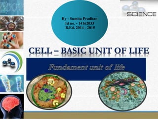
1. By - Sumita Pradhan
Id no. - 14162033
B.Ed. 2014 - 2015
2. SLIDES 3 – 11 ARE CONTENT SLIDES FOR
THE TOPIC FUNDAMENTAL UNIT OF LIFE.
PLEASE READ THE CONTENT
THOROUGHLY.
AFTER THAT, YOU HAVE TO ANSWER 10
MCQs.
TRY TO ANSWER ALL THE QUESTION.
( ANSWER WITH HONESTY)
ANSWER ONE QUESTION AT A TIME AND
GO ORDERLY.
WHENEVER FEEL DIFFICULTY GO TO THE
CONTENT PART.
ENJOY FUN LEARNING …!!
3.
4. Cell: Cell is called the fundamental unit of life.
The word cell is derived from the Latin word “cellula”
which means “a little room”
A cell is capable of independent existence and can
carry out all the functions which are necessary for a
living being. A cell carries out nutrition, respiration,
excretion, transportation and reproduction; the way an
individual organism does. Organisms may be broadly
classified into two
kinds: Unicellular and Multicellular.
Unicellular organisms are capable of independent
existence which shows a cell’s capability to exist
independently. Due to this, a cell is called the
fundamental and structural unit of life. All living
beings are composed of the basic unit of life, i.e. cell.
5.
6. • All living organisms are composed of one or more cells.
• The cell is the basic unit of structure, function, and organization in
all organisms.
• All cells come from preexisting, living cells.
HISTORY OF DISCOVERY OF CELLS
It was the British botanist Robert Hooke who, in 1664, while
examining a slice of bottle cork under a microscope, found its
structure resembling the box-like living quarters of the monks in a
monastery, and coined the word “cells”
The Dutch scientist A .V. Leeuwenhoek, in 1674, discovered the
minute forms of life such as bacteria and single celled animals in a
drop of water. Robert Brown discovered the nucleus (1831). Purkinje
coined the term ‘protoplasm (1839). Schleiden (1838) and Schwann
(1839) proposed the Cell Theory. Virchow (1855) made further
addition to the cell theory. The discovery of electron microscope (1940)
made it possible to study the structures of cell organelles.
7. • Shape and Size of Cells:- Cells come in all shapes and sizes. While most
of the cells are spherical in shape, cells of various other shapes are also
found. Most of the cells are microscopic in size, i.e. it is impossible to
see them with naked eyes. Some cells are fairly large, e.g. a neuron in
human body can be as long as 1 meter. The egg of an ostrich is the
largest known cell of a living animal and an average egg is 15 cm long
and 13 cm wide.
• A cell is enclosed in a membranous casing and is filled with a liquid
substance which is called the cytoplasm. There are many cell organelles
in a typical cell. Some of the main structures of a cell are as follows:
• Cell wall: Cell wall is made of cellulose. It is somewhat hard but
permeable to most of the substances. Cell wall is available in plant cells
and in cells of bacteria and fungi.
• Plasma membrane: Plasma membrane is a semi-permeable membrane.
It is composed of bilayer of lipid and protein.
• Functions of Plasma Membrane: Plasma membrane provides a
container to the cytoplasm. It facilitates passage of various substances in
and out of the cell.
8. • Nucleus: Nucleus is covered by double membrane; called nuclear
membrane. The fluid which is inside the nucleus is called nucleoplasm.
Nucleus contains chromosomes which are important for the functioning
of a cell. Chromosomes contain genes which are the carriers of genetic
information. Nucleus plays an important role during cell division.
Nucleus controls all the functions of the cell.
• Prokaryotes and Eukaryotes: Based on the level of organization of
nuclear material, a cell can be categorized as prokaryote or eukaryote.
9. Mitochondria: Mitochondrion is a capsule-like structure. It is a double
membrane structure. Its inner membrane is projected into numerous
finger-like structures; called cristae. Mitochondria are the sites of
cellular respiration. After cellular respiration, energy is stored in the
form of ATP (Adenosine triphosphate); in mitochondria. Mitochondria
have their own DNA and ribosomes and hence mitochondria can
produce their own protein.
Functions of Mitochondria: Cellular respiration; due to this,
mitochondria are also known as the ‘powerhouse of the cell’.
Endoplasmic Reticulum: Endoplasmic reticulum is a mesh-like structure
which is composed of numerous tubes. It extends from the plasma
membrane to the nuclear membrane. There are two kinds of
endoplasmic reticulum, viz. smooth ER and rough ER. Rough ER has
ribosomes on its surface which give it the rough appearance.
Function of ER: It serves as the transport channel in the cell. Substances
are transported from cell membrane to cytoplasm and to nucleus and
vice-versa. ER also serves the role of packaging many substances in the
cell.
Golgi apparatus: Golgi apparatus was discovered by Camillo Golgi. It is
composed of many sac-like structures which are stacked one above
another.
Functions of Golgi apparatus: Golgi apparatus is responsible for
packaging of various substances in the cell.
10. Lysosomes: Lysosomes are small sac-like structures and they are
derived from Golgi complex. Lysosomes contains digestive enzymes.
Functions of lysosomes: The enzymes in the lysosome digest foreign
particles and thus destroy them. Sometimes, the lysosome may burst
open and its content ends up digesting the contents of the cell. The cell
gets killed in the process. Due to this, lysosome is also called the
‘suicide bag of the cell’.
Ribosome: These are tiny dot like structures interspersed in the
cytoplasm and also on the surface of Rough ER. Ribosome is
responsible for protein synthesis.
Plastids: These are somewhat similar to mitochondria; in appearance.
Plastids are found in plant cells. They are of two types, chromoplast
and leucoplast. Colourful plastids are called chromomplast and
colourless plastids are called leucoplast. Chloroplast is green in colour
and is found in green parts of plants. Plastids too have their own DNA
and ribosome.
Functions of Plastids: Leucoplasts are responsible for storing food;
such as carbohydrates, protein and lipid. Chromoplasts impart various
colours to the plant parts. A leaf of a plant is green in colour because
of chloroplast. Chloroplast is the site of photosynthesis.
11. Vacuoles: These are fluid filled chambers and are often seen in
many cells. Vacuoles are very large in plant cells. A plant cell
usually has single but large vacuole. Such a vacuole fills almost
the entire space inside the cell. Vacuoles are much smaller and
very few in animal cells.