BRAIN ARTERIOVENOUS MALFORMATION
•Descargar como PPTX, PDF•
39 recomendaciones•11,797 vistas

Brain arteriovenous malformations (bAVM) are abnormal connections of arteries and veins in the brain, forming a tangled web of vessels instead of a normal capillary network treated with multimodalities including, SRS, embolisation and Microneurosurgery. This slides updates the management of AVM highlighting the importance of SM grading, Pollock radiation grading etc.

Recomendados
Más contenido relacionado
La actualidad más candente
La actualidad más candente (20)
Similar a BRAIN ARTERIOVENOUS MALFORMATION
Similar a BRAIN ARTERIOVENOUS MALFORMATION (20)
Más de suresh Bishokarma
Más de suresh Bishokarma (20)
Último
Último (20)
BRAIN ARTERIOVENOUS MALFORMATION
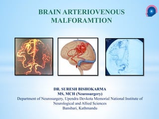
- 1. cka DR. SURESH BISHOKARMA MS, MCH (Neurosurgery) Department of Neurosurgery, Upendra Devkota Memorial National Institute of Neurological and Allied Sciences Bansbari, Kathmandu BRAIN ARTERIOVENOUS MALFORAMTION
- 2. Suspected vascular lesion Abnormal vessels in parenchyma Compact nidus appearance Single Brain AVM, Pial AVM Multiple CAMS Diffuse nidus appearance Early draining Proliferative type brain AVMs No early draining Proliferative angiopathy Caput medusae appearance DVA CM, VA No abnormal vessels in parenchyma Enlarged pial arteries Pial AVFs No enlarged pial arteries DAVFs Fine networks Moyamoya APPROACH TO VASCULAR LESION CAMS: Cerebrofacial arteriovenous metameric syndrome DVA: Developmental Venous Anomalies CM: Cavernous malformation VA: Venous angioma
- 3. 1. Venous anomalies 2. Capillary telangiectasias 3. Cavernous malformations 4. AVM AVM: Unique from other in there is presence of an arteriovenous shunt, which is defined as oxygenated blood passing directly into the venous system without gas exchange occurring in a capillary bed. These shunts are characterized by high flow and high pressure, which distinguish them from other vascular malformations. DYSPLASTIC VASCULAR MALFORMATIONS
- 4. AVMs were initially described in the mid- 1800s by Luschka1 (in 1854) and Virchow2 (in 1863). Three decades later, Giordano performed the first surgical exposure of an AVM in 1889.3 In the same year, Péan performed the first successful extirpation of an AVM.3 Multiple neurosurgeons attempted to treat AVMs in the early 20th century. Krause, for example, was the first to attempt a surgical elimination of an AVM by ligating its arterial feeders, without great success, however.4 Multiple techniques that were considered innovations at a certain point in the 20th century might look bizarre to the contemporary neurosurgeon. In 1951, Bilsland5 published a report in which he reviewed the use of preoperative exsanguination to decrease bleeding during intracranial surgery; of particular interest were AVMs because it was thought that exsanguination might help control intraoperative bleeding. In 1954, Brown6 supported this technique by illustrating a series of 133 patients. He stated that “[exsanguination] can be readily controlled more so than when specific vasodilator drugs are used to reduce bleeding. HISTORY OF AVM SURGERY
- 5. Brain arteriovenous malformations (AVMs) are dynamic, high pressure flow, dysplastic vascular lesion characterized by abnormal connections between arteries and veins leading to arteriovenous shunting with no capillary bed and intervening neural parenchyma in an intervening network of vessels—the so- called nidus. These usually are congenital lesions with a lifelong risk of bleeding of ≈ 2–4% per year. Small AVMs are now thought to have much higher pressure in the feeding arteries: so small AVMs are more lethal than larger ones and bleeds more frequent. S-m grade 1–3: annual risk of hemorrhage is 3.5%. S-m grade 4–5: annual risk of hemorrhage is 2.5%. The hemorrhage risk (but not the rate) may be higher in pediatrics or with posterior-fossa AVMs. DEFINITION
- 6. Unlike aneurysms, they are not thought to develop de novo, yet the vast majority of them remains clinically silent for decades. Unlike a neoplasm, it can grow along with the brain without necessarily displacing functional structures. Therefore, many never cause functional neurological compromise regardless of size AVM mimics
- 7. AVMs may be plexiform, fistulous, or both. A fistulous malformation may be an otherwise normal artery directly joining a vein. A plexiform malformation is defined as an artery connecting to a network of poorly differentiated, immature vessels before (nidus) passing into a vein. Types of AVM
- 8. True, cerebral, pial, parenchymal, cisternal, ventricular, medullary ADJECTIVE FOR AVM
- 9. Nidus Feeders Venous drainage MORPHOLOGY OF AVM
- 10. Size Location: proximity to eloquent structure Shape: Radiological or surgical planning. NIDUS
- 11. Number, size, Origin from part of COW, relative contribution to the nidus, location, association (Aneurysm) ICA or ECA origin. Supply from pial collaterals. Watershed AVM may have ACA and PCA Erratic feeders. Types of arterial feeders. 1. Direct feeders are the simplest to conceptualize: they end directly and exclusively in the nidus, and they are also known as terminal feeders. 2. Transit arteries or artery en passage are normal arteries that appear on angiogram to pass near or even through the nidus while going on to supply normal tissue. 3. The third type of feeding artery is the indirect feeder. This artery combines the previous two in that as it passes near the nidus it can contribute to the shunt before continuing on to supply normal brain. AVMs within the sylvian fissure usually harbor many en passage contributions from the distal middle cerebral arteries. FEEDERS
- 12. Number, size, and locations Tortuous course, ectasias, stenosis. Arterialization Draining into superficially via the cortical surface or deeply to the vein of Galen. Like transit arteries: normal veins may be draining functional cerebral tissue that may be adjacent to the lesion. VENOUS DRAINAGE
- 13. The arterial feeder vessels and venous drainage of a brain AVM will depend on the location of the nidus. Deep and ventricular locations will recruit perforator (lentriculostriate, thalamoperforator branches) and choroidal (anterior, medial, and lateral posterior choroidal arteries) supply, respectively, whereas venous drainage will typically be via the deep venous system. In more superficial or cortical locations, the main arterial supplies are through the pial arteries (branches of the anterior cerebral artery, MCA, and PCA), whereas venous drainage is mainly through the cortical veins. The absence of cortical venous drainage in a superficially located brain AVM may indicate thrombosis of the superficial outlets with subsequent rerouting into the deep system, which would suggest a more unstable lesion. Recruitment of the transdural supply is sometimes seen in large lesions. Vasculature of AVM
- 14. SUB-TYPES OF LOBAR AVM AVM Subtype Subtype Feeders Venous drain Frontal AVM 5 Subtypes 1 Lateral frontal, MCA branches that originate from the superior trunk. SSS Sylvian v 2 Medial frontal ACA Medial frontal veinSSS 3 Paramedian frontal Watershed: MCA, ACA lateral and medial frontal veins SSS 4 Basal frontal Watershed: MCA, ACA SSS via an anterior OrbFrV or FrPolV 5 Sylvian frontal M3 SupSylV, DeepSylV Temporal AVM 4 Subtypes 1 Lateral Temporal AVM ATA and MidTempA feeding anterior temporal AVMs and PosTempA and TempOccA feeding posterior temporal AVMs AntTempV, MidTempV, and PosTempV to the vein of Labbé and the TrvS 2 Sylvian temporal M3 opercular empSylV, SupSylV, and DeepSylV. 3 Basal temporal PCA >MCA TempBasV 4 Medial temporal. AChA and the P2 PCA BVR Parieto-Occipital AVM 4 subtypes 1 Lateral parieto- occipital, MCA cortical arteries: CenA, AntParA, PosParA, AngA, and TempOccA. SSS Sylvian v 2 Medial parieto-occipital PCA>ACA SSS, VOG 3 Paramedian parieto- occipital MCA, PCA, ACA SSS 4 Basal occipital TempA and inferior branches from CalcA. TrvS or TentS Michael T Lawton: Seven AVM: Tenets and Techniques for resection.
- 15. Deep AVM and subtypes Subtype Subtype Feeders Venous drain 1 Pure sylvian M2, M3 SupSylV > DeepSylV 2 Insular M2 Sylvian V. 3 Basal ganglia InsP and lLSA DeepSlyV (lateral variant) or CauV and ThaStrV (medial variant) 4 Thalamic avms Below: AntThaP (PCoA) and PosThaP (P1 PCA), and from above by lPChA and mPChA ICV Michael T Lawton: Seven AVM: Tenets and Techniques for resection.
- 16. SUBTYPES OF CEREBELLAR AVM Subtype Subtype Feeders Venous drain 1 Suboccipital cerebellar AVM SCA, AICA, and PICA IHemV and/or IVerV TrVs. 2 Tentorial Cerebellar AVM SCA SHemV that can bridge anteriorly to the VoG, posteriorly to Torc, or superiorly to TentS or StrS 3 Vermian Cerebellar AVM Superior: SCA Inferior: PICA SuperiorSVerV and VoG, inferior IVerV and Torc. 4 Tonsillar Cerebellar AVM PICA Medial and lateral TonsV as well as ReTonsV. 5 Petrosal Cerebellar AVM AICA AHemV and VCPonF, which then course to SPetrV and SPS. Michael T Lawton: Seven AVM: Tenets and Techniques for resection.
- 17. Rotational angiography and three-dimensional reconstruction has now replaced the old- fashioned stereoangiography, which previously provided excellent tracking of the course of feeding arteries and draining veins. Superselective angiography of a suspected vessel can show if it contributes to the nidus or not and help distinguish a transit artery. However, superselective catheterization of an artery en passage can have variable presentation on angiography depending on the hemodynamics of the vessel. AVM feeders is that they do not seem to follow the traditional pattern of progressive luminal narrowing as they flow distally. Arterial feeders can even exhibit pathological stenosis or even aneurysms. Angiogram
- 18. CTA Tangle of serpigenous vascular structures are seen at the left frontal lobe, supplied by the distal branches of the hypertrophied left anterior and middle cerebral arteries and drained by enlarged cortical veins draining into the superior sagittal sinus, few enlarged veins are also seen draining into the left transverse & sigmoid sinuses.
- 19. DSA
- 20. MRI
- 21. 1. Flow void on T1WIor T2WIwithin the AVM 2. Feeding arteries 3. Draining veins 4. Increased intensity on partial flip-angle (to differentiate signal dropout on T1WI or T2WI from calcium) 5. Significant edema around the lesion may indicate a tumor that has bled rather than an AVM 6. Gradient echo sequences (GRASS) help demonstrate surrounding hemosiderin which suggests a previous significant hemorrhage 7. A complete ring of low density (due to hemosiderin) surrounding the lesion suggests AVM over neoplasm MRI
- 22. Intracranial hemorrhage. Suspicion of an AVM. calcium deposition (25% to 30% ) Iso- to hyperdense serpiginous vessels that might be located at some distance from the hemorrhage. A CT angiogram (CTA) : delineate the nidus and associated vessels. CT SCAN
- 23. CT SCAN
- 24. A weakness common to both CT and MR modalities is the lack of temporal sequencing of images to determine the dynamic aspect of the malformation DIAGNOSTIC IMAGING
- 25. Use of adjunctive diagnostic studies is helpful in selective cases. Functional MRI (fMRI) has helped assess the proximity of language and motor function in relation to AVM but has limitations. Magnetic source imaging (MSI) can accurately localize sensory cortex and even visual and motor areas and the information can be overlaid on the MRI. MR Tractography should aid in assessing the relationship of deep white matter tracts to the AVM. The information derived from these studies is helpful in deciding the treatment and possibly assessing the neurological risk before intervention. Intraoperative functional mapping has limited value because one cannot do partial treatment in AVMs, in contrast to tumors. Angiotomography: Especially for spinal AVM SPECIAL TEST
- 26. Defect in Vasculogenesis lead to dysplastic vessels in embryonic period.: vasculogenesis process creates a haphazard network of immature cells that form angiocysts, which eventually fuse to form a primitive capillary plexus. AVM is a congenital lesions: HPE reveals embryonic blood vessels. Genetic association: Osler-Weber-Rendu, Wyburg-Mason syndromes, Heriditary Hemorrhagic Telengiactasis. Congenital theory: Stable vascular and capillary plexus: molecular basis PATHOPHYSIOLOGY
- 27. Steal: surrounding normal brain tissue: Lower vascular resistance: Lower local arterial pressures. This association may be attributable to higher feeding pressures in smaller AVMs. From this concept, two ideas have developed. Decrease vascular resistance from the arteriovenous shunt Dysautoregulation: lower perfusion pressures in adjacent tissue: ↑ NPPB. Arterialisation: tortuosity. Patho-anatomy in vascular bed
- 28. VEGF (vascular endothelial growth factor): ANGs (angiopoetins) regulate the recruitment of smooth muscle cells and pericytes to endothelial cells are thought to promote vascular stabilization during angiogenesis. FGFs (fibroblast growth factors): It has been reported that some endothelial and angiogenic molecular factors (angiopoetins), such as endoglin, Alk1, VEGF, SMAD1, and Notch signaling are related to the bAVM formation. Molecular biology
- 29. Characteristics VASCULOGENESIS ANGIOGENESIS ARTERIOGENESIS Definition Formation of endothelium from progenitor cells Formation and stabilization of capillary networks Growth and formation of arterial collaterals Stimulus Preprogrammed molecular factors Ischemia Shear stress on vascular endothelium Location Embryonic cell bed Post-vasculogenesis cell bed in embryo. Hypoxic cell bed in adult Sprouting from existing arteries Role in brain arteriovenous malformation Dysplasia in initiation Later remodeling and recurrence Growth, remodeling, and recurrence Role in dural arteriovenous fistulas Unlikely (noncongenital) Ischemia or trauma may initiate dysplasia. Later remodeling and recurrence Growth, remodeling, and recurrence Biological process suspected in AVM formation and re-modelling
- 30. The congenital nature of the AVM implies that the surrounding normal brain tissue has developed with relatively lower vascular resistance than what would have normally occurred. As a result, the vascular beds of normal brain parenchyma surrounding the AVM are perfused at lower local arterial pressures. This association may be attributable to higher feeding pressures in smaller AVMs. From this concept, two ideas have developed. Decrease vascular resistance from the arteriovenous shunt Dysautoregulation: lower perfusion pressures in adjacent tissue: ↑ NPPB. Genetics
- 31. 1. Hemorrhage: 2. Seizure 3. Headache 4. Neurological deficit PATHOLOGICAL SEQUELAE
- 32. Peak age for hemorrhage is between 15–20 yrs. Mortality: 10%, morbidity: 30–50% Hemorrhage location with AVMs 1. Intraparenchymal (ICH): 82% Note: ICH is usual presentation of deeper AVM. 2. Intraventricular hemorrhage: a) usually accompanied by ICH as the result of rupture of the ICH into the ventricle b) pure IVH (with no ICH) may indicate an intraventricular AVM A small, deep AVM may hemorrhage exclusively intraparenchymally, but a cortical AVM may have a subarachnoid blood component. 3. Subarachnoid: SAH may also be due to rupture of an aneurysm on a feeding artery; common withAVMs Note: SAH is usual presentation of superficial AVM or Intranidal aneurysm. 4. Subdural: uncommon. May be the source of a spontaneous SDH. 5. Small AVM associated with increased risk of bleed: High feeding pressure. 6. Spinal AVM: Excruciating low ow back pain with Headache and compressive myelopathy HEMORRHAGE
- 33. S-M grade 1–3: annual risk of hemorrhage is 3.5%. S-M grade 4–5: annual risk of hemorrhage is 2.5%. HEMORRHAGE RELATED TO SM GRADE Jayaraman MV et al. Hemorrhage rate in patients with Spetzler-Martin grades IV and V AVM: is treatment justified? Stroke. 2007
- 34. Annual and lifetime risk of hemorrhage and recurrent hemorrhage The average risk of hemorrhage from an AVM is ≈ 2–4%per year. Risk of bleeding at least once = 1- (annual risk of not bleeding) ^ expected years of remaining life. Risk of bleeding at least once in 25 years = 1-0.97^25 = 0.53 = 53% The cumulative probability can also be simply approximated with the linear formula of p = (105 − age)/100. RISK CALCULATION FOR HEMORRHAGE longevity is taken from insurance life-tables
- 35. Applying the law of multiplicative probability, one can construct a risk curve for hemorrhage based on the expected years of life remaining using the various annual risks reported.
- 36. Spetzler RF, et al. Relationship of Perfusion Pressure and Size to Risk of Hemorrhage from AVM. J Neurosurg. 1992
- 37. ANNUAL AVERAGE HEMORRHAGE RATES FOR VARIOUS AVM SUBGROUPS Stapf C, et al. Predictors of hemorrhage in patients with untreated bAVM. Neurology. 2006; 66:1350– 1355
- 38. LIFE TIME RISK OF HEMORRHAGE
- 39. The average interval between hemorrhages was noted to be 7 years. Finnish study: 2008: Laakso A et al. Long-term excess mortality in 623 patients with brain arteriovenous malformations. Neurosurgery
- 40. (1) Associated aneurysms (maybe an effect, but the magnitude of this effect is not quantifiable), (2) Deep venous drainage (maybe an effect, but the magnitude of this effect is not quantifiable), (3) venous outflow stenosis (a reasonable hypothesis that patients with a progressive restriction of venous outflow have an increased risk), (4) Increasing age (may increase the risk, but perhaps not independent of the correlation between age and aneurysms), (5) Pregnancy (more likely than not that there is no or minimal impact of pregnancy), (6) AVM size (unlikely to be of importance), and (7) Female gender (unlikely to be of importance). Future hemorrhage when patients present with ruptured bAVMs Morgan et al. Critical review of brain AVM surgery, surgical results and natural history. Acta Neurochir. 2017
- 41. Approximately 15% to 30% epileptogenic AVMs : Cortical (temporal or parietal) location of the nidus or feeding artery, feeding by the MCA/ECA, absence of aneurysms, presence of varices in the venous drainage, Pathogenesis: cortical irritation or remodeling from ischemia, altered hemodynamics, mass effect, or micro hemorrhage. Rx: Medical: Refractory: Radiosurgery/surgery. SEIZURE
- 42. A higher percentage of patients under the age of 20 and over the age of 60 present with hemorrhage, while seizure presentation is more likely between the ages of 20 and 60 years. USUAL PRESENTATION
- 43. Headaches are the presenting symptom in approximately 15% of patients without evidence of rupture. They can be characterized as similar to migraines with lateralization to one side, but they may have a more permanent nature. AVMs may occasionally be associated with migraines. Occipital AVMs may present with visual disturbance (typically hemianopsia or quadrantanopsia) and H/A that are indistinguishable from migraine. Headaches
- 44. Relatively rare. Spectrum of deficits varies with the morphological nature of the malformation. Transient, progressive, or permanent and the deficits can arise through a number of mechanisms. Mass effect Arterial steal: Redirecting flow toward the shunt at a cost to normal vascular beds to normal brain. Infact, steal is thought to be relatively rare because the surrounding tissue does manage to adapt. Large AVMs: increased physiological evidence of steal. NEUROLOGICAL DEFICIT
- 45. CHARACTERISTICS POINTS Size of AVM Small <3cm 1 Medium 3-6cm 2 Large >6cm 3 Location Non- eloquent site 0 Eloquent site * 1 Pattern of venous drainage Superficial only 0 Deep component 1 * Sensorimotor, language, visual cortex, hypothalamus, thalamus, internal capsule, brain stem, cerebellar peduncles, or cerebellar nuclei. GRADING SYSTEM THE SPETZLER-MARTIN SYSTEM
- 46. 1. It lacks the ability to assess risks for interventions other than exclusive microneurosurgery 2. The studies were carried out by a highly experienced vascular team and may not necessarily be applicable to a general neurosurgeon’s ability. 3. Deep perforator supply and nidus diffuseness parameters are not included. 4. Flow dynamics were not assessed 5. Posterior fossa AVM 6. Size measurement is taken as a linear parameter, which when taken in the context of volume, has tremendous variation within the range of dimension. Treatment with radiosurgery is volume dependent. As an example, the spherical volume of a 5.54-cm-diameter AVM is approximately four times greater than an AVM measuring 3.5 cm, even though both of them are assigned 2 points in the Spetzler-Martin scale. FALLACIES OF SM GRADING
- 47. SM Grading Points Supplementary Grading Size Age Small <3cm 1 <20 Medium 3-6cm 2 20-40 Large >6cm 3 >40 Venous Drainage Bleeding Superficial only 0 Yes Deep component 1 No Eloquence Compactness No 0 Yes Yes 1 No Total 5 SM-Supp point= 10 Lawton and young classification SM-Supplemantary grade of 6 is cut- off or boundary for AVM operability
- 48. “Considerations to be taken into account” 1. Age 2. Previous hemorrhage 3. Morphology 4. Grade 5. Eloquency 6. Associated aneurysms 7. Flow: High or low 8. Availability of interventional neuro-radiologist. 9. General medical condition of the patient. MANAGEMENT
- 49. 1. Medical or symptomatic management 2. Embolization 3. Microsurgery 4. Radiosurgery 5. Multimodality therapy CONSIDERATION FOR DEFINITIVE TREATMENT
- 50. It is best to have a multidisciplinary team consisting of a vascular neurosurgeon, neuroendovascular specialist, and radiation oncologist. Choice of therapy: Small AVM : Microsurgery is a choice: Role of embolisation is practically nil SM 2&3 : Radiosurgery SM 2&3 with; Size 3-6cm or 14cc volume : Radiosurgery Volume >14cc : Embolization SRS SM 4&5: NO SURGERY: SRS though inadequate: Volume * dose is complication. Dose staging: Small dose few year then high dose afterward Volume staging: Sequential irradiation of 15ml volume of target every 6months with full dose. A MULTIDISCIPLINARYAPPROACH
- 51. Medical management or nonintervention is indicated when the patient may have suffered a devastating neurological deficit. The malformation may be very extensive, located deep in the brain, with blood supply primarily from deep perforating vessels, which are not amenable to endovascular or radiosurgical treatment. Very advanced age Obviously poor medical condition, such as advanced heart disease, respiratory insufficiency, or cancer with metastasis. Symptomatic treatment refers to management of non-hemorrhagic sequelae such as AEDs for seizure control. MEDICAL MANAGEMENT
- 52. The advancement of applications in interventional neuroradiology has led to its application in the treatment of AVMs. Although the treatment of certain AVMs may be possible with interventional techniques alone and the technologies are evolving, embolization is not yet considered an exclusive general treatment for most AVMs for two reasons. First, AVMs that are embolized have a rate of permanent morbidity between 4% and 14%. Second, if the goal of AVM cure is considered to be complete obliteration of the nidus with no remaining arteriovenous shunting, then current techniques are not yet at the point where any vessel of any size and location can be safely embolized without risk of perforation or cerebral infarcts. Additionally, attempting to arrest all arteriovenous shunting visualized on angiography can result in eventual reconstitution of shunting by recanalization or de novo vascular development, depending on the embolized vessel’s reaction. Early microsurgery following embolization must be institute before recanalization, usually within a week. EMBOLISATION
- 53. Pre surgical: To control the feeders. Pre SRS: To reduce the size of nidus amenable for radiation. Goal of Embolization
- 54. Embolic agents generally can be classified into three categories: 1. Occlusive devices, 2. Microparticles, and 3. Liquids. EMBOLIC AGENTS
- 55. Balloons for large vessel occlusions, Braided silk threads which are highly thrombogenic, and coils. Fibered coils, in particular, are usually platinum coils with polyester fibers attached to increase thrombogenicity. Coils can be used to permanently occlude larger arterial feeders. OCCLUSIVE DEVICES
- 56. Polyvinyl alcohol (PVA): Radiolucent irregularly shaped: 50-μm-100 μm. Selected sizes : 45 to 1180 μm. Appropriate particle size selection: enough to lodge within the nidus. Exact sizes of the largest and smallest nidus vessels may be obscure. Particles must not reflux into en passage or transit vessels and occlude capillary beds of normal brain tissue Drawback: High recanalization rate after angiographically confirmed obliteration. A patent nidus may simply recruit new vasculature by the mechanisms of angiogenesis and arteriogenesis. Embospheres: Trisacryl gelatin microspheres: Better pack vessels than PVA particles. PARTICLES
- 57. N-Butyl cyanoacrylate (NBCA) and EVOH (ethylene and vinyl alcohol) (Onyx): The primary difference is in their mechanical properties for delivery. On contact with an ionic solution such as blood, NBCA polymerizes to form a solid cast that strongly adheres to surrounding structures such as the endothelium of a nidus vessel. Polymer flowing past the shunt into the venous circulation, risking pulmonary embolism Sometime may stick with catheter: conversion to microsurgery. Liquids
- 59. The microsurgical resection of an AVM must take into consideration the complex nature of its feeding arteries and draining veins. First, the ectatic, high blood flow and irregular pattering of AVM vessels generally give them a different appearance under direct visualization com- pared to normal cerebral vessels. Second, feeding arteries can usually be distinguished from draining veins not by sight, as the veins will usually have arterialized blood, but by noting if the distal vessel collapses with gentle occlusion. Third, unless there is definite evidence on an angiogram that an artery is a direct nidus feeder, the surgeon should always secure the feeding artery as close to the nidus as possible to ensure this is no en passage artery feeding normal brain. Microsurgery
- 60. 1. Wide exposure: Define the venous drainage. 2. Most often, proximal and distal vessels around the nidus is a venous drainage. Since artery will be feeding from the depth. 2. Occlude feeding (terminal) arteries before draining veins (lesions with a single draining vein can become impossible to deal with if premature blockage of the draining vein occurs, e.g. by kinking, coagulation) 3. Excision of whole nidus is necessary to protect against rebleeding (occluding feeding arteries is not adequate) 4. Identify and spare vessels of passage and adjacent (uninvolved) arteries 5. Dissect directly on nidus of AVM, work in sulci and fissures whenever possible 6. In lesions that are high-flow on angiography, consider preoperative embolization 7. Lesions with supplies from multiple vascular territories may require staging 8. Clip accessible aneurysms on feeding arteries. BASIC TENETS OF AVM SURGERY
- 61. Securing the individual vessels can be done with low power bipolar coagulation. However, the pathological nature of the AVM vessel wall (thin) may not allow it to be sufficiently coagulated to withstand arterial pressures. Excessive bipolar coagulation usage can cause retraction into eloquent tissue and significant neurological morbidity. Sundt vascular microclips (small feeder) and aneurysm clips (for large feeders) should always be loaded and ready for use. Planning: portion of the nidus should be inside the line of resection. With the arteries identified and secured, dissection of the nidus can begin One of the principles of AVM resection is that the draining veins be preserved at all costs to avoid an intranidal pressure increase, which may facilitate rupture. The last structures to be secured before removing the nidus are the draining veins. BASIC TENETS OF AVM SURGERY
- 62. Due to arterialization of vein, sometime its difficult to differentiate it from artery. 1. Size Vein dilates to considerable size due to lack of smooth muscle, any vessels >4mm is a vein unless proven otherwise. 2. Flow on occlusion On gentle compression of vein, it will collapse distally unlike artery. 3. Colour Though due to oxygenated blood both appear red, but on careful view under illuminant microscopy, vein appears bright red due to thin muscular layer and artery appears light pink. 4. Courses Arteries dive into sulci and fissure when veins rise to the surface toward sinuses. How to differentiate vein from artery in AVM
- 63. Lowest Cotton pressure to occlude bleeding Intranidal bleed exploration leads to multiple bleeding source: difficult Rapid isolation of the suspected vascular pedicle should be attempted while anesthesia is notified of the blood loss. Blood pressure reduction and packing of the surgical cavity may assist in localizing the hemorrhage, followed by more aggressive surgical exploration INTRAOPERATIVE BLEED
- 64. Patient should ideally be pre-treated with propranolol 20 mg PO QID for 3 days to minimize post-op normal perfusion pressure breakthrough (postulated cause of postoperative bleeding and edema. Labetalol has also been used peri-operatively to keep MAP 70–80 mm Hg Prevention of NPPB
- 65. Angioarchitectural Features with Associated Risks at Endovascular Treatment or Radiosurgery for Brain AVMs
- 66. It is generally advisable to undergo an angiogram to confirm total resection of the AVM prior to discharge. Post op Imaging
- 67. Delayed postoperative deterioration may be due to any of the following: Normal perfusion pressure breakthrough: characterized by post-op swelling or hemorrhage. Thought to be due to loss of autoregulation, although this theory has been challenged.26 Risk may be reduced by pre-op medication Occlusive hyperemia in the immediate post-op period probably due to obstruction of venous outflow from adjacent normal brain, in a delayed presentation may be due to delayed thrombosis of draining vein or dural sinus. Risk may be elevated by keeping the patient “dry” post-op re-bleeding from a retained nidus of AVM Seizures POST OP COMPLICATION
- 68. Stereotactic radiosurgery has been recognized as a major treatment modality for AVMs. Radiation induces a biological effect that is dependent on cellular mitosis The goal of radiosurgery : sufficient ionizing radiation to the complete nidus volume to obliterate arteriovenous shunting. Ideal alternative : comorbid patients, difficult AVMs to resect. Obliteration rates: 60% to 85%: dependent on size. Stereotactic radiosurgery (SRS)
- 69. Deep seated Eloquent area of brain Patient unsuitable of GA Residual AVM after surgery/Embolisation: But Size and volume constraints Take time to act- risk of bleed. Indications of Radiosurgery
- 70. Conventional: 25Gy Less than 18Gy not effective Ideal: 20-22Gy (Brainstem 18-20Gy) Re-radiation should be done only after 4 years Large: Embolisation SRS RADIATION DOSE
- 71. Delay of 2 to 5 years between treatment and nidus obliteration. Does not reduce bleeding risk: Applying the 2.7% annual rate leaves a 2- to 5-year actuarial risk of 5.3% to 12.7%. Risk: Post-radiation edema, cyst formation, and radionecrosis. Normal perfusion pressure breakthrough or premature venous occlusion: minimal than surgery/embolization. AVM is a biologically dynamic structure: compensate if the radiation dose is insufficient. DRAWBACKS
- 72. Successful AVM obliteration: 1. Size: <3 mL or a nidus or <2 cm, 2. Sharp delineation of the nidus border on imaging, 3. Younger age, 4. Fewer draining veins, and 5. Hemispheric location. 6. If unobliterated: 4 to 6 weeks to let the nidus achieve a stable geometry before re- radiosurgery is performed. OBLITERATION
- 73. Inaccurate Nidus DSA delineates the actual target nidus than MRA/CTA CAUSE OF FAILURE
- 74. To predict patient outcomes after radiosurgery. AVM score = (0.1)*(volume, mL) + (0.02)*(age, yr) + (0.3)*(location, hemispheric/corpus callosum/cerebellar = 0; basal ganglia/thalamus/brainstem = 1). Chance (in %) of excellent outcome (with 95% CI) AVM score ≤1.00: 89% (79-94) AVM score 1.01 - 1.50: 70% (59-79) AVM score 1.51 - 2.00: 64% (51-75) AVM score >2.00: 46% (33-60) Chance (in %) of modified Rankin Scale decline (with 95% CI) AVM score ≤1.00: 0 (0-8) AVM score 1.01 - 1.50: 13 %(7-22) AVM score 1.51 - 2.00: 20% (12-32) AVM score >2.00: 36% (24-50) Radiosurgery-based arteriovenous malformation (AVM) grading scale Pollock and Flickinger Ref: Pollock BE, Flickinger JC. Modification of the radiosurgery-based arteriovenous malformation grading system. Neurosurgery 2008; 63:239-4
- 75. After satisfactory complete angiographic obliteration Catheter angiogram (not CTA or MRA): 1 & 5 years. Follow-up of treated AVMs
- 76. Persistent arteriovenous shunting demonstrated on angiography 5 years after treatment. Options for therapy include revisiting multimodality treatment. Failure of SRS
- 77. Special scenario
- 78. Hemorrhage> seizures> congestive heart failure>neurological deficit. Congestive heart failure is a symptom CCF: left-to-right cardiovascular shunting (vein of Galen malformation) Poor prognosis: Critical care support and neuroendovascular advance. Aggressive treatment is warranted: Microsurgical resection with preoperative embolization, if necessary. Higher lifetime risk of recurring hemorrhage Better recovery from initial deficits. Surgically resected AVMs in the pediatric population have occasionally been observed to recur; these patients, therefore, need long-term follow-up. Hypothesis: microshunting: not apparent on angiography: enlarges over time. Arteriovenous Malformations in the Pediatric Population
- 79. AVMs are likely present during a woman’s reproductive years. Hemodynamic alterations during pregnancy. ↑ risk of ICH. TBV ↑ 40%; CO ↑ 60% to match ↑ vascular resistance. HTN near delivery. 2nd or 3rd trimester or early postpartum period. If a pregnant patient presents with a hemorrhage from an AVM, the risk of re-bleed during the same pregnancy can be up to 27%. This risk can be mitigated only with total resection of the AVM. Seizures: anticonvulsants: Teratogenicity (1st trimester): ensure safe AED. AVM and Pregnancy
- 80. Treating an AVM during pregnancy, however, entails risks to both mother and fetus. The patient needs to be followed in a high-risk pregnancy setting and delivered by cesarean section. Pregnant patients with unruptured AVMs should consider definitive treatment after parturition. If a patient presents with a life-threatening intracranial hemorrhage, immediate surgical evacuation is necessary for stabilization Radiosurgery: not a viable option. Risks to the fetus and its slow rate of occlusion, Endovascular therapies: Risk: radiation, contrast agent, and embolic solvents. Microsurgical resection: Patient wishes to have treatment before delivery. TREATMENT
- 81. The patient should be made aware that although these medications may pose a minimal teratogenic risk, especially in the first trimester, they provide the safest option for both mother and child as opposed to immediate surgical treatment
- 82. The increased flow associated with AVM vessels is thought to predispose them to aneurysm formation because their walls will experience higher shear stress. The pathogenesis of AVM and concomitant aneurysms may relate to a combination of factors such as underlying vascular defect, hemodynamic stress, vasoactive substances, and locally generated growth factors. Aneurysm with AVM
- 83. Categories of Aneurysm associated with AVM
- 84. Aneurysms with AVMs can be characterized into four types 1. Unrelated to flow vessels (remote); 2. Arising at the circle of Willis origin of a vessel supplying the AVM (proximal); 3. Arising from the midcourse of a feeding pedicle (pedicular); and 4. Arising from within the nidus itself (intranidal). The latter three types can be classified as “flow- related” aneurysms. Aneurysms with AVMs Proximal Peduncular Intranidal Remote COW Ellenbogen Principles of Neurological Surgery,4th ed
- 85. AVM + Aneurysm can present with ICH, SAH (R/o by CTA, MRA) Higher risk of aneurysm re- rupture Higher risk of morbidity and death. Flow related aneurysm associated with AVM may regress after AVM resection. Proximal flow-related aneurysms (ie, proximal on artery which supplies brain AVM) rarely regress with brain AVM treatment. Distal flow-related aneurysms (ie, distal on feeding artery): High prospect AVM is surgically treatable and the aneurysm can be secured Treat both in the same setting. Resection of the AVM is not feasible in acute setting. Treat the aneurysm by any means acutely and leave the nidus alone for later General Principles Proximal Peduncular Intranidal Remote COW
- 86. Resides within the nidus: Difficult to visualize. Difficult to diagnose its rupture as cause of ICH. Need to deal simultaneously. Un-resectable nidus: limited endovascular embolic therapy to occlude arterial pedicle feeding aneurysm. INTRANIDALANEURYSM
- 87. LANDMARK STUDIES IN AVM
- 88. Objective: Long-term follow-up studies in patients with brain arteriovenous malformations (AVM) have been scarce and without proper statistical estimates of mortality. We performed a retrospective survival study in 623 consecutive patients with AVMs admitted to the Department of Neurosurgery in Helsinki University Hospital between 1951 and 2005. Methods: Patients were followed from admission until death or the end of 2005. Patient survival was estimated using the relative survival ratio, which provides a measure of the excess mortality experienced by the patients compared with the general Finnish population matched by age, sex, and calendar time. Results: Median follow-up was 11.9 years,. Treatment was conservative in 155 patients. Total AVM occlusion was attained in 356 patients, and partial occlusion was obtained in 94 patients. Overall, 206 deaths were observed. Of these, 100 were related to AVMs. Diagnosis of AVM was associated with significant long-term excess mortality, with cumulative relative survival ratios of 0.85 (95% confidence interval, 0.81-0.88) and 0.69 (95% confidence interval, 0.62-0.75) at 10 and 30 years after admission, respectively. Men had higher excess mortality than women. The excess in mortality was highest in conservatively treated patients, intermediate in patients with partially occluded AVMs, and lowest in those with totally occluded AVMs. The subgroup with the best outcome consisted of those with totally occluded unruptured AVMs, which did not demonstrate excess mortality after the first year. Conclusion: AVMs are associated with long-term excess mortality that may be reduced by active, even partial, treatment. Male patients have a higher excess mortality rate than female patients. FINNISH TRIAL 2008: 55 YEAR F/U Finnish study. Laakso A et al. Long-term excess mortality in 623 patients with brain arteriovenous malformations. Neurosurgery 2008.
- 89. Methods: Data were collected on 640 consecutively enrolled AVMs in a database that included all patients not considered for surgery Surgical risk: SM 1,2: 0.7% SM 3 to 4 in non-eloquent cortex: 17% SM grade 3 to 5 AVMs in eloquent cortex (n = 168): 21% CONCLUSION: The results of this series suggest that it is reasonable to offer surgery as a preferred treatment option for SM grade 1 to 2 AVMs. This study also reinforces the predictive value of SM grading system, with some caveats. How Safe Is Arteriovenous Malformation Surgery? A Prospective, Observational Study of Surgery As First-Line Treatment Davidson AS et al. How Safe Is Arteriovenous Malformation Surgery? A Prospective, Observational Study of Surgery As First-Line Treatment for Brain Arteriovenous Malformations. Neurosurgery. 2010.
- 90. Steiger et al. also demonstrated that microsurgical removal of unruptured Spetzler-Martin grade 1 and 2 AVMs produced more favorable long-term results than the modeled natural history, while surgical treatment of Spetzler-Martin grades 3 and 4 AVM did not. Microsurgical removal of unruptured AVM Steiger HJ et al. Microsurgical resection of Spetzler-Martin grades 1 and 2 un-ruptured brain arteriovenous malformations results in lower long- term morbidity and loss of quality-adjusted life-years (QALY) than conservative management–results of a single group series. Acta Neurochir. 2015.
- 91. Colombian multicenter RCT study on management of unruptured AVMs. Medical management alone VS medical management and prophylactic interventional therapy (endovascular, surgical, and radiation therapy, alone or in combination) The primary end point was the composite measure of death from any cause or any stroke The secondary analysis :functional status and quality of life at a minimum of 5 years. An interim analysis performed after 6 years led to discontinuation of the study due to excessive morbidity in the treatment arm as compared with the conservatively treated cohort. 223 patients; Mean F/U 33.3 months. April 4, 2007 and ended on April 15, 2013 after following 223 patients for 33 months on average. An interim analysis performed after 6 years led to discontinuation of the study due to 3-fold increase in their risk of death or stroke than those who only received pharmacological treatment. Results: Stroke or death: medical management group (10.1%:) and Interventional therapy group (30.7%) ARUBA TRIAL A Randomized Trial of Unruptured Brain Arteriovenous Malformation
- 92. ARUBA trial argues that the best treatment for these patients is solely medical management, using anticonvulsants if the patient has seizures, and analgesics if the patient experiences headaches. However, the ARUBA trial has received plenty of criticism concerning its study design and the credibility of its findings. Arguments
- 93. 1.Even prior to commencement of enrollment, the design of the ARUBA study had been heavily criticized, particularly in regard to the proposed 5-year follow-up period, which many argued would unfairly detect all procedure related complications but would be too short to detect the potential long-term benefits of prophylactic intervention. 2. Methodology failure: Problem with ARUBA study had to do with the trial hypothesis: that conservative management improves patient outcomes as compared to prophylactic intervention. They explain how this hypothesis is not only unusual but illogical because intervention should have been the experimental arm that needed to be tested against conservative management, which should have been the control group, and not vice versa. In fact, it is unethical to presume that intervention is harmful and inferior to medical therapy and then decide to prove it. Inadequate analysis: Inappropriately drawn conclusions were the most frequently cited critiques, followed by the lack of subgroup analyses and the lack of detail regarding the treatment results. Generalisation: Microsurgery occurred in only 14.9% of ARUBA intervention cases, raising concerns about the study’s generalizability. Ethical consideration: Is it ethical, at least in the mind of a competent cerebrovascular surgeon, to randomize a young patient with a small anterior frontal pole AVM to conservative management and thus expose him/her to the definite, albeit small, risk of hemorrhage, and deny him/ her the chance of a cure through resection, which can be performed with minimal morbidity? Conversely, would we randomize an older patient with a Spetzler-Martin (S-M) Grade V AVM knowing that any intervention in this setting is ineffective, very dangerous, or both? Do we disregard personal experience and the wealth of literature accumulated over the years because it is being not obtained through RCTs? Critiques
- 94. We must keep in mind that cerebral AVMs are uncommon complex lesions and that no two patients are similar. There are many confounding factors that influence both the natural history and the risk of intervention, and thus it becomes impossible and impractical to conduct a study that can account for every possible combination of variables. Final
- 95. Thank you NATIONAL INSTITUTE OF NEUROLOGICAL AND ALLIED SCIENCES, BANSBARI, KATHMANDU BRAIN ARTERIOVENOUS MALFORMATION UPENDRA DEVKOTA MEMORIAL NATIONAL INSTITUTE OF NEUROLOGICALAND ALLIED SCIENCES, BANSBARI, KATHMANDU References: 1.Ellenbogen Principle of Neurological surgery 4th edition 2.Greenberg Handbook of Neurosurgery; 8th edition 3.Michael T Lawton: Seven AVM: Tenets and Techniques for resection. 4.Beijnum JV et al. Treatment of Brain Arteriovenous Malformations A Systematic Review and Meta-analysis. JAMA. 2011. 5.The ARUBA Trial: A Randomized Trial of Unruptured Brain Arteriovenous Malformations. http://arubastudy.org/ 6.Finnish study: Laakso A et al. Long-term excess mortality in 623 patients with brain arteriovenous malformations. Neurosurgery.2008 7.Meling TR.To treat or not to treat brain AVMs—that’s still the question. Acta Neurochir. 2017. 8.Davidson A et al. How Safe Is Arteriovenous Malformation Surgery? Neurosurgery. 2010. 9.Pollock BE, Flickinger JC. Modification of the radiosurgery-based arteriovenous malformation grading system. Neurosurgery 2008; 63:239-4
Notas del editor
- Suspected vascular lesion Abnormal vessels in parenchyma Compact nidus appearance Single Brain AVM, Pial AVM Multiple CAMS Diffuse nidus appearance Early draining Proliferative type brain AVMs No early draining Proliferative angiopathy Caput medusae appearance DAV No abnormal vessels in parenchyma Enlarged pial arteries Pial AVFs No enlarged pial arteries DAVFs Fine networks Moyamoya
- terrified neurosurgeons for decades.
- Embolization or surgical ligation of an unrecognized en passage artery can lead to infarcts of normal tissue.
- Vascular development is believed to be a concerted mechanism that is a successive combination of three processes: vasculogenesis, angiogenesis, and arteriogenesis. Vasculogenesis is defined as a process of differentiation of vascular progenitor cells into angioblastic cells which eventually form the endothelial layer of all vessels. The vasculogenesis process creates a haphazard network of immature cells that form angiocysts, which eventually fuse to form a primitive capillary plexus. The next process, angiogenesis, involves selective apoptosis along with the migration of supporting vascular smooth muscle cells to form a stable vascular bed. The interplay of these two processes during the embryonic stage requires multiple steps for cell proliferation, migration, differentiation, and programmed destruction. Any misstep during this stage could be the initiating factor in the formation of a congenital vascular malformation.
- First, as the vascular resistance from the arteriovenous shunt decreases (possibly as a result of nidus enlargement), arterial blood will preferentially flow toward the shunt unless the normal brain tissue can reduce its resistance as well. This situation can progress until the normal brain can no longer match the arteriovenous shunt and becomes underperfused, causing neurological dysfunction. This phenonemenon, known as vascular steal, has been difficult to definitively demonstrate However, there are studies showing that patients with AVMs harbor neuropsychological dysfunction possibly as a result of this phenomenon The second idea to develop from the arteriovenous shunting is that as adjacent tissues develop with lower perfusion pressures, they lose their ability to autoregulate their blood flow in response to normal pressure changes. Consequently, when the arteriovenous shunt is removed iatrogenically, overall systemic vascular resistance is increased, allowing for normal physiological perfusion pressures. As these capillary beds are not used to higher pressures, they tend to become hyperemic with increased risk for postresection hemorrhage. This phenomenon is known as normal perfusion pressure breakthrough (NPPB).
- First, as the vascular resistance from the arteriovenous shunt decreases (possibly as a result of nidus enlargement), arterial blood will preferentially flow toward the shunt unless the normal brain tissue can reduce its resistance as well. This situation can progress until the normal brain can no longer match the arteriovenous shunt and becomes underperfused, causing neurological dysfunction. This phenonemenon, known as vascular steal, has been difficult to definitively demonstrate However, there are studies showing that patients with AVMs harbor neuropsychological dysfunction possibly as a result of this phenomenon The second idea to develop from the arteriovenous shunting is that as adjacent tissues develop with lower perfusion pressures, they lose their ability to autoregulate their blood flow in response to normal pressure changes. Consequently, when the arteriovenous shunt is removed iatrogenically, overall systemic vascular resistance is increased, allowing for normal physiological perfusion pressures. As these capillary beds are not used to higher pressures, they tend to become hyperemic with increased risk for postresection hemorrhage. This phenomenon is known as normal perfusion pressure breakthrough (NPPB).
- In the absence of treatment, the authors estimate the cumulative risk of future hemorrhage to be approximately 5%, 16%, and 29% at 5, 10, and 20 years after diagnosis of an unruptured bAVM the cumulative risk of a new permanent neuro- logical deficit or death after a re-hemorrhage in untreated pa- tients is 70% (95% CI: 63–76%) and the risk of death is 42% (95% CI: 36–49%).
- If one assumes a life expectancy of 78 years, the linear formula of “105 − Age” approximates the 2.7% per year risk profile between the ages of 25 to 55 when most arteriovenous malformations present themselves.46
- Abstract Objective: Long-term follow-up studies in patients with brain arteriovenous malformations (AVM) have been scarce and without proper statistical estimates of mortality. We performed a retrospective survival study in 623 consecutive patients with AVMs admitted to the Department of Neurosurgery in Helsinki University Hospital between 1951 and 2005. Methods: Patients were followed from admission until death or the end of 2005. Patient survival was estimated using the relative survival ratio, which provides a measure of the excess mortality experienced by the patients compared with the general Finnish population matched by age, sex, and calendar time. Results: Median follow-up was 11.9 years, and total follow-up was 10,165 person-years. Treatment was conservative in 155 patients. Total AVM occlusion was attained in 356 patients, and partial occlusion was obtained in 94 patients. Overall, 206 deaths were observed. Of these, 100 were related to AVMs. Diagnosis of AVM was associated with significant long-term excess mortality, with cumulative relative survival ratios of 0.85 (95% confidence interval, 0.81-0.88) and 0.69 (95% confidence interval, 0.62-0.75) at 10 and 30 years after admission, respectively. Men had higher excess mortality than women. The excess in mortality was highest in conservatively treated patients, intermediate in patients with partially occluded AVMs, and lowest in those with totally occluded AVMs. The subgroup with the best outcome consisted of those with totally occluded unruptured AVMs, which did not demonstrate excess mortality after the first year. Conclusion: AVMs are associated with long-term excess mortality that may be reduced by active, even partial, treatment. Male patients have a higher excess mortality rate than female patients.
- An ideal classification system would have the flexibility to cover many pathological variants, the sim- plicity to be used in a bedside fashion, and the utility to prog- nosticate. Th
- Age of patient. History of previous hemorrhage. Size and compactness of nidus. Grading of AVM Associated aneurysms: on feeding vessels, draining veins or intra-nidal. Flow: high or low. Availability of interventional neuroradiologist. General medical condition of the patient.
- Using transfemoral access, the intracerebral vasculature is accessed with a micro- catheter. Selected feeding arteries are isolated and various agents are delivered in an attempt to reduce the arteriovenous shunt. An overall danger to avoid throughout the procedure is the avoidance of premature venous outflow occlusion, which can cause AVM rupture. This is especially difficult in endovas- cular cases because a well-occluded shunt may not allow for adequate visualization of the draining veins and the lack of surgical access precludes quick control of a bleed.
- N-Butyl cyanoacrylate (NBCA) ismonomer stabilized in a hydrophobic ethiodol solution mixed with tantalum for radiopacity ONYX: copolymer that is mixed with tantalum powder and dissolved in DMSO (dimethyl sulfoxide). The outer portion remains non-adhesive but solidifies as the central core continues to flow. This pliable effect can be used in a slower, more controlled manner to selectively embolize the vasculature over a longer period of time.
- Cortical incisions risk injury to eloquent cortex and epileptogenesis. Interhemispheric routes offer limited, deep exposure of medial structures with risks to bridging veins and complications asso- ciated with callosotomy. Transventricular routes can cause bleeding into the CSF circulation, risking hydrocephalus and chronic shunt dependence.
- Bear in mind that angiographic pictures only relay two-dimensional projections and a good neurosurgeon must use his or her mind’s eye to visualize that structure from a different perspective.
- During pregnancy total blood volume can increase by up to 40% while cardiac output can increase by up to 60% to match the increasing vascular bed demanded by the fetus. Arterial pressures can vary during pregnancy; however, hypertension can occur near delivery.