
Recomendados
Más contenido relacionado
La actualidad más candente
La actualidad más candente (20)
Similar a Hydatid cyst
Similar a Hydatid cyst (20)
Más de syed ubaid
Último
Último (20)
Hydatid cyst
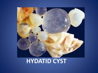
- 1. HYDATID CYST
- 2. DR.SYED UBAID M.S(KEM MUMBAI) FMAS LAPAROSCOPIC AND GENERAL SURGEON ASSOCIATE PROFESSOR OF SURGERY IIMSR,JALNA
- 3. HYDATID DISEASE Echinococcosis (hydatid disease) is a zoonosis caused by the larval stage of Echinococcus granulosus. Humans are accidental intermediate hosts, whereas animals can be both intermediate hosts and definitive hosts.
- 4. HYDATID DISEASE The two main types of hydatid disease are caused by E. granulosus and E. multilocularis. The former is commonly seen in the Mediterranean, South America, the Middle East, Australia, and New Zealand, and is the most common type of hydatid disease in humans. In humans, 50–75% of the cysts occur in the liver, 25% are located in the lungs, and 5–10% distribute along the arterial system. Infection with echinococcal organisms is the most common cause of liver cysts in the world.
- 6. Etiology The life cycle of E. granulosus has two hosts. The definitive host is usually a dog or some other carnivore. The adult worm of the parasite lives in the proximal small bowel of the definitive host attached by hooklets to the mucosa. Eggs are released into the host's intestine and excreted in the feces. Sheep are the most common intermediate host, and these animals ingest the ovum while grazing.
- 7. Etiology The ovum loses the protective chitinous layer and is digested in the duodenum. The released hexacanth embryo (oncosphere) passes through the intestinal wall into the portal circulation and develops into cysts within the liver. The definitive host eats the viscera of the intermediate host and the cycle is completed.
- 8. Etiology Humans may become intermediate hosts through contact with the definitive host (usually a dog) or by ingestion of contaminated water or vegetables. Once in the liver, cysts grow to 1 cm in the first 6 months and 2–3 cm annually thereafter, depending on host tissue resistance. Once the parasite passes through the intestinal wall into the portal venous or lymphatic system, the liver acts as the first line of defense, and thus is the most frequently involved organ. The right lobe of the liver is the most commonly involved.
- 9. Life Cycle of E granulosus
- 10. Incidence Hydatid liver disease affects all age groups, both sexes equally, and no predisposing pathologic conditions are associated with infection. Washing hands after contact with canines, eliminating the consumption of vegetables grown at ground level from the diet, and stopping the practice of feeding entrails of slaughtered animals to dogs have all aided in decreasing the incidence of the disease.
- 11. Pathology
- 12. Pathology • Hydatid liver cysts tend to expand slowly and without symptoms and are thus frequently very large on presentation. Single lesions are noted in 75% and are predominantly located within the right lobe (80%). Even though the lesion is single, half contain daughter cysts and are multilocular.
- 13. Pathology • The typical hydatid cyst has a three-layer wall surrounding a fluid cavity. The outer layer is the pericyst, a thin, indistinct fibrous tissue layer representing an adventitial reaction to the parasitic infection. The pericyst acts as a mechanical support for the hydatid cyst and is the metabolic interface between the host and the parasite. As the cyst grows, bile ducts and blood vessels stretch and become incorporated within this structure, which explains the biliary and hemorrhagic complications of cyst growth and difficulties with resection. Over time, the pericyst calcifies.
- 14. Hydatid Cyst Structure • The hydatid cyst has three layers: (a) the outer pericyst, composed of modified host cells that form a dense and fibrous protective zone; (b) the middle laminated membrane, which is acellular and allows the passage of nutrients; (c) the inner germinal layer, where the scolices (the larval stage of the parasite) and the laminated membrane are produced.
- 15. • Daughter vesicles (brood capsules) are small spheres that contain the protoscolices and are formed from rests of the germinal layer. Before becoming daughter cysts, these daughter vesicles are attached by a pedicle to the germinal layer of the mother cyst. At gross examination, the vesicles resemble a bunch of grapes.
- 16. Cyst layers and contents
- 18. Clinical Presentation • The clinical features of hydatid liver disease depend on the site, size, stage of development, whether the cyst is alive or dead, and whether the cyst is infected or not.Pain in the RUQ or epigastrium is the most common symptom, whereas hepatomegaly and a palpable mass are the most common signs.
- 19. Clinical Presentation SYMPTOMS Percentage Asymptomatic 75% Abdominal pain 20% Dyspepsia 13 % Fever and chills 8 % Jaundice 6%
- 20. Clinical Presentation • SIGNS Right upper quadrant mass 70% Right upper quadrant tenderness 20% LABORATORY DATA Eosinophilia 35% Bilirubin >2 mg/Dl 20% WBC count <10,000/mm3 10%
- 21. Casoni or intradermal test oIndirect hemagglutination test and enzyme-linked immunosorbent assay are the most widely used methods for detection of anti-Echinococcus antibodies (immunoglobulin G [IgG]).These tests give false positive results in cases of schistosomiasis and nematode infestations that is why they are not specific for diagnosing hydatidosis.
- 22. • Immunoelectrophoresis : depends on the formation of specific arc of precipitation ( called arc 5 ) which is highly specific and can be used to exclude cross-reactions caused by noncestode parasites
- 23. Radiology • Chest radiographs may show an elevated diaphragm and concentric calcifications in the cyst wall, but are of limited value. • Classic findings of hydatid cysts are calcified thick walls, often with daughter cysts.
- 24. X RAY
- 26. Radiology • Ultrasound and CT are considered the first choice for imaging. Ultrasound defines the internal structure, number, and location of the cysts and the presence of complications. The specificity of ultrasound in hydatid disease is around 90%. • The classification Type I Pure fluid collection. Type II Fluid collection with a split wall (floating membrane). Type III Fluid collection with septa (honeycomb image). Type IV Heterogenous echographic patterns Type V Reflecting thick walls.
- 29. Computed tomography Computed tomography gives similar information to ultrasound, but more specific information about the location and depth of the cyst within the liver. Daughter cysts and exogenous cysts are also clearly visualized, and the volume of the cyst can be estimated. CT is imperative for operative management, especially when a laparoscopic approach is utilized.
- 33. MRI &ERCP • MRI provides structural details of the hydatid cyst, but adds little more than ultrasound or CT, and is more expensive. Endoscopic retrograde cholangiopancreatography (ERCP) may show communication between the cysts and bile ducts and can be used to drain the biliary tree before surgery.
- 36. Treatment • Most echinococcal cysts are asymptomatic on presentation, but complications such as pulmonary infection, cholangitis, rupture, and anaphylaxis give good reason to consider treatment for all. • Medical, surgical, and percutaneous approaches may be part of the treatment armamentarium. • Small cysts (<4 cm) located deep in the parenchyma of the liver, if uncomplicated, can be managed conservatively. Basic principles of treatment ar (1) eradication of the parasite within the cyst, (2) protection of the host against spillage of scoleces, and (3) management of complications.
- 37. Anthelmintics • Medical therapy for echinococcosis is limited to the benzimidazoles (mebendazole and albendazole) and used alone is only 30% successful. • Albendazole is readily absorbed from the intestine and metabolized by the liver to an active form. • Mebendazole is poorly absorbed and is inactivated by the liver.
- 38. Anthelmintics Albendazole is thus the drug of choice for medical therapy. Given for at least 3 months preoperatively, albendazole reduces the recurrence rate when cyst spillage, partial cyst removal, or biliary rupture has occurred.Duration of therapy in these instances is at least 1 month Postoperatively. Albendazole is administered in several 1-month oral doses (10-15 mg/kg/d) separated by 14-day intervals.
- 39. The PAIR technique (percutaneous aspiration, injection and re-aspiration) The most frequently utilized protoscolecidal agents 1. 15–20% saline, 2. 95% ethanol, 3. a combination of 30% saline and 95% ethanol, and 4. mebendazole solution. Complications o Spillage, o anaphylaxis, and o recurrence
- 41. Surgery • Surgery is still the treatment of choice for uncomplicated hydatid disease of the liver. • The objectives of surgical treatment are to: (1) inactivate the scoleces, (2) prevent spillage of cyst contents, (3) eliminate all viable elements of the cyst, and (4) manage the residual cavity of the cyst.
- 42. Scolecidal Agents • Early on, surgical management of hydatid cysts via cyst evacuation resulted in a high rate of peritoneal implantation. This problem prompted the use of scolecidal agents for injection into the cyst and for use in the surrounding peritoneum. Formalin, hypertonic saline, cetrimide, hydrogen peroxide, polyvinylpurrolidone-iodine, silver nitrate, and ethyl alcohol are among some of the many agents that have been used.
- 47. OMENTOPLASTY
- 48. MARSUPILIZATION
- 49. CAPETONAGE
- 50. PERICYSTECTOMY
- 51. HEPATECTOMTY
- 52. LAPAROSCOPY
- 53. Complications • Ruptures • Secondary infection, • Anaphylactic shock, and • liver replacement, in order of decreasing frequency.
- 54. THANK YOU