Blotting techniques
•Descargar como PPTX, PDF•
84 recomendaciones•22,354 vistas

1. The document discusses various blotting techniques including Southern blotting, Northern blotting, and Western blotting. It provides a brief history and overview of each technique. 2. Southern blotting is used to detect DNA, Northern blotting detects RNA, and Western blotting detects proteins. The techniques involve separating biomolecules by electrophoresis, transferring them to a membrane, then using a probe for detection. 3. The document outlines the basic methodology for each type of blotting. This includes separation, transfer to a membrane, blocking, hybridization with a probe, washing, and detection. Each technique allows for the analysis of biomolecules and has various applications in research.

Recomendados
Más contenido relacionado
La actualidad más candente
La actualidad más candente (20)
Similar a Blotting techniques
Similar a Blotting techniques (20)
Último
Último (20)
Blotting techniques
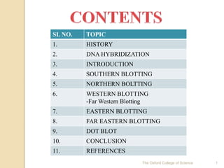
- 1. 1The Oxford College of Science SL NO. TOPIC 1. HISTORY 2. DNA HYBRIDIZATION 3. INTRODUCTION 4. SOUTHERN BLOTTING 5. NORTHERN BOLTTING 6. WESTERN BLOTTING -Far Western Blotting 7. EASTERN BLOTTING 8. FAR EASTERN BLOTTING 9. DOT BLOT 10. CONCLUSION 11. REFERENCES
- 2. HISTORY 2The Oxford College of Science
- 3. 1963-Nygaard and Hall: Shown that single stranded DNA can be immobilized on nitrocellulose filters. 1966 &1965-Denhardt and Spiegelman: Nucleic acid thus fixed can be detected with exquisite sensitivity by hybridization to radiolabelled probes. 1970s: Possibility of mapping whole genomes arose along with the era of rDNA and gene cloning. Thus need to find a single gene among thousands of fragments of DNA was needed. 1975-Edward Southern: Powerful DNA transfer and probing techniques(Southern Blotting). 1977-George Stark & colleagues: Detection of RNA (Northern Blotting) 1979-Stark developed early protein blotting. Harry Towbin gave faster and simpler approach. W. Neal Burnette’s (1981) technique named Western Blotting. 3The Oxford College of Science
- 4. 4The Oxford College of Science
- 5. 5The Oxford College of Science
- 6. Formation of stable base pairs between probe and target DNA. PREPARE TARGET DNA PREPARE PROBE DNA HYBRIDISATION Probe (labelled) Target Sequence Hybrid 6The Oxford College of Science
- 7. 7The Oxford College of Science
- 8. Blotting is a method of putting DNA, RNA or Proteins onto a membrane for further studies and detection. Southern Blotting: DNA is detected with a hybridization DNA or RNA probe. Northern Blotting: RNA is detected with a hybridization DNA or RNA probe. Western Blotting: Protein is detected with a complementary antibody. The three blotting techniques have similar methodology. Molecules separated by electrophoretic procedures are transferred to membranes that is specially suited to support the detection of fragments with a particular DNA sequence, single species of RNA or proteins. 8The Oxford College of Science
- 9. 9The Oxford College of Science
- 10. The first type of blotting to be discovered. Plays significant role in Recombinant DNA Technology as well as Molecular Biology. Used to detect target DNA in a sample. Sample Target DNA 10The Oxford College of Science
- 11. FLOW CHART:- Preparation of sample & running the agarose gel. Southern Transfer. Probe preparation. Prehybridization. Hybridization. Post Hybridization Washing. Signal Detection. Isotope Non-isotope 11The Oxford College of Science
- 12. Schematic Diagram. 12 The Oxford College of Science
- 13. Gel Electrophoresis (Size Separation): Size based separation in an electric field Sample Preparation Isolation of DNA by common extraction protocol Purified DNA partially digested by restriction endonuclease Linearized DNA loaded. Aragose Electrophoresis •Pretreatment of gel with -Concentrated HCl -Alkaline solution • DNA is negatively charged thus moves from cathode to anode. •Sorter fragments move faster. •Agarose 0.5% to 2%. •Voltage of about 100mV •Buffers used are TAE and Sodium Borate •P32 labeled marker as ladder 13The Oxford College of Science
- 14. Step II Southern Transfer: Transfer DNA from gel to solid support. SOLID SUPPORT Nitrocellulose Membrane -Nucleic acids more than 400 bases are inefficiently bound. - Attachment by hydrophobic interactions. -Become brittle while baking in vacuum. - Care required for storing. Nylon -Buffers of low ionic strength can be used. - Transfer can be carried out electrophoretically. -Two types a)Neutral b)Positively charged (amines). 14The Oxford College of Science
- 15. NITROCELLULOSE MEMBRANE NYLON MEMBRANE Hydrophobic binding. Covalent binding. Fragile Durable >200-300 bp probe length <200-300 bp probe can be used Lower background noise Higher. Cannot be exposed to basic solution Can be exposed. Not easily reprobed Can be easily reprobed several times. 15The Oxford College of Science
- 16. Transfer of electrophoretically separated DNA from gel to a 2D support is the key step. 1.Upward Capillary Action. Rate of transfer depends on size of DNA and concentration of gel. Gel dehydrates eventually. DEPURINATION -dilute HCl and then strong base. 16The Oxford College of Science
- 17. 2. Downward Capillary Action. •Alkaline buffer (NaOH) •Nylon membrane (generally charged) •Rapid •More efficient 3.Simultaneous Transfer To Two Membranes. •Target DNA fragments higher in concentration. •Transfer buffer is just the liquid trapped in gel •Efficiency of transfer poor. •Used for plasmids, bacteriophages, cosmids and genome of simple organisms 17The Oxford College of Science (Saccharomyces cerevisiae & Drosophilla) •Not sensitive enough to analyze complex mammalian genome.
- 18. 4. Electrophoretic Transfer. •Charged nylon membrane •Analysis of small fragments of DNA(~50kb) separated through polyacrylamide gels •Gel neutralised •1X TBE buffer used. 5.Vacuum Transfer. •Rapid •Gel is placed in contact with membrane supported on a porous screen over a vacuum chamber. •Buffer drawn from an upper reservoir, elutes nucleic acids from the gel & deposits them on the membrane. 18The Oxford College of Science
- 19. Step III Blocking (Prehybridization) After the southern transfer the membrane is generally baked at 80ºC for 2 hours or UV (in nylon) for permanent attachment. The membrane has high affinity for proteins and nucleic acids. Thus non specific binding between probes and the material has to be prevented by blocking. This is done by soaking the membrane in a solution containing high concentrations of DNA (example herring or salmon sperm DNA) or ficoll. 19The Oxford College of Science
- 20. Step IV Hybridization Single stranded DNA to be detected forms hybrid with complementary single stranded probe that can be detected either by radioactivity (P32) or by chemiluminiscence (digoxigenin). Quantitative analysis: -Darker band: complete hybridization -Lighter band: Incomplete hybridization Stringency is determined by the hybridization temperature and salt concentration in buffer. Under gentle agitation, with a small amount of detergent hybridization is allowed for hours(in a closed bag at about 68ºC). 20The Oxford College of Science
- 21. Step V Step VI Washing Detection Membrane is rinsed several times with different buffers to remove any unbound nucleic acid probe, in order to avoid unspecific background signals. •If probe is radioactive, visualization is done on x-ray film by autoradiography. •Membrane is pressed against the film, which in turn is exposed, for a few minutes to weeks. •Non-radioactive methods are safer, quicker & cheaper. 21The Oxford College of Science
- 22. 1. Detection of an RFLP by Southern Blotting. 22The Oxford College of Science
- 23. 2.Detection of DNA defect & DNA segment (Gene Defect) Eg-Detection of the sickle cell globin gene TYPE OF Hb AMINO ACID SEQUENCE NUCLEOTIDE SEQUENCE A -Pro-Glu-Glu- -CCT-GAG-GAG- S -Pro-Val-Glu- -CCT-GTG-GAG- Using restriction enzymes we can prove that they are different types of genes. •Normal DNA- 2 segments •Sickle cell- 1 23The Oxford College of Science
- 24. 24The Oxford College of Science
- 25. 3. Used for homology based cloning on the basis of amino acid sequence of protein product of the gene. Oligonucleotides are designed similar to target sequence. Chemically synthesized, radio labeled and used to screen a DNA library. Sequences that hybridize with the hybridization probe are further analyzed. 4. To identify methylated sites in a particular gene. Msp I and Hpa II recognize and cleave the same sequence. Msp I DNA unmethylated Hpa II C has to methylated 5. To isolate specific DNA in a DNA sample (for RDT). 6. Used in phylogenetic analysis. 7. Diagnosis of infectious diseases. 25The Oxford College of Science
- 26. 8. In DNA fingerprinting (Paternity and maternity testing, Criminal identification and Forensics and personal identification). 9. To follow inheritence of certain selected genes. 10. To know restriction sites for restriction endonucleases. 26The Oxford College of Science
- 27. 1) Laborious 2) Time consuming 3) More amount of DNA required 4) 3µl DNA per sample is costly. 27The Oxford College of Science
- 28. 28The Oxford College of Science
- 29. DETECTION OF RNA USING SS PROBE HYBRIDIZATION TECHNIQUE •Most of the procedure remains same as Southern Blotting but the only problem that arises is that the single stranded RNA tend to re-anneal themselves in a 2º & 3º structures. •These structures are resolved using formaldehyde or formalin. •No alkali treatment required. •Using this technique, we can come to know about the presence of our transcripts & expression of gene in DNA, along with looking for RNA splicing, maturation and degradation. 29The Oxford College of Science
- 30. FLOW CHART:- Preparation of RNA sample & running the agarose gel. Northern Transfer. Probe preparation. Prehybridization. Hybridization. Post Hybridization Washing. Signal Detection. Isotope Non-isotope 30The Oxford College of Science
- 31. Sample Preparation: Pretreated with formaldehyde to prevent the formation of base paired secondary structure. NOTE • Care should be taken as RNA is less stable • No solution should contain RNAse • DNAse should be added 31The Oxford College of Science
- 32. PROBLEM •Amount of sample to be loaded may vary as there are different types of RNAs •If mRNA is abundant, we can take the whole RNA in count Or else we take poly(A) content as mRNA contains poly(A) tails. •This should be followed or the band may vary. 32The Oxford College of Science
- 33. 33The Oxford College of Science •Electroblot- reliable and efficient. •Hybridization 60ºC for less than 2 hours. •EDC cross linking (UV cross linking)
- 34. 34The Oxford College of Science APPLICATIONS 1)To understand gene expression at the level of mRNA. 2)mRNA transcript size 3)RNA degradation 4)RNA splicing 5)RNA half life 6)Used to confirm or check transgenic or knockout mice.
- 35. The Oxford College of Science 35 LIMITATIONS 1) Standard Northern Blotting is relatively ineffective than nuclease protection assay or RT-PCR (Reverse Transcription). 2) Detection with multiple probes become a problem 3) RNA is fragile and can be easily degraded by RNAse. Thus the quality of data & quantification of expression quite negatively affected.
- 36. 36The Oxford College of Science
- 37. 37The Oxford College of Science •Used for the detection of proteins. •Also called Immunoblotting •4-5 days to get complete result. •Two types -Direct: Single antibody -Indirect: 1º and 2º antibody used (in practice) . Steps: Sample Preparation SDS-PAGE Blocking Probing Washing Detection
- 38. The Oxford College of Science 38 1. Sample preparation: Cells lysed in extraction buffer containing proteinase inhibitors Samples cooled or frozen & homogenized using mechanical force. Centrifugation employed for protein purification. Samples have boiled for one to five minutes in a denaturing buffer (eg. Laemmli’s buffer)
- 39. The Oxford College of Science 39 2. SDS-PAGE Separation of proteins by electrophoresis. 3.Electrotransfer Transfer of proteins to PVDF (Polyvinyldienefluoride) membrane. 4.Blocking • 5% nonfat dry milk or 3% BSA. • Presence of detergent (Tween 20) at 0.05% is also very important.
- 40. The Oxford College of Science 40 5. Probe • Primary (rabbit anti human β actin) and Secondary antibody (HRP-conjugated anti rabbit) employed • Monoclonal antibody • Primary antibody concentration 0.5-5µg/ml 6. Enzyme Luminol is degraded by secondary antibody HRP to give luminescence(425 nm) 7. Storing Washed and exposed to x-ray film so as to study later
- 41. 41The Oxford College of Science
- 42. 42The Oxford College of Science APPLICATIONS 1) Verify the presence of a protein. 2) Find the relative amount of a protein in different samples. 3) Analyze protein-protein interactions.
- 43. The Oxford College of Science 43 1) Highly sensitive. As little as 1.5ng of an average sized protein can be detected. 2) Comparatively quick methodology. 3) Quantification can be done by densitometries and integrating areas under the peak.
- 44. The Oxford College of Science 44 LIMITATIONS 1) Many steps where errors may occur. 2) Significant amount of sample needed. 3) Accurate quantification is vey difficult. 4) Time consuming protocol. 5) Tertiary structure destroyed and therefore relevant epitope recognized by primary antibody may not be understood.
- 45. The Oxford College of Science 45 •Detects protein-protein interactions in vitro. • Proteins in a cell lysate containing prey proteins are firstly separated by SDS -PAGE. Transferred to a membrane (as in a standard WB) Proteins on the membrane are then denatured and renatured. Membrane is then blocked and probed, usually with purified bait protein(s). The bait proteins are detected as spots in the membrane where a prey protein is located. (As bait proteins and the prey protein form a complex)
- 46. The Oxford College of Science 46 •Compared with other biochemical binding assays, Far WB allows prey proteins to be endogenously expressed without purification. •Application includes: 1) receptor-ligand interactions study 2) screen libraries for interacting proteins and 3) identify protein-protein interactions without using antigen- specific antibodies. •Typically, 2-3 days are required to carry out the experiment.
- 47. 47The Oxford College of Science
- 48. 48The Oxford College of Science •Used to analyze protein post translational modifications (PTM) such as lipids, phosphomoieties and glycoconjugates. It is most often used to detect carbohydrate epitopes. • Considered an extension of the of Western blotting. •Multiple techniques have been described by the term Eastern blotting, most use proteins blotted from SDS-PAGE gel on to a PVDF or nitrocellulose membrane. •Transferred proteins are analyzed for post-translational modifications using probes that may detect lipids, carbohydrate, phosphorylation or any other protein modification.
- 49. 49The Oxford College of Science APPLICATIONS Detection of protein modifications in two bacterial species Ehrlichia- E. muris and IOE. Cholera toxin B subunit (which binds to gangliosides), Concanavalin A(which detects mannose- containing glycans) and nitrophospho molybdate-methyl green (which detects phosphoproteins) were used to detect protein modifications. The technique showed that the antigenic proteins of the non-virulent E.muris is more post-translationally modified than the highly virulent IOE
- 50. 50The Oxford College of Science Post-translational modifications occurring at the N- terminus of the amino acid chain play an important role in translocation across biological membranes. These include secretory proteins in prokaryotes and eukaryotes and also proteins that are intended to be incorporated in various cellular and organelle membranes such as lysosomes, chloroplast, mitochondria and plasma membrane. Expression of post translated proteins is important in several diseases.
- 51. The Oxford College of Science 51 Developed in 1994 by Taki and colleagues at the Tokyo Medical and Dental University, Japan for the analysis of lipids separated by high-performance thin layer chromatography (HPTLC). The lipids are transferred from the HPTLC plate to a PVDF membrane for further analysis, for example by enzymatic or ligand binding assays or mass spectrometry in minutes. Name in dual reference to Southern blot & Japan.
- 52. The Oxford College of Science 52 •Purification of glycosphingolipids and phospholipids. •Structural analysis of lipids in conjunction with direct mass spectrometry. •Binding study using various ligands such as antibodies, lectins, bacterium, viruses, and toxins. •Enzyme reaction on membranes.
- 53. The Oxford College of Science 53 •Simplification of blotting techniques. • In a dot blot the biomolecules to be detected are not first separated by electrophoresis. Instead, a mixture containing the molecule to be detected is applied directly on a membrane as a dot, and then is spotted through circular templates directly onto the membrane or paper substrate. • It offers no information on the size of the target biomolecule. Furthermore, if two molecules of different sizes are detected, they will still appear as a single dot. •Can only confirm the presence or absence of a biomolecule.
- 54. 54The Oxford College of Science
- 55. 55The Oxford College of Science • Blotting is a powerful and sensitive technique for identifying the presence of specific biomolecules within a sample. •The first of these techniques developed was the Southern blot to detect specific DNA sequences by Dr. Edwin Southern, 1975. •Subsequently, the method was modified to detect other targets. >Northern blot (for detection of RNA) >Western blot (for detection of protein) >Far western blot (Protein-protein interaction) >Eastern blot (for detection of post translationally modified proteins) >Far eastern blot (lipid analysis) >Southwestern blot (for detection of DNA binding proteins)
- 56. The Oxford College of Science 56 • Blotting techniques have been widely employed for more than 30 years and have provided the foundation of our understanding of molecular biology. • However, these techniques have been largely—and in some cases completely—usurped by new technologies . • Southern blots have been replaced by multiple techniques. 1. Real-time PCR boasts incredible sensitivity; theoretically, this method is able to detect even a single copy of the target sequence and compare relative copy numbers across samples rapidly and reliably, with little technical expertise required. 2. Fluorescent in situ hybridization (FISH) allows detection of specific sequences within a tissue sample with high sensitivity and precise localization. • Northern blots have given way to reverse-transcription PCR, again a more sensitive and more user-friendly technique
- 57. 57The Oxford College of Science BOOKS 1. Molecular cloning, A laboratory Manual; Volume I; Third Edition; Sambrook and Russel; Cold Spring Harbour Laboratory Press, New York; Pages 6.33-6.58 and 7.42-7.46 WEBSITES 1. file:///F:/blotting/for%20conclusion.htm 2. http://en.wikipedia.org/wiki/Dot_blot 3. http://en.wikipedia.org/wiki/Far-Eastern_blotting 4. http://en.wikipedia.org/wiki/Eastern_blot#cite_note-thomas-14 5. http://www.ncbi.nlm.nih.gov/pubmed/18079728 6. http://books.google.co.in/books?id=ZDgh_TaLzNEC&pg=PA48&lpg=PA48&dq =blotting+techniques+history&source=bl&ots=BUsrvNI6_K&sig=I1NbWeJ5R Ux6SQ3LTo3k4aL- Kwg&hl=en&sa=X&ei=E4kAVL6zKIehugT39YL4BQ&ved=0CF4Q6AEwBw# v=onepage&q=blotting%20techniques%20history&f=false 7. http://en.wikipedia.org/wiki/Reverse_transcription_polymerase_chain_reaction# Application
- 58. 58The Oxford College of Science