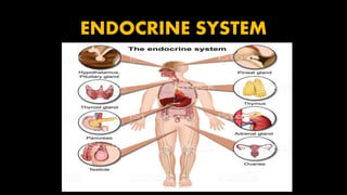
The document discusses the endocrine system and different types of glands. There are three types of glands: exocrine glands which secrete products into ducts like sweat and salivary glands; endocrine glands which secrete hormones directly into the bloodstream, such as the pituitary, thyroid, and adrenal glands; and heterocrine glands. The pituitary gland and hypothalamus work together to regulate growth, metabolism, and other bodily functions through the release of hormones. The pituitary has an anterior and posterior lobe; the anterior lobe secretes hormones that target various organs and tissues while the posterior lobe stores and releases oxytocin and vasopressin which are produced in the hypothalamus
2. GLANDS:
An organ which secretes particular
chemical substances for use in the
body or for discharge into the
surroundings.
There are three types of
glands in our body:
Endocrine glands
Exocrine glands
Heterocrine glands
3. EXOCRINE GLANDS
Exocrine glands are glands that secrete their products
into ducts
EXAMPLE:
Sweat glands
Salivary glands
Mammary glands
Digestive glands
4. ENDOCRINE GLANDS
Glands that secrete their product (hormones) directly into the
blood rather than through a duct
EXAMPLE:
Pituitary gland
Thyroid gland
Parathyroid gland
Adrenal glands
Pineal gland
5. • in addition several organs and tissues are not endocrine glands exclusively but
contain cells that secrete hormones include
• Hypothalamus
• Thymus
• Pancreas
• Ovaries
• Testes
• Kidneys
• Stomach
• Liver
• Small intestine
• Skin
• Heart
• Adipose tissue
• placenta
6. Pituitary gland & Hypothalamus
• Pituitary gland or Hypophysis was called the master endocrine gland.
• Pituitary gland itself has a master – Hypothalamus
• Hypothalamus is the major link between the nervous and endocrine
systems.
• Cells in the hypothalamus synthesize at least nine different
hormones.
• Pituitary gland secretes seven
• These two combine together play important roles in the regulation of
virtually all aspects of growth, developmet, metabolism and
homeostasis
7. • The pituitary gland is a pea shaped structure measuring 1- 1.5cm in diameter
• The pituitary gland or hypophysis lies in the hypophyseal fossa of the sella turcica
of the sphenoid bone.
• It attaches to the hypothalamus by a stalk, the infundibulum.
• It has anatomically and physiologically separate portions
Anterior pituitary
Posterior pituitary
• The anterior pituitary lobe also called the adenohypophysis, accounts for about
75% of the total weight of the gland and is composed of epithelial tissue.
• The anterior pituitary consists of two parts in an adult;
• Pars distalis- is the larger portion
• Pars tuberalis – forms a sheath around the infundibulum
8.
9. • Posterior pituitary lobe also called the neurohypophysis, is composed of neural
tissue
• It also consists of 2 parts
oPars nervosa – the larger bulbar portion
o infundibulum.
o a third region of the pituitary gland is the pars intermedia atrophies during
human fetal development and ceases to exit as a separate lobe in adults.
10. Anterior pituitary
• The anterior pituitary or adenohypophysis secretes hormones that regulate a
wide range of bodily activities from growth to reproduction.
• Release of anterior pituitary hormones is stimulated by releasing hormones and
suppressed by inhibiting hormones by hypothalamus
11. Hypophyseal Portal system
• Hypothalamic hormones reach the anterior pituitary through a portal
system.
• In portal system blood flows from
one capillary network
Portal vein
Second capillary network
12. • The superior hypophyseal arteries, the branch of the internal carotid arteries
bring blood into the hypothalamus.
• At the junction of the median eminence of the hypothalamus and the
infundibulum, these arteries divide into a capillary network called the primary
plexus of the hypophyseal portal system.
• From the primary plexus the blood flows into the hypophyseal portal vien that
passes down the outside of the infundibulum.
• In the anterior pituitary, the hypophyseal portal veins divide again and form
another capillary network called the secondary plexus of the hypophyseal portal
system
13. • Above the optic chiasm are clusters of specialized neurons called neurosecretory
cells
• They synthesize the hypothalamic releasing and inhibiting hormones in their cell
bodies and package the hormones inside vesicles, which reach the axon terminals
by axon transport.
• Nerve impulses stimulate the vesicles to undergo exocytosis.
• The hormones diffuse into the primary plexus of the hypophyseal portal system
• Hypothalamic hormones flow with the blood through the portal vein into the
secondary plexus.
• Hypothalamic hormones act on anterior pituitary cells
• hormones secreted by anterior pituitary cells pass into the secondary plexus
capillaries, which drain into the hypophyseal veins and out into general
circulation.
• Anterior pituitary hormones travel to target tissues throughout the body.
14.
15.
16. Types of anterior pituitary cells and their
Hormones
Types of
anterior
pituitary cells:
Somatotrophs Thyrotrophs Gonadotrophs Lactotrophs corticotrophs
17. Somatotrophs:
• Secrete hGH also known as somatotropin
• Human growth hormone in turn stimulates several tissues to secrete insulin like
growth factors , hormones that regulate general body growth and regulate
aspects of metabolism
Thyrotrophs:
• Secrete TSH also known as thyrotropin
• TSH controls the secretions and other activities of the thyroid gland
Gonadotrophs:
• Secrete 2 gonadotropins; FSH (follicle stimulating hormone), LH (luteinizing
hormone)
• They stimulates the secretion of estrogen and progesterone and the maturation
of oocytes in the ovaries
• they stimulate sperm production and secretion of testosterone in the testes
18. Lactotrophs:
• Secrete prolactin (PRL), which initiates milk production in the
mammary glands.
• Corticotrophs : secrete adrenocorticotropic hormone, (ACTH), also
known as corticotropin
• stimulates cortex to secrete glucocorticoids such as cortisol
• Some corticotrophs remnants of the pars intermedia, also secrete
Melnocyte- stimulating hormone(MSH)
19.
20. Posterior Pituitary
• Posterior pituitary or neurohypophysis does not synthesize hormones, it does
and store 2 hormones
• It consists of axons and axon terminals of more than 10,000 hypothalamic
neurosecretory cells.
• The cell bodies of the neurosecretory cells are in the paraventricular and
supraoptic nuclei of the hypothalamus
• Their axons form hypothalamohypophyseal tract.
• This tract begins in the hypothalamus and ends near blood capillaries in the
posterior pituitary
• The neuronal cell bodies in the supraoptic nucleus produce antidiuretic hormone
also called vasopressin
21.
22. • The axon terminals in the posterior pituitary are associated with specialized
neuroglia called pituicytes
• After their production the cell bodies of neurosceretory cells oxytocin and
antidiuretic hormone are packaged into secretory vesicles, which moves by fast
axonal transport to the axon terminals in the in the posterior pituitary
• Where they are stored until nerve impulses trigger exocytosis and release of the
hormones
• Blood is supplied to the posterior pituitary by the inferior hypophyseal arteries,
which branch from the internal carotid arteries.
• In the posterior pituitary, the inferior hypophyseal arteries drain into the
capillary plexus of the infundibular process
• A capillary network that receives secreted oxytocin and ADH
• From this plexus, hormones pass into the posterior hypophyseal veins for
distribution to target cells in other tissues.