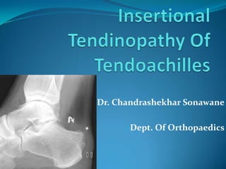
Insertional tendinopathy of tendoachilles
•Descargar como PPTX, PDF•
17 recomendaciones•7,020 vistas


Recomendados
Más contenido relacionado
La actualidad más candente
La actualidad más candente (20)
Destacado
Destacado (20)
Similar a Insertional tendinopathy of tendoachilles
Similar a Insertional tendinopathy of tendoachilles (20)
Más de Mahatma Gandhi Hospital Parel Mumbai
Más de Mahatma Gandhi Hospital Parel Mumbai (11)
Último
Último (20)
Insertional tendinopathy of tendoachilles
- 1. Dr. Chandrashekhar Sonawane -Dept. Of Orthopaedics
- 2. 47 years old male with left heel pain since 15 days pain more in the morning, aggravated by weight bearing, relieved with medications and rest. no H/O trauma, swelling, fever, any other joint pain, no H/O DM , koch’s
- 3. O/E - tenderness over posterior part of left heel and painful dorsiflexion of foot
- 5. conservative treatment anti-inflammatory drugs along with heel support No improvement after 6 months Surgery haglund bump excision by a central tendon splitting method
- 7. Total no. of cases in last year - 24 Conservative management - 22 Oprative management - 02
- 9. The largest and strongest tendon in the human body Formed from the tendninous contributions of the gastrocnemius and soleus muscles The tendons converge appr. 15 cm proximal to the insertion at the posterior calcaneus
- 10. The right Achilles tendon appears to spiral counterclockwise 30‐150º toward its insertion at the calcaneus The spiraling allows for elongation and elastic recoil within the tendon, facilitating storage and release of energy during movement
- 12. posterior tibial artery and its contributions to the musculotendinous junction, as well as vessels which cross the paratenon. The watershed zone is an area 2‐6 cm proximal to the calcaneus, in which the blood supply is less abundant and becomes even sparser with age
- 13. The osteotendinous junction of the Achilles tendon is made up of 1) Bone, 2) Fibrocartilage, and 3) Tendon.
- 14. -Complex interlocking between calcified fibrocartilage and bone at the insertion site -Interlocking is of fundamental importance in anchoring the tendon to the bone.
- 15. The triad of Pain, Swelling (diffuse or localized), and Impaired Performance constitutes tendinopathy.
- 16. Clain and Baxter classified Achilles tendon disorders into Noninsertional and Insertional Tendinopathy in 1992
- 17. The exact incidence is unclear. Often diagnosed in older, less athletic, and overweight individuals as well as in older athletes , those wearing improper footwear
- 20. Obesity
- 21. Older athletes
- 23. Insertional tendinopathy could be considered an overuse injury, but with predisposition caused by preexisting weakening of the tendon.
- 24. Repetitive Traction Forces flat foot, pes cavus, obesity, overuse, poor training Degeneration , attrition , mechanical and chemical irritation chronic inflammatory response spur formation and calcification
- 25. Edema, mucoid degeneration, disruption of collagen bundles, necrosis, small hemorrhages, and calcification are noted Also, areas with proliferating blood vessels with lymphocytes and histiocytes suggesting a reparative process
- 26. -Increased activity of NADP-diaphorase, LDH, β- glucuronidase, and alkaline phosphatase. -Submicroscopic calcification and fibrillar degeneration. -Increased levels of type II and III collagen and decreased levels of type I collagen
- 27. Early morning stiffness, Pain that deteriorates after exercise Thickening or nodularity at the insertion. Range of motion of the ankle may or may not be limited
- 29. Insertional tendinopathy of the Achilles tendon seems to present more often as a triad rather than as a solitary pathology.
- 30. Insertional tendinopathy of the Achilles tendon, Retrocalcaneal bursitis, Haglund’s deformity, the prominent posterosuperior calcaneal process
- 31. HAGLUND’S DEFORMITY ( PUMP/ HUMP DEFORMITY) Two bursae are appreciated in relation to distal attachment of the Achilles tendon . Retrocalcaneal bursae. Tendoachilles bursae
- 32. COMPONENTS OF HAGLUNDS DEFORMITY RETROCALCANEAL BURSITIS MARROW EDEMA IN THE CALCANEUM THICK ACHILLES TENDON WITH PARTIAL TEAR TENDOACHILLES BURSITIS
- 33. Systemic affections - Gout, Sarcoidosis, Systemic corticosteroids, Oral fluoroquinolones, Diffuse idiopathic skeletal hyperostosis, and Seronegative spondyloarthropathies
- 34. Haglund’s deformity, Retrocalcaneal bursitis, Os trigonum, Posterior talar process fracture, Flexor hallucis longus tendinopathy, Peroneal tendinopathy, Tibialis posterior tendinopathy, Osteochondral lesions of talus
- 35. -Blood investigations to rule out systemic conditions -(MRI scan and US scan) can help to confirm the diagnosis -Radiographs help identify ossification of insertion of the Achilles tendon or a spur (fishhook osteophyte) on the superior portion of the calcaneum
- 37. Radiopacities of the Achilles tendon were classified into three types by Morris et al.
- 38. Type I - Radiopacities at the Achilles insertion or superior pole of the calcaneus. Bony changes to the calcaneus are often seen in type I lesions. Insertional tendinopathy of Achilles tendon causes type I abnormality
- 39. Type II -Radiopacities are intratendinous and are Located 1–3 cm proximal to the Achilles insertion, and are separated from calcaneal surface
- 40. Type III. Radiopacities are located proximal to the insertion zone, upward to 12 cm above the insertion zone. Type III is subdivided into IIIA (partial tendon calcification) and IIIB (complete tendon calcification).
- 41. Classification based on ultrasonographic changes at the Achilles tendon insertion was introduced by Paavola et al.
- 42. Classification Insertional Changes No alteration No calcification. Homogeneous fiber structure in the insertional area. Mild abnormality Insertional calcification, length 10 mm or less and thickness less than 2 mm. Homogeneous fiber structure in the insertional area. Moderate Insertional calcification, length more than 10 mm abnormality and thickness less than 2 mm. Slight alterations in the echo structure of tendon in the insertional area. Severe abnormality Insertional calcification, length more than 10 mm or thickness more than 2 mm. Moderate to severe variety in the echo structure of tendon in the insertional area.
- 43. -Success rates of 85% to 95% have been reported with simple measures like rest, ice, modification of training, heel lift, and orthoses -stretching and strengthening exercises can also be effective. -Tendon loading stimulates collagen fiber repair and remodeling. Therefore, complete rest of the injured tendon is not advisable
- 44. Surgical options are considered after 3 to 6 months of conservative management -Debridement of the calcific or diseased portion, Excision of the retrocalcaneal bursa, and Resection of the Haglund’s deformity, if present.
- 45. Various surgical procedures have been described We prefer to reattach the Achilles tendon using bone anchors if one-third or more of the insertion is disinserted.
- 46. A midline posterior skin incision combined with a central tendon-splitting approach for debridement, retrocalcaneal bursectomy, and removal of the calcaneal bursal projection as described by McGarvey
- 54. Calcified areas being probed with needle
- 55. Haglund’s bump excised after splitting tendo achilles
- 56. First two weeks- Protected weight bearing along with leg elevation as much as possible
- 57. 2 weeks to 4 weeks- A synthetic anterior below-knee slab is applied, with the ankle in neutral and secured to the leg with three or four removable Velcro straps for 4 weeks
- 58. After 6 weeks- the anterior slab is removed. -Stationary cycling and swimming from 8th week -gentle training -Gradual progression to full sports activity at 20 to 24 weeks
- 59. Follow-up - Patients are reviewed at 3, 6, and 9 months from the operation, and at 6-month intervals thereafter.
- 60. Insertional tendinopathy of the Achilles tendon is a degenerative rather than an inflammatory lesion, though the accompanying bursitis may paint an inflammatory picture
- 61. Type I collagen contributes to the tensile strength in tendons, allowing them to resist force and tension and to stretch. Therefore, tendons with an increased type III and a reduced type I collagen content are less resistant to tensile stresses
- 62. The diagnosis is mainly clinical, and radiographs help in confirming the diagnosis as do ultrasound scan or MRI scan